www.cbpv.org.br/rbpv ISSN 0103-846X (Print) / ISSN 1984-2961 (Electronic)
Doi: http://dx.doi.org/10.1590/S1984-29612017030
Ehrlichia canis
and
Rickettsia
spp. in dogs from urban areas in
Paraiba state, northeastern Brazil
Ehrlichia canis
e
Rickettsia
spp. em cães de áreas urbanas da Paraíba, nordeste do Brasil
Tereza Emmanuelle de Farias Rotondano1*; Felipe da Silva Krawczak2; Werona de Oliveira Barbosa3; Jonas Moraes-Filho2; Fernanda Nieri Bastos2; Marcelo Bahia Labruna2; Sérgio Santos de Azevedo4;
Marcia Almeida de Melo4; Alzira Maria Paiva de Almeida5
1 Departamento de Ciências Veterinárias, Centro de Ciências Agrárias, Universidade Federal da Paraíba – UFPB, Areia, PB, Brasil 2 Departamento de Medicina Veterinária Preventiva e Saúde Animal, Faculdade de Medicina Veterinária e Zootecnia, Universidade de
São Paulo – USP, São Paulo, SP, Brasil
3 Departamento de Ciência Animal, Universidade Federal do Semi-Árido – UFERSA, Mossoró, RN, Brasil
4 Unidade Acadêmica de Medicina Veterinária, Centro de Saúde e Tecnologia Rural, Universidade Federal de Campina Grande –
UFCG, Patos, PB, Brasil
5 Centro de Pesquisas Aggeu Magalhães, Fundação Oswaldo Cruz – FIOCRUZ-PE, Recife, PE, Brasil
Received March 19, 2017 Accepted April 26, 2017
Abstract
The aims of our study was to identify Ehrlichia canis and antibodies against Rickettsia spp. belonging to the spotted fever group (SFG) in dogs sampled from Paraiba state, northeastern Brazil. Blood and serum samples collected by convenience from dogs in urban areas of five municipalities were analyzed by real-time PCR for the detection of E. canis
DNA and by immunofluorescence assay test (IFAT) for the identification of antibodies against Rickettsia rickettsii, R. felis, R. parkeri, R. amblyommii and R. rhipicephali antigens. E. canis DNA was detected in 8.9% (64/719) of the blood samples, whereas 5.63% (43/763) of the serum samples were positive for at least one of the Rickettsia antigens tested by IFAT. This study showed for the first time the occurrence of E. canis and suggested the circulation of SFG Rickettsia in dogs in the study region of Paraiba state, northeastern Brazil.
Keywords:Ehrlichia spp., Rickettsia spp., IFAT, PCR, Brazil.
Resumo
Os objetivos do nosso estudo foram identificar Ehrlichia canis e anticorpos contra Rickettsia spp. pertencentes ao Grupo da Febre Maculosa (GFM) em cães amostrados no estado da Paraíba, nordeste do Brasil. As amostras de sangue e soro, coletados por conveniência, de cães em áreas urbanas de cinco municípios foram analisadas por PCR em tempo real para a detecção de DNA de E. canis e pela Reação de Imunofluorescência Indireta (RIFI) para identificação de anticorpos contra Rickettsia rickettsii, R. felis, R. parkeri, R. amblyommii e R. rhipicephali. O DNA de E. canis foi detectado em 8,9% (64/719) das amostras de sangue, enquanto que 5,63% (43/763) das amostras de soro foram positivas para pelo menos um dos antígenos de Rickettsia testados por RIFI. Este estudo mostrou pela primeira vez a ocorrência de
E. canis e sugere a circulação de Rickettsia do GFM em cães na região em estudo do estado da Paraíba, Nordeste do Brasil.
Palavras-chave:Ehrlichia spp., Rickettsia spp., RIFI, PCR, Brasil.
*Corresponding author: Tereza Emmanuelle de Farias Rotondano. Departamento de Ciências Veterinárias, Centro de Ciências Agrárias, Universidade Federal da Paraíba – UFPB, Rodovia BR 079, Km 12, CEP 58397-000, Areia, PB, Brasil. e-mail: [email protected]
Introduction
Rickettsioses are vector-borne diseases caused by bacteria of the Rickettsiales order, which include two families of obligate intracellular pathogens, Rickettsiaceae and Anaplasmataceae (DUMLER et al., 2001). Rickettsial infections are widely distributed and are
transmitted by a variety of species of ticks. Thus, the epidemiology of several rickettsial infections is determined by the specific geographic distribution of the tick vector (DANTAS-TORRES, 2007; PAROLA et al., 2013).
E. canis strain and the occurrence of co-infections, especially with other arthropod-borne pathogens, such as Babesia vogeli and
Hepatozoon canis (VIEIRA et al., 2011). The diagnosis of CME
is based on the observation of the presence of morulae within leucocytes in blood smears, isolation of the bacterium in cell culture, immunofluorescence assay test (IFAT), and molecular detection (PCR) (ROTONDANO et al., 2012).
Brazilian spotted fever (BSF), caused by the bacterium Rickettsia rickettsii, a member of the Spotted Fever Group (SFG), has long been recognized in Brazil, mainly in the southern and southeastern regions (ANGERAMI et al., 2009; PAROLA et al., 2013). More recently, the occurrence of other human pathogenic Rickettsia
species such as R. felis, R. parkeri and Rickettsia spp. strain Atlantic rainforest has been increasingly reported in Brazil (HORTA et al., 2006; SILVEIRA et al., 2007; SPOLIDORIO et al., 2010). While R. felis was detected in Rhipicephalus sanguineus ticks in the state of Paraiba, northeastern Brazil (TANIKAWA et al., 2013), there was a recent report of a R. parkeri-like strain infecting Amblyomma nodosum ticks collected from birds in Paraíba (LUGARINI et al., 2015). The IFAT is the most common test and serological gold standard for the diagnosis of rickettsial infection in humans and animals (DANTAS-TORRES, 2007).
The knowledge of the occurrence of tick borne-agents among canine populations is crucial for early diagnosis, treatment and control of these diseases, preventing their spread to humans and other canine populations. This study aimed to investigate the occurrence of E. canis DNA and Rickettsia spp. antibodies in urban dogs in the state of Paraiba, northeastern Brazil.
Materials and Methods
Samples and collection area
Blood samples were obtained from 719 domiciled dogs for detection of E. canis DNA and sera samples from 763 dogs for the detection of anti-Rickettsia spp. antibodies (Table 1). The samples were collected by cephalic or jugular venipuncture into Vacutainer
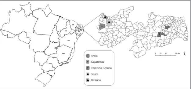
tubes containing sodium citrate. The animals were clinically healthy and were sampled by convenience of the owner residences at urban areas of the five municipalities in the state of Paraiba: Campina Grande (7°13’S, 35°52’W), Areia (6°57’S, 35°41’W), Uiraúna (6°57’S, 38°24’W), Cajazeiras (6°53’S, 38°33’W) and Sousa (6°45’S, 38°13’W) (Figure 1).
Detection of Ehrlichia canis DNA by real-time
PCR-DNA extraction and amplification
DNA was extracted from whole blood samples using a commercial DNA extraction kit (Wizard kit for DNA extraction, Promega)
(ROTONDANO et al., 2012). The detection of E. canis was performed by real-time PCR to amplify 378 base pairs (bp) of the dsb gene encoding a disulfide bond formation protein, using the primers Dsb321 and Dsb671 and a species-specific probe, as previously described (DOYLE et al., 2005). Positive and negative DNA samples were extracted from E. canis cultured in DH82 cells and uninfected cultures of DH82 cells, respectively. Controls were included for all PCR assays.
Serological tests to detect antibody against Rickettsia
spp.
Canine sera were tested by immunofluorescence assay test (IFAT) using crude antigens derived from five Brazilian Rickettsia isolates (R. rickettsii strain Taiaçu, R. parkeri strain At24, R. amblyommii
strain Ac37, R. rhipicephali strain HJ5, and R. felis strain Pedreira) as previously described (LABRUNA et al., 2007).
Briefly, serum samples were serially diluted in phosphate-buffered saline (PBS) in two fold dilutions from 1/64 up to 1/1280, and instilled on glass slides coated with the antigens. A commercial fluorescein, isothiocyanate-labelled rabbit anti-dog IgG (Sigma, St Louis, MO, USA), was used as secondary antibody. In each slide, a known non-reactive (negative control for all tested antigens) and a known reactive serum (positive control for all tested antigens) from
the work of Krawczak et al. (2016) were tested in a 1/64 dilution. For each serum, the endpoint titer reacting with each Rickettsia
antigen was determined, and the results were categorized as follows per Labruna et al. (2007): serum showing for a Rickettsia
species titer at least four-fold higher than that observed for any other Rickettsia species was considered homologous to the first
Rickettsia species or to a very closely related genotype.
Statistical analysis
A chi-square test was used to compare the prevalence of the pathogens and the geographic origin of the dogs analyzed. A p value <0.05 was considered statistically significant. All analyses were performed using the SPSS program version 13.0 for Windows.
Ethical considerations
This study was approved by the Ethics Committee of the Federal University of Campina Grande (PB) protocol number 07/2012.
Results
Detection of E. canis DNA by real-time PCR
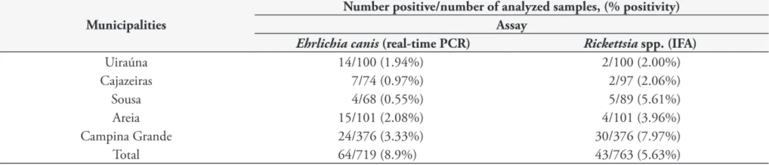
Ehrlichia canis DNA was amplified in 8.9% (64/719) of the dog blood samples analyzed by real-time PCR. The distribution of positive samples and the percentage of positivity per municipality are shown in Table 1.
Detection of anti-Rickettsia spp.antibodies by IFAT
Among the 763 dog serum samples analyzed by IFAT 5.63% (43/763) were reactive to at least one of the five rickettsial antigens tested, with titers ≥64. The titers of positive sera for the different species ranged from 64 to 512 for R. rickettsii and R. felis,
128 to 2048 for R. amblyommii, and 64 to 1024 for R. parkeri
and R. rhipicephali. Three animals showed homologous reactions: one for R. amblyommii, one for R. felis, and one for R. rickettsii, which was four-fold higher than those for the other antigens tested (Table 2).
There was no significant difference among the municipalities in the frequency of the serological reactivity to different Rickettsia
species: R. rickettsii (p = 0.319), R. parkeri (p = 0.089), R. rhipicephali
(p = 0.501), R. amblyommii (p = 0.263) and R. felis (p = 0.734). Only 0.55% (4/719) of the samples were positive for both agents tested in the present study. Four samples from Campina Grande municipality showed antibodies against at least one Rickettsia spp. antigens tested and had also DNA of E. canis.
Discussion
The present study assessed the occurrence of E. canis and exposure to other rickettsial agents in dogs from urban areas in the state of Paraiba in northeastern Brazil. Of the blood samples analyzed by real-time PCR, 8.9% yielded E. canis DNA. These results reveal a comprehensive picture of the CME situation in the region since real-time PCR reveals not only the exposure to the pathogen
Table 1. Distribution by municipality of the number of dogs from Paraiba, Brazil, analyzed for Ehrlichia canis DNA by real-time PCR and
Rickettsia spp. by indirect immunofluorescence assay test (IFAT) showing number of positives and percentages.
Municipalities
Number positive/number of analyzed samples, (% positivity) Assay
Ehrlichia canis (real-time PCR) Rickettsia spp. (IFA)
Uiraúna 14/100 (1.94%) 2/100 (2.00%)
Cajazeiras 7/74 (0.97%) 2/97 (2.06%)
Sousa 4/68 (0.55%) 5/89 (5.61%)
Areia 15/101 (2.08%) 4/101 (3.96%)
Campina Grande 24/376 (3.33%) 30/376 (7.97%)
Total 64/719 (8.9%) 43/763 (5.63%)
Table 2. Distribution by municipality of the number of dogs from Paraiba state, Brazil, analyzed for Rickettsia spp. by indirect immunofluorescence assay test (IFAT) showing the number of positives and percentages.
Municipalities Nº Tested
dogs
Nº Positive dogs (%)
Number of dogs reactive to each species of Rickettsia
(% positivity)
PAIHR
R. rickettsii R. parkeri R.
rhipicephali
R.
amblyommii R. felis
Uiraúna 100 2 (2) 0 (0) 1 (1) 0 (0) 1 (1) 0 (0) 0
Cajazeiras 97 2 (2.06) 1 (1.03) 0 (0) 2 (2.06) 1 (1.03) 0 (0) 0
Sousa 89 5 (5.62) 2 (2.24) 4 (4.49) 1 (1.12) 2 (2.24) 0 (0) 0
Areia 101 4 (3.96) 1 (0.99) 2 (1.98) 2 (1.98) 3 (2.97) 1(0.99) 2:1 R. amblyommii1 ,
R. felis
Campina Grande 376 30 (7.98) 9 (2.39) 13(3.45) 7 (1.86) 16 (4.25) 2(0.23) 1 R. rickettsii
Total 763 43 (5.63) 13 (1.7) 20 (2.62) 12 (1.57) 23 (3.01) 3(0.39)
but also the infection process as ehrlichial DNA was detected. Even though Rotondano et al. (2015) had detected DNA of E. canis
by real-time PCR in 25% (25/100) of the dogs sampled in Paraiba state, their samples comprised a veterinary hospital population, in contrast to random health dogs in the present study. This fact could be one of the reasons for the higher frequency of infected dogs in the study of Rotondano et al. (2015).
Here, we found 5.63% (43/763) of the canine serum samples reactive to at least one of the Rickettsia antigens. According to Piranda et al. (2008) the presence of seropositive animals indicates the circulation of Rickettsia of the SFG in an area at least 6-18 months earlier. As common hosts of tick-vectors for BSF, dogs are an important marker for disease in surveillance studies, and the occurrence of serologically positive dogs in a geographical area indicates the threat of human infection (ARAES-SANTOS et al., 2015).
This study provides evidence for exposure to Rickettsia spp. in urban dogs in the state of Paraiba. In the serum samples from three animals, it was possible to determine the probable antigen involved in homologous reaction (PAIHR) -R. amblyommii, R. felis
and R. rickettsii. Among the other reactive animals, it was not possible to discriminate the infecting agent (Table 2). R. rickettsii
is a known canine pathogen, whereas the role of pathogenicity by R. amblyommii in dogs is limited to serological evidence only. So far, the only tick-transmitted agents involved in human disease in Brazil are R. rickettsii and Rickettsia sp. strain Atlantic rainforest (ANGERAMI et al., 2009; SPOLIDORIO et al., 2010). The role of R. amblyommii in pathogenicity is not yet fully elucidated (BREITSCHWERDT et al., 1988). However, some Rocky Mountain Spotted Fever (RMSF) cases in the U.S. that were attributed to R. rickettsii could be due to R. amblyommii
instead (APPERSON et al., 2008).
In Brazil, two species of ticks are implicated in transmission of
R. rickettsii to humans and other animals, Amblyomma sculptum
(published as A. cajennense) and A. aureolatum (KRAWCZAK et al., 2014; SARAIVA et al., 2014). While A. aureolatum is not present
in northeastern Brazil (SARAIVA et al., 2014), A. sculptum is a very rare or absent tick species in the Caatinga biome of northeastern Brazil (MARTINS et al., 2016). Therefore, it is unlikely that
A. sculptum acts as an important vector of R. rickettsii within this biome.
R. sanguineus is the main ectoparasite in dogs in the dry Agreste and Sertão mesoregions of the Paraiba state (ROTONDANO et al., 2015). Although Tanikawa et al. (2013) had detected R. felis DNA in 4,5% (1/22) of R. sanguineus collected from Paraiba state, the role of R. sanguineus as vector of R. rickettsii in Brazilis not totally elucidated, further studies are needed to confirm its role as a vector among dogs in state of Paraiba.
In spite of the fact that Ctenocephalides felis, a host for
R. felis, is the most common flea species infesting dogs in Brazil
(OLIVEIRA et al., 2002), only three animals of the present study displayed R. felis antibodies and one of them showed antibody against R. felis four-fold higher than that observed for any other
Rickettsia species antigens, being thus considered homologous to R. felis. The potential presence of R. felis in dogs in the study region has important implications since R. felis has already been
considered as a human pathogen (SCHRIEFER et al., 1994; LABRUNA et al., 2007).
A recent study detected two Rickettsia species infecting bird ticks in the Atlantic forest of Paraíba state: Rickettsia sp. strain NOD (a R. parkeri-like agent) in Amblyomma nodosum, and
R. amblyommii in A. longirostre (LUGARINI et al., 2015). Since these two ticks species have occasionally been reported infesting dogs in Brazil (MORAES-FILHO et al., 2009; SABATINI et al. 2010), it is possible that they could be involved in the seropositivity of some of the dogs in the present study; however, we have no data on the history of tick infestations of our canine sample.
Conclusions
This study is the first to detect both antibodies to SFG
Rickettsia and DNA of E. canis in dogs from the state of Paraiba in northeastern Brazil. The finding of infection by E. canis in the whole blood of the animals analyzed reveals the endemic status of CME in the region. The study detected, for the first time, a positive serological reaction indicating Rickettsia spp. antibodies in forty-three samples, and the probable antigen involved in a homologous reaction (R. amblyommii, R. felis and R. rickettsii)
was identified in three samples. Taking into account that the dogs can serve as important indicators of tick-borne diseases, further studies to better understand the dynamics of rickettsiae diseases and their vectors in this region are extremely important to know about the real situation the circulation the rickettsias in the Paraiba state, northeastern Brazil.
Acknowledgements
The CNPq (Conselho Nacional de Pesquisas) provided a TEFR doctoral scholarship and Gilvan Mariano from the Informatics Sector of CPqAM/ FIOCRUZ-PE provided the art work in Figure 1.
References
Angerami RN, Silva AMR, Nascimento EMM, Colombo S, Wada MY, Santos FCP, et al. Brazilian spotted fever: two faces of a same disease? A comparative study of clinical aspects between an old and a new endemic area in Brazil. Clin Microbiol Infect 2009; 15(Suppl 2): 207-208. PMid:19392896. http://dx.doi.org/10.1111/j.1469-0691.2008.02160.x.
Apperson CS, Engber B, Nicholson WL, Mead DG, Engel J, Yabsley MJ, et al. Tick-borne diseases in North Carolina: is “Rickettsia amblyommii” a possible cause of rickettsiosis reported as Rock Mountain spotted fever?
Vector Borne Zoonotic Dis 2008; 8(5): 597-606. PMid:18447622. http:// dx.doi.org/10.1089/vbz.2007.0271.
Araes-Santos AI, Moraes-Filho J, Peixoto RM, Spolidorio MG, Azevedo SS, Costa MM, et al. Ectoparasite infestations and canine infection by Rickettsiae and Ehrlichiae in a Semi-Arid Region of Northeastern Brazil.
Vector Borne Zoonotic Dis 2015; 15(11): 645-651. PMid:26565771. http://dx.doi.org/10.1089/vbz.2015.1786.
in female dogs inoculated with Rickettsia rickettsii and Rickettsia montana. Am J Vet Res 1988; 49(1): 70-76. PMid:3128147.
Dantas-Torres F. Rocky Mountain spotted fever. Lancet Infect Dis 2007; 7(11): 724-732. PMid:17961858. http://dx.doi.org/10.1016/S1473-3099(07)70261-X.
Doyle CK, Labruna MB, Breitschwerdt EB, Tang YW, Corstvet RE, Hegarty BC, et al. Detection of medically important Ehrlichia spp. by quantitative multicolor TaqMan real-time Polymerase Chain Reaction of the dsb gene. J Mol Diagn 2005; 7(4): 504-510. PMid:16237220. http://dx.doi.org/10.1016/S1525-1578(10)60581-8.
Dumler JS, Barbet AF, Bekker CP, Dasch GA, Palmer GH, Ray SC, et al. Reorganization of genera in the families Rickettsiaceae and Anaplasmataceae in the order Rickettsiales: unification of some species of Ehrlichia with
Anaplasma, Cowdria with Ehrlichia and Ehrlichia with Neorickettsia, descriptions of six new species combinations and designation of Ehrlichia equi and ‘HGE agent’ as subjective synonyms of Ehrlichia phagocytophila. Int J Syst Evol Microbiol 2001; 51(6): 2145-2165. PMid:11760958. http://dx.doi.org/10.1099/00207713-51-6-2145.
Horta MC, Labruna MB, Durigon EL, Schumaker TTS. Isolation of
Rickettsia felis in the mosquito cell line C6/36. Appl Environ Microbiol
2006; 72(2): 1705-1707. PMid:16461734. http://dx.doi.org/10.1128/ AEM.72.2.1705-1707.2006.
Krawczak FS, Binder LC, Oliveira CS, Costa FB, Moraes-Filho J, Martins TF, et al. Ecology of a tick-borne spotted fever in southern Brazil. Exp Appl Acarol 2016; 70(2): 219-229. PMid:27392739. http://dx.doi. org/10.1007/s10493-016-0070-1.
Krawczak FS, Nieri-Bastos FA, Nunes FP, Soares JF, Moraes-Filho J, Labruna MB. Rickettsial infection in Amblyomma cajennense ticks and capybaras (Hydrochoerus hydrochaeris) in a Brazilian spotted fever-endemic area. Parasit Vectors 2014; 7(1): 7. PMid:24387674. http://dx.doi. org/10.1186/1756-3305-7-7.
Labruna MB, Horta MC, Aguiar DM, Cavalcante GT, Pinter A, Gennari SM, et al. Prevalence of Rickettsia infection in dogs from the urban and rural areas of Monte Negro Municipality, western Amazon, Brazil. Vector Borne Zoonotic Dis 2007; 7(2): 249-255. PMid:17627445. http://dx.doi. org/10.1089/vbz.2006.0621.
Lugarini C, Martins TF, Ogrzewalska M, Vasconcelos NCT, Ellis VA, Oliveira JB, et al. Rickettsial agents in avian ixodid ticks in northeast Brazil. Ticks Tick Borne Dis 2015; 6(3): 364-375. PMid:25800099. http://dx.doi.org/10.1016/j.ttbdis.2015.02.011.
Martins TF, Barbieri ARM, Costa FB, Terassini FA, Camargo LMA, Peterka CRL, et al. Geographical distribution of Amblyomma cajennense
(sensu lato) ticks (Parasitiformes: Ixodidae) in Brazil, with description of the nymph of A. cajennense (sensu stricto). Parasit Vectors 2016; 9: 186. PMid:27036324. http://dx.doi.org/10.1186/s13071-016-1460-2.
Moraes-Filho J, Pinter A, Pacheco RC, Gutmann TB, Barbosa SO, Gonzáles MA, et al. New epidemiological data on Brazilian spotted fever in an endemic area of the state of São Paulo, Brazil. Vector Borne Zoonotic
Dis 2009; 9(1): 73-78. PMid:18847319. http://dx.doi.org/10.1089/ vbz.2007.0227.
Oliveira RP, Galvão MAM, Mafra CL, Chamone CB, Calic SB, Silva SU, et al. Rickettsia felis in Ctenocephalides spp. fleas, Brazil. Emerg Infect Dis 2002; 8(3): 317-319. PMid:11927031. http://dx.doi.org/10.3201/ eid0803.010301.
Parola P, Paddock CD, Socolovschi C, Labruna MB, Mediannikov O, Kernif T, et al. Update on Tick-Borne Rickettsioses around the World: a Geographic Approach. Clin Microbiol Rev 2013; 26(4): 657-702. PMid:24092850. http://dx.doi.org/10.1128/CMR.00032-13. Piranda EM, Faccini JL, Pinter A, Saito TB, Pacheco RC, Hagiwara MK, et al. Experimental infection of dogs with a Brazilian strain of Rickettsia rickettsii: clinical and laboratory findings. Mem Inst Oswaldo Cruz 2008; 103(7): 696-701. http://dx.doi.org/10.1590/S0074-02762008000700012.
Rotondano TEF, Almeida AMP, Lustosa EMC, Cordeiro AA, Camboim EKA, Azevedo SS, et al. An assessment of whole blood and fraction by nested PCR as a DNA source for diagnosing canine ehrlichiosis and anaplasmosis. Sci World J 2012; 2012: 605743. PMid:22973174. http:// dx.doi.org/10.1100/2012/605743.
Rotondano TEF, Almeida HK, Krawczak FS, Santana VL, Vidal IF, Labruna MB, et al. Survey of Ehrlichia canis, Babesia spp. and Hepatozoon spp. in dogs from a semiarid region of Brazil. Rev Bras Parasitol Vet 2015; 24(1): 52-58. PMid:25909253. http://dx.doi.org/10.1590/S1984-29612015011.
Sabatini GS, Pinter A, Nieri-Bastos FA, Marcili A, Labruna MB. Survey of ticks (Acari: Ixodidae) and their rickettsia in an Atlantic rain forest reserve in the State of São Paulo, Brazil. J Med Entomol 2010; 47(5): 913-916. PMid:20939390. http://dx.doi.org/10.1093/jmedent/47.5.913.
Saraiva DG, Soares HS, Soares JF, Labruna MB. Feeding period required by Amblyomma aureolatum ticks for transmission of Rickettsia rickettsii to vertebrate hosts. Emerg Infect Dis 2014; 20(9): 1504-1510. PMid:25148391. http://dx.doi.org/10.3201/eid2009.140189.
Schriefer ME, Sacci JB Jr, Dumler JS, Bullen MG, Azad AF. Identification of a novel rickettsial infection in a patient diagnosed with murine typhus.
J Clin Microbiol 1994; 32(4): 949-954. PMid:8027348.
Silveira I, Pacheco RC, Szabó MPJ, Ramos HGC, Labruna MB. Rickettsia parkeri in Brazil. Emerg Infect Dis 2007; 13(7): 1111-1113. PMid:18214195. http://dx.doi.org/10.3201/eid1307.061397.
Spolidorio MG, Labruna MB, Mantovani E, Brandão PE, Richtzenhain LJ, Yoshinari NH. Novel Spotted Fever Group Rickettsiosis, Brazil.
Emerg Infect Dis 2010; 16(3): 521-523. PMid:20202436. http://dx.doi. org/10.3201/eid1603.091338.
Tanikawa A, Costa FB, Labruna MB, Azevedo SS. A survey for rickettsial agents on Rhipicephalus sanguineus (Ixodida, Ixodidae) ticks in Northeastern Brazil. Braz J Vet Res Anim Sci 2013; 50(5): 414-417. http://dx.doi. org/10.11606/issn.2318-3659.v50i5p414-417.