Major Article
Corresponding author: Dra. Alzira Almeida. e-mail: [email protected] Received 31 July 2017
Accepted 13 December 2017
Seroprevalence and spatial distribution dynamics
of
Yersinia pestis
antibodies in dogs and cats from plague
foci in the State of Ceará, Northeastern Brazil
Larissa Leão Ferrer de Sousa
[1],[2], Carlos Henrique Morais de Alencar
[2],
Alzira Maria Paiva de Almeida
[3]and Luciano Pamplona de Góes Cavalcanti
[2][1]. Secretaria da Saúde do Estado do Ceará, Fortaleza, CE, Brasil.
[2]. Departamento de Saúde Comunitária, Universidade Federal do Ceará, Fortaleza, CE, Brasil. [3]. Centro de Pesquisa Aggeu Magalhães, Fundação Oswaldo Cruz, Recife, PE, Brasil.
Abstract
Introduction: In Brazil, the plague is established in several foci located mainly in the northeastern part of the country, where
it alternates between active and quiescent periods. These foci in the State of Ceará have high epidemiological importance. In addition to other plague detection activities, plague areas can be monitored through serological surveys of dogs and cats (domestic carnivores), which, following feeding on plague-infected rodents, can develop mild to severe forms of the disease and produce long-lasting antibodies. This study aimed to characterize the circulation dynamics and spatial distribution of Yersinia pestis antibodies in dogs and cats in plague foci areas of Ceará. Methods: An ecological study was conducted to analyze
the temporal series and spatial distribution of secondary data obtained from domestic carnivore serum surveillance in Ceará’s plague areas from 1990 to 2014. Results: Joinpoint analysis revealed that the overall trend was a reduction in antibody-positive
animals. The mean proportion of antibody-positivity during the whole study period was 1.5% (3,023/203,311) for dogs, and 0.7% (426/61,135) for cats, with more than 4% antibody-positivity in dogs in 1997 and 2002. Antibody titers ranging from 1/16 to 1/64 were frequent. Despite luctuations and a signiicant reduction, in recent years, there were antibody-positive animals annually throughout the study period, and the localities containing antibody-positive animals increased in number. Conclusion: Yersinia pestis is actively circulating in the study areas, posing a danger to the human population.
Keywords: Yersinia pestis. Domestic carnivores. Reservoirs. Time series. Surveillance.
INTRODUCTION
The plague is a vector-borne disease found primarily in wild rodents, which can affect humans and other mammals, such as wild or domestic carnivores. Plague persists in natural foci in many countries worldwide. As yet, the accumulated technological advances and scientiic knowledge have not been enough to eradicate it1,2. Human cases of the disease are recorded annually in several countries in Africa, Asia, and the Americas3.
Currently, the plague is considered a reemerging disease by the World Health Organization (WHO), with a tendency to increase in case number and geographical dispersion4. The plague can spread quickly, signiicantly impacting public health. Pneumonic plague can generate an international public health emergency, thus requiring WHO notiication in accordance with current International Health Regulations5.
Introduced to Brazil in 1899, the infection was established in several natural foci, and spread throughout the States of Piauí, Ceará, Rio Grande do Norte, Paraíba, Pernambuco, Alagoas, Bahia, Minas Gerais, and Rio de Janeiro. The zoonosis remains in these foci, alternating between quiescent periods and epizootics, a phenomenon expressed by high rodent mortality and sporadic human cases6.
Until the 1970s, human plague notiication numbers in Brazil varied from 20 to 100 cases per year. Since 1990, human plague cases have been rare and sporadic. In the State of Ceará, Northeastern Brazil, the last laboratory-conirmed human cases occurred in the 1990s, with the exception of one case in 2005, which was initially misdiagnosed as dengue7.
Brazilian focal plague areas are continuously monitored through serological surveys of domestic carnivores [dogs (Canis lupus familiaris) and cats (Felis catus)], and the serological analyses are performed at the Central Laboratory of Public Health (LACEN) in the focal area states6,7.
after recovery, they produce antibodies that remain detectable for several months. This justiies their use as sentinels/detectors of Y. pestis circulation between the rodents/reservoirs in the foci areas8-11. In nature, the time of antigen exposure is unknown; thus, it is generally accepted that high proportions of antibody-positive animals with high antibody titers indicate recent plague activity. It is estimated that sampling a few dogs is equivalent to sampling hundreds of rodents in serological surveys to detect anti-plague antibodies12.
This study characterized the circulation dynamics and spatial distribution of Y. pestis antibodies in dogs and cats in the plague focal areas of the State of Ceará in Northeastern Brazil between 1990 and 2014.
METHODS
Ethical considerations
The project was approved by the Research Ethics Committee of the Federal University of Ceará (UFCE), registry CAAE 51371215.2.0000.5054, number 1 383 310.
Study design
The study was conducted using a time series analysis of sero-surveys among domestic carnivores in plague focal areas of Ceará from 1990 to 2014.
Study area
The study area included 44 (23.9%) of the state’s 184 municipalities, located in seven focal plague areas: Serra de Ibiapaba, Serra de Baturité, Serra da Uruburetama, Serra das Matas; Serra da Pedra Branca, Serra do Machado, and Chapada do Araripe. The total area comprises 29,855 km², 20.4% of the state’s surface6,7.
Municipality inclusion criteria
Municipalities that performed plague serological surveillance of dogs and cats for at least one year between 1990 and 2014 were included in the study. Duplicate records in the Sistema de Informação do Programa de Peste (SISPESTE) were excluded.
Data collection
Secondary data were collected from Plague Control Program (PCP) reports in Ceará, SISPESTE, and the Plague Regional Laboratory in Fortaleza (CE), from 1990 to 2005 and from LACEN-CE from 2005 to 2014.
The ield surveillance program was conducted during annual cycles of health agent visits and blood sample collections from dogs and cats for serological analyses13.
Serological analyses
Passive Hemagglutination Assays/Hemagglutination-Inhibition (HA/HI) assays were used to detect speciic Y. pestis anti-F1 antibodies13. In brief, the tests were performed using
96-well U-shaped polystyrene plates. Twenty-ive microliters of each serum was serially diluted (1/4 to 1/8,192) into 25µL of HA buffer containing sterile normal saline (NS) and 1/100 normal rabbit sera (NRS). The sera were simultaneously diluted
into the HI buffer containing 100µg/mL of F1-speciic Y. pestis protein antigen suspension, as described above. Next, 25µL of a saline suspension (NS plus 1/250mL NRS) of sheep red blood cells (SRBCs) sensitized with 100ug/mL of F1 (SRBCs/ F1) was added to each well. Samples were incubated overnight at 4-10°C. The test was considered positive when the HA end-point titer was depressed by three or more dilutions in the HI test. A titer of >1/16 was considered antibody-positive. The
NRS used to prepare the buffers and all sera tests were previously inactivated at 56°C for 30 min in a water-bath and adsorbed for 30 min at room temperature with fresh sheep red blood cells13.
Data analysis
A database was built using MS Excel 2010 to organize, separate and categorize the information. The data were exported and analyzed using Stata software, version 11.2. Time trend analysis was performed using the Joinpoint Regression Program, version 4.5.0.1. The trend analysis included dog surveillance data from 1997 to 2014, and cat surveillance data from 1997 to 2009. To calculate the annual percentage change (APC) in the antibody-positive animal proportions, a trend analysis was performed using continuous linear regression with inlection points (Joinpoint method). This method allowed the time series data to be adjusted from the minimum number of joinpoints (i.e., 0 joinpoints, which is a straight line) and determined whether the inclusion of more joinpoints was statistically signiicant. The signiicance is represented by graphs and two indicators, the APC and the average annual percentage change (AAPC).
The trend analysis of the signiicant variables from Ceará and for each plague focus was determined. Each signiicant point that indicated a change in the increase or decrease (if any) was maintained in the inal model14. The independent
variable (Y) was the study year, and the dependent variables (X) were the proportion of antibody-positive dogs, the proportion of positive cats, and the proportion of antibody-positive animals (dogs and cats). Differences were considered statistically signiicant at a p value < 0.05.
The joinpoint analysis was performed using the number of antibody-positive animals as the numerator, and the number of tested animals as the denominator, multiplied by 100. Data were converted logarithmically. Errors were considered heteroskedastic, and the regression coeficients were estimated by weighted least squares. For both the proportion of antibody-positive animals, and the temporal evaluation of the data, an adjusted autocorrelation model of the errors based on the data was also used.
Year
Dogs Cats*
tested positives proportion of
positives tested positives
proportion of positives
1997 8,367 417 4.98 5,970 67 1.12
1998 10,191 45 0.44 6,469 17 0.26
1999 10,491 131 1.25 6,132 31 0.51
2000 8,714 65 0.75 4,991 6 0.12
2001 9,581 378 3.95 4,995 170 3.40
2002 6,581 313 4.76 3,596 30 0.83
2003 9,096 183 2.01 5,159 23 0.45
2004 9,466 176 1.86 4,640 16 0.34
2005 11,135 387 3.48 4,535 28 0.62
2006 12,261 139 1.13 4,362 7 0.16
2007 13,258 207 1.56 5,137 19 0.37
2008 12,153 125 1.03 4,689 11 0.23
2009 14,342 109 0.76 460 1 0.22
2010 14,586 51 0.35 - -
-2011 14,005 109 0.78 - -
-2012 12,476 63 0.50 - -
-2013 13,257 69 0.52 - -
-2014 13,351 56 0.42 - -
-Total 203,311 3,023 1.49 61,135 426 0.70
TABLE1: Distribution of antibody-positive dogs and cats from the plague foci in Ceará from 1997 to 2014.
*Surveillance of cats was discontinued after 2009. RESULTS
From 1997 to 2014, serum samples from 203,311 dogs from seven plague foci in Ceará were analyzed for Y. pestis anti-F1 antibodies using HA-HI testing, and 3,023 (1.49%) were antibody-positive. Of the serum samples from 61,135 cats analyzed from 1997 to 2009, 426 (0.7%) were HA-HI-positive
(Table 1).
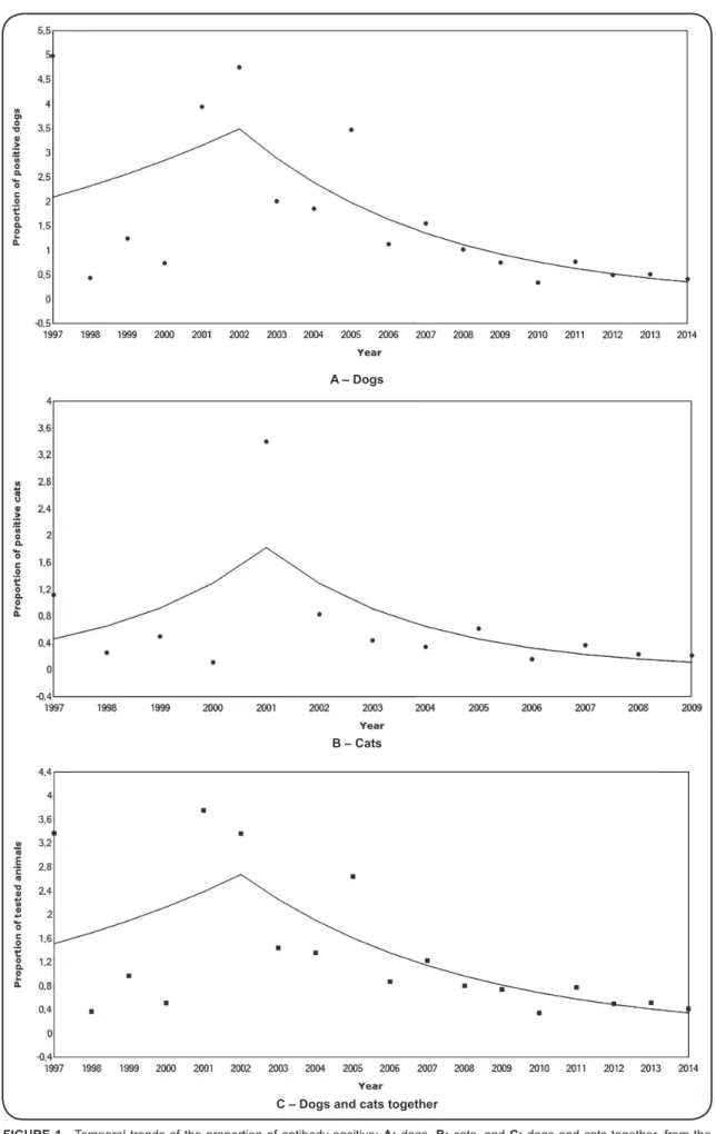
Joinpoint analysis showed two different trends, both for dogs and for cats (Figure 1A, Figure 1B and Figure 1C).
The proportion of antibody-positive dogs displayed a non-signiicant positive trend (APC = 10.7, 95% CI: -14.2 to 42.9, p = 0.402) in the period of 1997 to 2002. From 2002 to 2014, the trend changed, and the proportion of antibody-positive dogs decreased signiicantly (APC = -17.2, 95% CI: -24.6 to -9.1, p < 0.001). The AAPC for the entire period displayed a signiicant decreasing trend (AAPC = -9.8, 95% CI: -18.4 to -4.4) (Table 2 and Figure 1A). Joinpoint analysis showed that
the proportion of antibody-positive cats increased signiicantly from 1997 to 2001 (APC = 40.5, 95% CI: 10.3 to 78.9, p = 0.011), then decreased signiicantly from 2001 to 2009 (APC
= -29.1, 95% CI: -35.6 to -22.1, p < 0.001). The AAPC for the entire period displayed an overall downward trend, but the model was not statistically signiicant (AAPC = -11.0, 95% CI: -38.1 to 3.4) (Table 2 and Figure 1B).
In the accumulated period from 1997 to 2014, 16 (36.4%) municipalities had up to 25 antibody-positive dogs, 19 (43.2%) had 25 to 100, five (11.4%) had 100 to 200, and four (9%) municipalities had over 400 antibody-positive dogs. Over the years, both antibody-positive dogs per locality and the number of localities with antibody-positive dogs decreased. Therefore, after 2003, no municipality had more than 40 antibody-positive dogs. The number antibody-positive cats per municipality also decreased, as did the number of municipalities with antibody-positive cats. Throughout the entire study period, 11 (25%) municipalities had no positive cats, 15 (34%) had up to ive antibody-positive cats, 10 (22%) had ive to 15, six (14%) had 15 to 40 and two (5%) municipalities had 40 to 140 antibody-positive cats.
FIGURE 1 - Temporal trends of the proportion of antibody-positive: A: dogs, B: cats, and C: dogs and cats together, from the plague foci in Ceará from 1997 to 2014, using joinpoint analysis.
A – Dogs
B – Cats
Segment Lower endpoint Upper endpoint APC Lower CI Upper CI p-value
Dogs
1 1997 2002 10.7 -14.2 42.9 0.402
2 2002 2014 -17.2 -24.6 -9.1 <0.001
AAPC 1997 2014 -9.8 -18.4 -4.4
Cats
1 1997 2001 40.5 10.3 78.9 0.011
2 2001 2009 -29.1 -35.6 -22.1 <0.001
AAPC 1997 2009 -11.0 -38.1 3.4
Dogs and cats together
1 1997 2002 12.1 -12.6 43.7 0.340
2 2002 2014 -15.6 -23.8 -6.5 0.003
AAPC 1997 2014 -8.2 -17.2 -3.0
TABLE2: Joinpoint regression analysis of the proportion of positive animals in Ceará from 1997-2014 (dogs, and dogs and cats together) and from 1997-2009 (cats).
APC: annual percentage change; CI: conidence interval; AAPC: average annual percentage change.
Foci Minimum 1st Quartile Median 3rd Quartile Maximum
Chapada do Araripe 0.31 0.35 0.87 1.01 1.47
Serra da Ibiapaba 0.34 0.45 0.48 0.65 1.02
Serra da Pedra Branca 0.26 0.29 0.43 0.63 0.8
Serra das Matas 0 0.23 0.7 1.52 3.33
Serra de Baturité 0.32 0.35 0.5 1.11 1.54
Serra de Uruburetama 0.23 0.29 0.81 1.05 1.34
Serra do Machado 0 0.54 0.85 1.26 1.77
Ceará State 0.35 0.42 0.52 0.78 1.03
TABLE3: Minimum, median, and quartiles of the proportion of antibody-positive dogs in Ceará from 2008 to 2014. (Table 3). During the study period, the proportion of infected
dogs decreased over time in the seven plague foci. However, there was a notably high proportion of antibody-positive dogs in Serra do Machado almost every year (Table 4).
Regarding the HA/HI titer distribution in the historical series from 1990 to 2014, a total of 3,631 dogs and cats had anti-plague antibodies, with titers ranging from 1/16 to 1/8,192. The most frequent titers were 1/32 (n = 1,100 animals), 1/64 (n = 997) and 1/16 (n = 916), representing 30%, 27% and 25% of the total, respectively. The highest titers were less frequent or rare: 1/128 (n = 290) 1/256 (n = 154), 1/512 (n = 148), 1/1 1,024 (n = 15), 1/2,048 (n = 8), 1/4,096 (n = 2) and 1/8,192 (n = 1). This may be due to the laboratory routine, which usually
tests up to 6 dilutions (1/128) and only occasionally up to 12 dilutions (1/8,192).
DISCUSSION
Dogs and cats participate in the epidemiological cycle of the plague and are good indicators of infection activity in the focal areas. They can carry infected rodents and leas to human premises, thus increasing the risk of human infection15.
Dogs in highly enzootic areas may be repeatedly exposed to plague-infected rodents and leas and develop high Y. pestis antibody titers16. In China, serological surveys of dogs are an
Year
Plague foci
Ceará State Chapada do
Araripe
Serra da Ibiapaba
Serra da Pedra Branca
Serra das Matas
Serra de Baturité
Serra de Uruburetama
Serra do Machado
2008 0.87 1.02 0.80 3.33 1.11 1.34 1.77 1.03
2009 1.47 0.45 0.26 1.52 1.54 1.05 0.85 0.76
2010 0.31 0.34 0.29 0.00 0.35 0.44 0.54 0.35
2011 0.98 0.65 0.43 0.70 0.76 0.93 1.26 0.78
2012 1.01 0.48 0.43 0.90 0.48 0.29 0 0.50
2013 0.35 0.51 0.63 0.23 0.50 0.81 1.18 0.52
2014 0.53 0.48 0.48 0.41 0.32 0.23 0.59 0.42
Total 0.75 0.57 0.46 0.56 0.67 0.59 0.78 0.62
TABLE 4: Distribution of the proportion of antibody-positive dogs from the plague foci in Ceará from 2008-2014.
All animals in our study were domesticated, living in direct contact with humans. In the rural areas included in this study, dogs are commonly used for hunting, and cats help to control the rodents in homes, and in grain and food storage buildings. Therefore, they are exposed to Y. pestis through contact with rodents and their leas.
The historical time series analysis in our study revealed similar trends for both positive dogs and cats, with a reduction in positivity from 2001 among cats and from 2002 among dogs. The proportion of positive dogs increased from 1997 to 2002 (not signiicantly) and declined signiicantly from 2002 to 2014. Among the cats, the trend increased from 1997 to 2001 and decreased from 2001 to 2009, both being statistically signiicant
(Table 2, Figure 1A, Figure 1B and Figure 1C).
The human plague prevalence has also declined in the Ceará plague foci since the 1990s7. Our indings corroborate those of other
studies on temporal associations with human plague incidence18-20.
Parmenter et al.21 found that the cyclical pattern of the annual
distribution of plague cases in New Mexico (USA) was associated with luctuations in the rodent population or in the rodents’ responses to environmental variations such as food and shelter availability.
During our study period, the prevalence of antibody-positive dogs was 1.49% (3,023/203,311), with more than 4% antibody-positivity in 1997 and 2002, and the overall prevalence for cats was 0.70% (426/61,135), with higher antibody-positivity (3.40%) in 2001 (Table 1). Previous studies found that
antibody-positivity prevalence among dogs and cats from the Ceará plague foci, and from Chapada da Borborema (PB), were similar13,22.
During a plague outbreak in a village in Yunnan Province, China serum-positivity rates in domestic cats and dogs were 23.5% and 26.5%, respectively23. This could be because many diseased
cats do not survive the infection, while dogs are more resistant and more likely to recover10.
Because it was assumed that plague antibody detection is more sensitive in dogs than in cats and rodents, the plague
surveillance of cats was discontinued in the Brazilian foci areas24. Despite the low antibody-positive prevalence (< 1%)
during the study period (with the exception of 1997 and 2001), cats tested antibody-positive in all years investigated. Therefore, serological surveillance in cats should not be discontinued, as it may be valuable for the plague monitoring program. Almeida
et al.22 recorded 76% positive samples from cats, and 49.1% from
dogs in Serra de Baturité (CE) during a human plague outbreak. The high positivity among cats was likely due to animals being infected and then surviving the disease during those events. Therefore, to improve the monitoring of Y. pestis in plague areas, implementing surveillance for cats is recommended.
In the USA, serological surveillance is performed on a wide variety of mammals, as it is thought that this procedure is more sensitive, and allows better assessment of plague behavior and circulation in focal areas25-26. Lowell et al.27 state that
surveillance involving many species provides better coverage of plague activity than monitoring a single species. Plague surveillance programs monitoring die-offs and serum prevalence in reservoir animal populations can provide an early and timely warning of possible human cases10,27.
In the surveillance routine of sentinel animals, including coyotes (Canis latrans), badgers (Taxidea taxus), and raccoons
(Procyon lotor) in American plague areas, an overall prevalence
of 16% (3,481/21,826) was found, varying by region and species. The prevalence rate was higher in mustelids (43% - 504/1,173), followed by bears (23% - 95/459) and canids (15% - 1,079/7,291)28. Malmlov et al. reported a 28% (7/25) serum
prevalence in coyotes26.
result from eco-epidemiological factors favoring dissemination, such as the densities of natural vectors and rodent hosts, in addition to possible climatic changes, such as temperature and precipitation19. Comparing the proportional distribution of
antibody-positive dogs in the seven plague foci, the prevalence was reduced in all but Pedra Branca, where antibody-positivity was instead increased. Serra do Machado had the highest prevalence in dogs during the study period.
Analyzing the focal associations, statistically signiicant correlations were found between Serra da Uruburetama and the year; Chapada do Araripe and Serra das Matas; Serra de Ibiapaba, Serra da Pedra Branca and Serra do Machado; Serra de Baturité and Serra de Uruburetama and between Serra de Uruburetama and Serra do Machado (Table 3 and Table 4). The
prevalence rates among the different plague focal areas may have been inluenced by factors common to the seven foci. Thus, we assume a common pattern exists for actively circulating Y. pestis in the focal areas. These results agree with those of Baltazard29, who considered that the geographical massifs
of Ibiapaba, Baturité, Machado, Uruburetama, Pedra Branca and Matas constitute a single focus. Giles et al.30 conirmed
that plague is focal in northeast Brazil, with a discontinuous distribution. These studies showed that those foci depend on a broad set of conditions, and are not necessarily correlated with their elevation, but likely to the presence of the hairy-tailed bolo mouse, Necromys lasiurus, which is the primary plague host in these foci, and is responsible for epizootics.
The proportion of antibody-positive animals luctuated in all foci, however, these luctuations were only statistically signiicant in Serra das Matas. A prevalence higher than 1% was observed in some years, which increases the risk for human cases to emerge12.
Antibody titers among dogs and cats in the study period showed a higher frequency of 1/32, followed by 1/64 and 1/16. Titers >1/64 suggest recent infections and recent contact with
Y. pestis, signaling a potential risk for human infection12-13,22.
Paul-Murphy et al.31 reported anti-Y. pestis antibody titers
≥1/128 in 15 pumas, Felis concolor, and a range of 1/1,024 to 1/4,096 in four pumas in rural areas of California (USA). In coyotes from urban areas in California, titers ranged from 1/16 to 1/1,02426. In plague areas of China, titers varying from 1/40
to 1/1,280 were found in 49 domestic dogs23.
In conclusion, during the study period, the prevalence of antibody-positive dogs and cats luctuated, with a signiicant reduction in recent years. However, our results also revealed annual antibody-positive animals, providing evidence that Y. pestis is persisting in active circulation. Thus, even the quiescent areas with no human cases have the potential for transmission risk, and despite the decrease in antibody-positive domestic carnivores, continual surveillance is needed in these areas. Due to the plague’s cyclical characteristics - alternating periods of activity and quiescence depending on a series of complex factors - plague can reemerge and reach the human population. Hence, surveillance, monitoring, and control actions must be continued.
Acknowledgements
We would like to thank the Secretaria da Saúde do Estado do Ceará (SESA), Núcleo de Controle de Vetores (NUVET) and Laboratório Central de Saúde Pública (LACEN).
Conlicts of interest
The authors declare that there is no conlict of interest.
REFERENCES
1. Butler T. Plague gives surprises in the irst decade of the 21st
century in the United States and worldwide. Am J Trop Med Hyg. 2013;89(4):788-93.
2. Butler T. Plague history: Yersin's discovery of the causative bacterium in 1894 enabled, in the subsequent century, scientiic progress in understanding the disease and the development of treatments and vaccines. Clin Microbiol Infect. 2014;20(3):202-9.
3. World Health Organization (WHO). Plague around the world,
2010-2015. Wkly Epidemiol Rec. 2016;91(8):89-104.
4. Stenseth NC, Atshabar BB, Begon M, Belmain SR, Bertherat E, Carniel E, et al. Plague: past, present and future. PLoS Med. 2008;5(1):e3.
5. World Health Organization (WHO). International Health Regulations (2005). 2nd edition Geneva: WHO; 2008. 74p.
6. Tavares C, Aragão AI, Leal NC, Leal-Balbino TC, de Oliveira MB, de Oliveira GFGM, et al. Plague in Brazil: from now and then. Adv Exp Med Biol.2012;954:69-77.
7. Aragão AI, Pontes RJS, Seoane ACM, Nascimento OJ, Tavares C, Almeida AMP. Tendência secular da peste no Estado do Ceará, Brasil. Cad Saúde Pública. 2007;23(3):715-24.
8. Rust Jr JH, Cavanaugh DC, O’Shita R, Marshall Jr JD. The role of domestic animals in the epidemiology of plague, I: Experimental infection of dogs and cats. J Infect Dis. 1971;124(5):522-6.
9. Rust Jr JH, Miller BE, Bahamnyar M, Marshall Jr JD, Purnaveja S, Cavanaugh DC, et al. The role of domestic animals in the epidemiology of plague. II. Antibody to Yersinia pestis in sera of dogs and cats. J Infect Dis. 1971;124(5):527-31.
10. Barnes AM. Surveillance and control of bubonic plague in the United States. Symp Zool Soc Lond. 1982;50:237-70.
11. Gasper PW, Barnes AM, Quan TJ, Benziger JP, Carter LG,
Beard ML, et al. Plague (Yersinia pestis) in cats: description of experimentally induced disease. J Med Ent. 1993;30(1):20-6. 12. Dennis DT, Gage KL, Gratz N, Poland JD, Tikhomirov E. Plague
manual: epidemiology, distribution, surveillance and control. Geneva: World Health Organization. 1999. Available from: www. who.int/csr/resources/publications/plague/WHO.../en]
13. Aragão AI, Pinheiro KMA, Seoane ACM, Tavares C, Almeida
AMP. Prevalência de anticorpos contra Yersinia pestis em
carnívoros domésticos nos focos pestosos do Estado do Ceará. Rev Soc Bras Med Trop. 2009;42(6):711-5.
14. Kim HJ, Fay MP, Feuer EJ, Midthune DM. Permutation tests for joinpoint regression with applications to cancer rates. Stat Med. 2000;19(3):335-51.
15. Gage KL. Factors affecting the spread and maintenance of plague. In: Almeida AMP, Leal NC, editors. Advances in Yersinia Research. New York: Springer Science Business Media. 2012. p. 79-94. 16. Gould LH, Pape J, Ettestad P, Grifith KS, Mead PS. Dog-associated
17. Li B, Guo Y, Guo Z, Liang Y, Zhu Z, Zhou Q, et al. Serologic survey of the sentinel animals for plague surveillance and screening for complementary diagnostic markers to F1 antigen by protein microarray. Am J Trop Med Hyg. 2008;79(5):799-802.
18. Enscore RE, Biggerstaff BJ, Brown TL, Fulgham RF, Reynolds PJ, Engelthaler DM, et al. Modeling relationships between climate and the frequency of human plague cases in the southwestern United States, 1960-1997. The Am J Trop Med Hyg. 2002;66(2):186-96. 19. Ben Ari T, Gershunov A, Gage KL, Snall T, Ettestad P, Kausrud
KL, et al. Human plague in the USA: the importance of regional and local climate. Biol Lett. 2008;4(6):737-40.
20. Kreppel KS, Caminade C, Telfer S, Rajerison M, Rahalison L, et al. A non-stationary relationship between global climate phenomena and human plague incidence in Madagascar. PLoS Negl Trop Dis. 2014;8(10):e3155.
21. Parmenter RR, Yadav EP, Parmenter CA, Ettestad P, Gage KL. Incidence of plague associated with increased winter-spring precipitation in New Mexico, Am J Trop Med Hyg. 1999;61(5): 814-21.
22. Almeida AMP, Brasil DP, Melo MEB, Leal NC, Almeida CR. Importância dos carnívoros domésticos (cães e gatos) na epidemiologia da peste nos focos do Nordeste do Brasil. Cad Saúde Pública. 1998;4(1):49-55.
23. Wang H, Cui Y, Wang Z, Wang X, Guo Z, Yan Y, et al. A dog-associated primary pneumonic plague in Qinghai Province, China. Clin Infect Dis. 2011;52(2):185-90.
24. Costa ECV, Sobreira M, Leal NC, Almeida AMP. Rodents and other small mammal reservoirs in plague foci in northeastern Brazil. J Infect Dev Ctries. 2017;11(5):426-30.
25. Gage KL, Kosoy MY. Natural history of plague: perspectives from more than a century of research. Annu Rev Entomol. 2005;50: 505-28.
26. Malmlov A, Breck S, Fry T, Duncan C. Serologic survey for cross-species pathogens in urban coyotes (Canis latrans), Colorado, USA. J Wildl Dis. 2014;50(4):946-50.
27. Lowell JL, Eisen RJ, Schotthoefer AM, Xiaocheng L, Montenieri JA, Tanda D, et al. Colorado animal-based plague surveillance systems: relationships between targeted animal species and prediction eficacy of areas at risk for humans. J Vector Ecol. 2009;34(1):22-31. 28. Salkeld DJ, Stapp P. Seroprevalence rates and transmission of
plague (Yersinia pestis) in mammalian carnivores. Vector Borne Zoonotic Dis. 2006;6(3):231-9.
29. Baltazard M. Research on plague in Brazil. Bull Soc Pathol Exot. 2004;T97:93-117.
30. Giles J, Peterson AT, Almeida A. Ecology and geography of plague transmission areas in Northeastern Brazil. PLoS Negl Trop Dis. 2011;5(1) e925.
31. Paul-Murphy J, Work T, Hunter D, McFie E, Fjelline D. Serologic