141 141 141 141 141 Mem Inst Oswaldo Cruz, Rio de Janeiro, Vol. 93, Suppl. I: 141-151, 1998
Histoarchitecture of Schistosomal Granuloma Development
and Involution: Morphogenetic and Biomechanical
Approaches
Henrique L Lenzi/
+,
Eitan Kimmel
*/
++,
Helio Schechtman
**,
Marcelo
Pelajo-Machado
+++,
Waldemiro S Romanha
,
Ronaldo G Pacheco
***,
Mario Mariano
****,
Jane A Lenzi
Departamento de Patologia, Instituto Oswaldo Cruz, Av. Brasil 4365, 21045-900 Rio de Janeiro, RJ, Brasil *Department of Agricultural Engineering, Technion-Israel Institute of Technology, Technion City, Haifa 32000, Israel **Programa de Computação Científica, Presidência, Fiocruz, Av. Brasil 4365, 21045-900 Rio de Janeiro, RJ, Brasil ***Departamento de Medicina Geral, Hospital Universitário Gaffrée e Guinle, Universidade do Rio de Janeiro, Rua Mariz e Barros 775, 20270-004 Rio de Janeiro, RJ, Brasil ****Departamento de Imunologia,
Instituto de Ciências Biomédicas, Universidade de São Paulo, Av. Prof. Lineu Prestes 2415, 05508-900 São Paulo, SP, Brasil
The authors present morphogenetic and biomechanical approaches on the concept of the Schisto-soma mansoni granulomas, considering them as organoid structures that depend on cellular adhesion and sorting, forming rearrangement into hierarchical concentric layers, creating tension-dependent structures, aiming to acquire round form, since this is the minimal energy form, in which opposing forces pull in equally from all directions and are in balance. From the morphogenetic point of view, the granulomas function as little organs, presenting maturative and involutional stages in their develop-ment with final disappearance (pre-granulomatous stages, subdivided in: weakly and/or initial reactive and exudative; granulomatous stages: exudative-productive, productive and involutional). A model for the development of granulomas was suggested, according to the following stages: encapsulating, focal histolysis, fiber production, orientation and compacting and involution and desintegration. The authors concluded that schistosomal granuloma is not a tangled web of individual cells and fibers, but an orga-nized structure composed by host and parasite components, which is not formed to attack the miracidia, but functions as an hybrid interface between two different phylogenetic beings.
Key words: Schistosoma mansoni - granuloma - biomechanics - fibrosis - extracellular matrix - laser confocal microscopy
There are various definitions for the term granu-loma, creating a nosological chaos, which com-bines etiologic, mechanistic and anatomical ap-proaches (Epstein 1983). Some of these definitions, as well as the classification and pathogenesis of granulomas, have been discussed in reviews by Adams (1976, 1983), Warren (1976), Epstein (1977), Boros (1978), Williams and Williams (1983), Hirsh and Johnson (1984), De Britto and Franco (1994) and Mariano (1995). Most of them stress the etiology, or the main cellular component, or the participation or not of the immune system,
+Corresponding author. Fax: +55-21-598.4466. E-mail:
[email protected] ++CNPq Visiting Professor +++CAPES fellowship
Received 4 May 1998 Accepted 31 August 1998
or the differences in terms of the relative number of the new macrophages entering the granuloma (low and high turnover granulomas), or the par-ticipation or not of tissue injury. Although the con-cept of the granuloma has been dogged by many confusions, there are some universal accepted convergences, or diagnostic criteria such as: (i) granuloma is a focal chronic inflammatory reac-tion; (ii) it is characterized by the collection of cells of the “mononuclear phagocyte series”, with or without the addition of other cell types (Turk 1992); (iii) the collection of mononuclear cells is com-pact and organized (Adams 1983); and (iv) epi-thelioid cells define the typical granulomatous le-sion (Mariano 1995).
struc-142 142 142 142
142 Histoarchitecture of Schistosomal Granuloma Henrique L Lenzi et al.
tures, aiming to acquire round form, since this is the minimal energy form, in which opposing forces pull in equally from all directions and are in bal-ance (Ingber 1993). From the morphogenetic point of view, the granulomas function as little organs, presenting maturative and involutional stages in their development, with final disappearance. The dynamism of the granulomatous process depends on the characteristic of the etiologic agent, which could be intracellular, intra and extracellular or extracellular (Lenzi et al. 1991). Schistosomal granuloma is the best example of the late category and is the subject of this paper.
PHASES OF SCHISTOSOMA MANSONI GRANULOMA DEVELOPMENT
The evolution of the host response to schisto-some eggs in experimental animals has been di-vided in various stages, according to the authors: florid and cicatricial stages (Grimaud 1986); cel-lular, productive and regressive stages (Junqueira et al. 1986); nonreactive or weakly reactive, exu-dative-productive, productive (subdivided in early and advanced phases) and involutional stages (Li Hsü et al. 1972).
In patients, the granuloma stages were divided by Coelho (1955) in three stages: first, character-ized by focal histolysis and cellular exudation; second, productive or encysted histiocytic reac-tion, and third, repair or healing, with fibrous sub-stitution, forming a collagenous nodule. On the other hand, Raso and Bogliolo (1970) considered the following stages in the evolution of human schistosomal granulomas: necrotic-exudative, exu-dative, productive and cure by fibrosis. We pro-pose here a little modification of the Li-Hsü et al. (1972) classification, dividing the maturative and involutional stages of murine S. mansoni granu-loma in the following stages:
I. Pre-granulomatous stages
I.A. weakly and/or initial reactive (IR) (Fig. 1) I.B. exudative (E) (Fig. 2)
II. Granulomatous stages
II.A. exudative-productive (EP) (Fig. 3) II.B. productive (P) (with early and advanced
or fibrotic (Fig. 4) phases (PF)
II.C. involutional (I), subdivided in the follow ing subtypes:
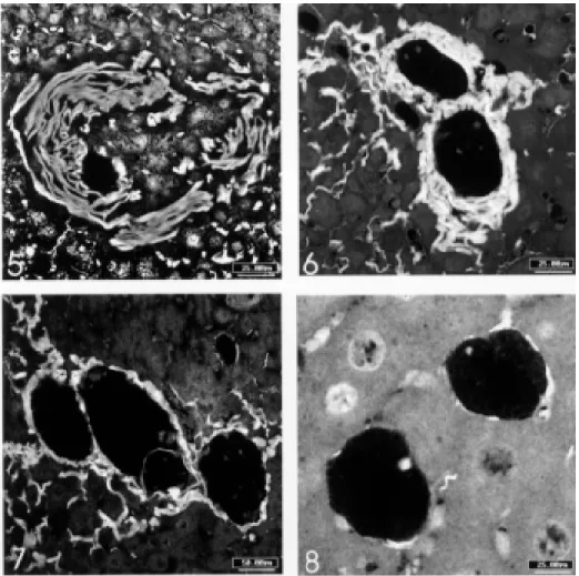
II.C.a. with dissociation (and disintegration) of the collagen fibers (Fig. 5) II.C.b. with thick collagen layer (Fig. 6) II.C.c. with thin collagen layer (Fig. 7) II.C.d. pigmented macrophagic type (Fig. 8).
The modification of Li-Hsü et al. (1972) classifi-cation was founded on conceptual basis, i.e., the weakly/initial reactive (Figs 1, 9-11) and
exuda-tive stages (Figs 2, 12, 13) do not fit to the funda-mental requirement of the granuloma concept: they are not chronic inflammatory reaction; they do not constitute compact or organized collection of monocyte/macrophage. It should be emphasized that epithelioid cells, which are secretory modi-fied macrophages with large areas of Golgi, a rich display of rough endoplasmic reticulum, and little or no phagocytic potential, is not an absolute re-quirement to the diagnosis of schistosomal granu-loma. Actually this type of granuloma, according to the Hirsh and Johnson (1984) concept, is not a pure epithelioid, but a mixed type of granuloma-tous inflammation, where the epithelioid cells can or can not be present.
In murine S. mansoni granulomas, the giant cells are of two types: one appears randomly, usu-ally located far from the eggs, showing cytoplasm rich in schistosomal pigment (pigmented giant cells); the second type of giant cells are dominant and lay on the top of the egg shells or are egg scav-engers, appearing more in the productive and in-volutional stages of the granulomas (foreign- body-type giant cells and, rarely, Langhans’ giant cells). Usually epithelioid and giant cells in hepatic, in-testinal and pancreatic granulomas first appear around 45 to 55 days being more constant from 80 to 100 days after infection.
The pre-granulomatous exudative stage (Figs 2, 12, 13) is very important in the pathogenesis of the schistosomal infection, causing histolysis and microthrombosis, specially in the liver, and taking part in the “metastatic capability” of the S. mansoni
eggs to cross the vessels, allowing them to settle in the justa-vascular tissues (Lenzi et al. 1991). It also participates in the parasite host co-evolution, op-erating upon parasite transmission, favoring the egg release to the feces (Lenzi et al. 1987, 1997). To better study the early phase of cellular influx to the eggs we have used the air pouch model in Swiss Webster mice, prepared as described by Edwards et al. (1981) with modifications (Pacheco & Lenzi, unpub. data). After injection of S. mansoni
purified eggs in endotoxin-free saline, in the air pouch, the cells were collected at 4, 6, 8, 12, 24, 36, 48, 72 and 96 hr and were counted in Neubauer and Fuchs-Rosenthal chambers and in cyto-centrifuged slides. After stimulus, the absolute number of neu-trophils, monocytes/macrophages, eosinophils and lymphocytes predominated, in that order, and reached higher values at 6-8 hr, decreasing steadily with normalization after 36-48 hr. We showed that TNF-a, ICAM-1 and CD18 took part in this
pro-cess, which should be considered as an inflamma-tory and non-immune event. Then the S. mansoni
pre-granu-143 143143 143143 Mem Inst Oswaldo Cruz, Rio de Janeiro, Vol. 93, Suppl. I, 1998
lomatous phase is not very well studied and prob-ably takes part in the definition of the infection out-come. It represents an antique memory resulted from millenary experience between S. mansoni and the hosts, that preceded the beginning of the lymphoid system (Lenzi et al. 1997).
In recent years, a growing body of evidence shows that neutrophils, eosinophils, and not only monocytes/macrophages can synthesize and re-lease a range of immunoregulatory cytokines. Be-yond the classical cytokines, like IL-1, TNF and others, monocyte/macrophage lineage expresses
144 144 144 144
144 Histoarchitecture of Schistosomal Granuloma Henrique L Lenzi et al.
Fig. 5: involutional granuloma with dissociation and disintegration of the collagen fibers, mainly in the right side of the picture (phosphomolibdic acid-picrosirius, confocal laser scanning microscopy, bar = 25 mm). Fig. 6: two involutional granulomas with thick collagen layer, surrounding internal zone composed by pigmented macrophages (the macrophages are not seen in this stain) (phosphomolibdic acid-picrosirius, confocal laser scanning microscopy, bar = 25 mm). Fig. 7: three involutional granulomas, with thin collagen layer in the periphery. Residual egg shell is seen in the central granuloma (phosphomolibdic acid-picrosirius, confo-cal laser scanning microscopy, bar = 50 mm). Fig. 8: two involutional pigmented macrophagic granulomas showing only fragments of residual collagen fibers in the periphery (phosphomolibdic acid-picrosirius, confocal laser scanning microscopy, bar = 25 mm).
on this finding, it was hypothesized that, by deac-tivating activated cells which migrate to the lesion, MRP-14 could contribute to the persistence of the lesion. Neutrophils can also produce interleukin 1b (IL-1b), tumor necrosis factor a (TNF-a),
granulocyte-macrophage colony-stimulating fac-tor (GM-CSF), macrophage colony-stimulating factor (M-CSF), interleukin 6 (IL-6), interleukin 8 (IL-8) and interleukin 1 receptor antagonist (IL-1 ra) (review by Lloyd & Oppenheim 1992).
Otherwise, there are two principal areas in
which recent evidence has suggested collaborative roles for eosinophils: (i) eosinophils as cytokine (“eokine”)-producing cells (IL-1a, 3, 5,
IL-6, IL-8, GM-CSF, TGF-a, TGF-b, TNF-a, MIP-a
145 145145 145145 Mem Inst Oswaldo Cruz, Rio de Janeiro, Vol. 93, Suppl. I, 1998
146 146 146 146
146 Histoarchitecture of Schistosomal Granuloma Henrique L Lenzi et al.
et al. 1991). These aspects provide strong support for the participation of acute cellular components within T-cell networks during inflammation, and may be relevant to inflammatory-cell interactions in the granuloma process.
A summary about semi-quantitative participa-tion of some cellular and extracellular matrix com-ponents, during maturation and involution of S. Webster hepatic schistosomal granulomas are pre-sented in the Table.
HISTOARCHITECTURE OF THE SCHISTOSOMAL GRANULOMA
In the exudative pre-granulomatous stages there is focal destruction of the involved vessel walls and/or adjacent parenchyma (Figs 1, 9-11). The anatomical scaffold of extracellular matrix of the involved tissue is generally completely destroyed, but scarce residual fibers remain, which are mainly of vascular origin. The murine peri-ovular reac-tion may occur inside or outside the vessels, as was also observed in human cases (Raso & Bogliolo 1970), and in the Calomys callosus model (Lenzi JA et al. 1995). As infection goes on, little by little, a random reticular scaffold or mesh (see Table, Fig. 15) intermingled with the cells begins to appear, which later on intensifies, presenting trellis-like, or storiform (Fig. 16), or concentric (Figs 14, 17, 18), or sometimes radial arrangements, defining clear zones inside the exudative-productive and productive granulomas: inner or internal or paucifibrillar zone, which consists of macrophages, with or without epithelioid transformation and oc-casionally giant cells; the middle or paracentral zone, rich in fibroblasts (and myofibroblasts?) with or without mast cells and the outmost or external zone, which, depending on the time of infection, presents exuberant extramedullary hematopoiesis (Fig. 18) and, in the more chronic phase of the in-fection, is rich in lymphocytes (T and B) and plasma cells. The perigranulomatous hematopoie-sis is a property of liver granulomas, appearing rarely in pulmonary granulomas, being absent in other places (spleen, intestines, pancreas, etc.) and varies in intensity depending on the mouse strain.
HEMODYNAMIC REPERCUSSIONS OF THE GRANULOMA IN LIVER PARENCHYMA
Bloch et al. (1972), using video-microscopy to study living liver in mice infected with S. mansoni, defined the functional unit of the liver, i.e., the smallest mass of tissue which contains all the struc-tures that participate in the functions of the organ. The unit, which is not the liver lobule, consists of a mass of tissue whose axis is the center of any sinusoid that joins an interlobular portal venule and a central or sublobular venule. The volume of the unit is delimited by a radius extending from the
center of such a sinusoid, to the center of the im-mediately adjacent hepatic cells. Thus a mass of hepatic tissue is delimited whose radius consists of a sinusoid with half of a hepatic cell on every side of the vessel. The length of the unit is a cylin-der. The volume of such a unit in mice is approxi-mately 158,200 m3. The unit may, at its origin, be
joined by an arterio-sinus twig, the terminal por-tion of the hepatic arterial system. Bloch et al. (1972) observed that the eggs used to lodge in ter-minal segments of interlobular portal venules, and each egg was able to obstruct the blood flow of 10 to 20 functional units. Five weeks after the onset of the infection with 20 cercariae, which were sub-cutaneously injected, 830 functional units could have been involved, while 176,230 and 273,600 might have been involved at the 10th and 20th weeks respectively. Assuming that the mouse liver contains approximately 4.5 x 10⁶ functional units, at the 20th week of the infection the maximum ef-fect that would have been produced solely by eggs would have involved about 6% (273,600) of the total number of units. These authors observed that ligation of the hepatic artery stopped blood flow in the vessels of the granuloma and adjacent sinu-soids, within 1 to 2 sec, and also in interlobular portal venules and in central and sublobular venules. This predominance of arterial supply in livers that suffer schistosomal embolization was also showed by Andrade and Cheever (1971). They studied the post-vital vascular system in human liv-ers infected with S. mansoni and found a marked increase in the number and size of the intrahepatic arterial branches, which was more pronounced in those areas in the liver where the portal branches were obliterated.
However, there is an intriguing question: why during the pre-granulomatous exudative phase of the periovular region, which is characterized by micro-abscess formation, with frequent vascular destruction, there is no hemorrhage inside the pe-riovular reaction? One possible explanation for this event is the production of local Thromboxane A2 (TxA2) by the periovular cells, as observed by Tripp et al. (1988). The TxA2 is a potent vasocon-strictor, which may contribute to decrease of the granuloma vascularization, resulting in confine-ment of the egg antigens. Another explanation can be the occurrence of endothelial cell aggregation, by unknown mechanisms, in sinusoid ends, that touch or are nearby the periovular reaction, as shown by Lenzi et al. (1988) (Fig. 3).
BIOMECHANICAL ASPECTS OF THE SCHISTO-SOMAL GRANULOMA
direc-1
4
7
1
4
7
1
4
7
1
4
7
1
4
7
M
e
m
In
s
t O
s
w
a
ld
o
C
ru
z
,
R
io
d
e
Ja
n
e
ir
o
, V
o
l.
9
3
, S
u
p
p
l.
I,
1
9
9
8
TABLE
Sequencial composition of murine hepatic granulomas during Schistosoma mansoni infection
Time of infection (days) 25 30 35 40 45 50 55 60 70 80 90 100 110 120 160 Type of periovular reaction IR E E/ EP EP EP EP EP/ EP/ EP/ EP/ EP/ EP/ EP/
EPt P P PF/I PF PF PF/I PF/I
Macrophages - ++ +++ +++ +++ +++ +++ +++ +++ +++ +++ ++ ++ ++ ++
Epithelioid cells - - - - +/- + + + + + + + + + +
Giant cells - - - +/- +/- +/- +/- +/- +/- +/- + + + + ++
Neutrphils - - - +/- +/- + ++ + + + + + +/- + ++
Eosinophils - ++ +++ +++ +++ +++ +++ +++ +++ +++ +++ ++ ++ ++ ++
Mast cells - - - + + ++ + +/- +/- ++ + + + + +
“Fibroblasts” - - - + + + + + + ++ ++ +++ ++ ++ ++
Lymphocytes - - - + + + + + + + + + + + +
Plasma cells - - - + + + ++ + ++ ++ ++ ++ ++ ++ ++
Reticular fibers - - - +M +MCo +CoM +CoM +CoM +Co +Co +Co +Co +Co +Co +Co
PAS - - - + + +/- +/- +/- +/- ++ - + +/- + +
Alcian blue pH 1.0 - - - ++ + + + + + ++ ++ ++ + + ++
Alcian blue pH 2.5 - - - + +/- + + ++ ++ +++ +++ ++ ++ ++
+/-Pigment - - - +D +D +Pe +Pe +Pe ++Pe +Pe ++Pe ++Pe ++Pe ++Pe +Pe
148 148 148 148
148 Histoarchitecture of Schistosomal Granuloma Henrique L Lenzi et al.
149 149149 149149 Mem Inst Oswaldo Cruz, Rio de Janeiro, Vol. 93, Suppl. I, 1998
tion of the mechanical stresses. Another possible pattern is when the fibers develop in a preferred direction according to the stress. The role of the collagen structure might be the protection of the sealing cellular envelope, reinforcing it against any kind of mechanical impairment such as a sudden rise of the internal pressure. On the other hand, its role might be just to hold in place the enclosing tissues of the new organoid structure. In fact, the huge amount of extracellular matrix (ECM) pro-duction by the granuloma cells, not dependent on surrounding pressure forces (data not published), emphasizes more the biological than the mechani-cal role of the ECM molecules on the granuloma histogenesis.
We suggest a model for the development of granulomas, according to the following stages: (1) encapsulating (EN); (2) focal histolysis (FH); (3) fiber production (FP); (4) orientation and compact-ing (OC), and (5) involution and disintegration (ID). First, during few days, the egg is trapped in a vein of about its size. Then, a very quick process occurs when many cells such as monocytes and eosinophils, with a few lymphocytes, make a cel-lular cushion around the egg (EN) (Figs 1, 9-11). Some cells form bridges from the egg to the en-dothelium, and then penetrate through the vessel wall, creating a small perivenular sheath (Figs 1, 9-11), and later on, the number of infiltrating cells increase and intensely diffuse to the perivascular tissue (outward cellular wave of FH), and together with cells that are chemo-attracted from the sinu-soids (liver) or other adjacent vessels (inward cel-lular wave of FH), form a loose cluster of many exudative cells with dimensions that can reach up to five times the size of the egg, while pushing the wall fibers away (Figs 12, 13). These exudative aggregate of cells occupies the space caused by focal histolysis. Next, the paracentral layer is built, rich in fibroblasts and myofibroblasts from the original vessel wall, and new collagen fibers (FP) (Figs 13, 14). The build up of the fibrous mesh of the paracentral layer starts usually from innermost part of the residual vascular wall, in the interface between the internal layer and the beginning of the paracentral layer, proceeding toward the outside (centrifugal direction) (Figs 13, 14). But this is not the only mode of development. In the case of the egg trapped in a portal space, fibers are produced over a huge area, all at about the same time and faster, namely the FP process might start early dur-ing EN stage. In some cases, the build up starts near the egg and the development is outwards. Si-multaneously, scattered fibers are generated in an external layer. The fibers of the paracentral layer are not stretched and are oriented in all directions at random at this stage - the early stage of the
exu-dative/productive phase. The mesh has some or-der in the form of a star-shape nodal points, which might be associated with fiber production sites (Figs 15, 16). If the granuloma would have been formed in a zone of stressed tissue, the fibers would take, from the start, a preferred orientation parallel to the direction of stretching of the tissue. Granu-lomas, in general, start to develop in non stressed tissues such as small venules in the liver. Radial orientation and/or mesh-like pattern of reticular fibers are usually seen in external layer, expanded by active hematopoietic metaplasia (Figs 14, 17, 18) where is common to see pigmented macroph-ages (Figs 19, 20). Only in few cases, radial orien-tation of fibers is observed in pulmonary granulo-mas, which might be associated with radial stretch-ing forces actstretch-ing upon the granuloma. In a later stage of the exudative-productive phase, the fibrous mesh of the paracentral layer becomes more com-pact, parallel and concentric, acquiring a circum-ferential arrangement (OC) (Figs 4, 17, 18). In very few cases the internal layer of the granuloma con-tinues to grow against the fibrous structure of the paracentral layer, and may press from inside out-wards, causing circumferential stretching stresses in the central layer. The collagen fibers then would be stretched and reoriented slowly until they would take the concentric form around the egg. After the spontaneous death of the miracidia (Reis & Andrade 1987), the internal layer shrinks, while the paracentral layer is then pushed inwards, due to the pressure from the surrounding tissues. In-flammation subsides, maintaining almost only pig-mented macrophages, and degradation of extra-cellular matrix begin to predominate over ECM formation, tending to reestablish a normal or near-normal stroma/parenchyma ratio (Figs 5-8) (Andrade & Grimaud 1986, 1988, Andrade 1989, 1992) (ID). At the end of ID stage, the granuloma disappears.
DISCUSSION
150 150 150 150
150 Histoarchitecture of Schistosomal Granuloma Henrique L Lenzi et al.
pre-granulomatous and a granulomatous stage. The former has a lytic character, which prepares the space through destruction of the parenchyma to the establishment and organization of the later one (granulomatous stage). In the granulomatous stage, cellular adhesion and sorting play an important role. Various types of cells exhibit different degrees of adhesion, mainly heterotypic (data not shown). Cells, influenced by chemical and biomechanical forces, arrange themselves into specific patterns, including the sorting out of different types of cells from each other. The effects of aggregation and sorting create different zones in the granuloma structure, where the combination of different types of cells consistently form an internal layer (peri-ovular), enveloped by the paracentral and external layers. Probably the cells with stronger mutual adhesion aggregate in the center, whereas cells with weaker attraction remain at the surface, creating a differential cellular adhesiveness. Cells appear to migrate from the less adhesive (external layer) to the more adhesive layer (internal layer) through an adhesive gradient (haptotaxis) provoked by egg products. The ECM presents, during the matura-tion and involumatura-tional phases of the granuloma, three basic patterns: provisional (rich mainly in fibronectin); quasi-definitive (predominantly col-lagenic) and involutional (degradation or ECM-lysis predominate over synthesis or ECM-genesis). The tissue architecture of the granuloma ECM-fi-bers are initially arranged in a mesh pattern, evolv-ing to a final and compact concentric arrangement. Probably the pattern of the three-dimensional struc-ture of the granuloma matrix has a particularly strong influence on the phenotype of the cells, mainly of the fibroblasts and myofibroblasts as observed in wound repair (Gailit & Clark 1994). Although it is not widely appreciated, the fibro-blast phenotype changes radically during wound repair (Welch et al. 1990). Fibroblast could initially differentiate into myofibroblast and in a second step get on a form of programmed cell death triggered by contraction of the collagen matrix (Lin & Grinnell 1993) presenting evidence that mechani-cal interactions between cells and their surround-ing matrix can modulate the autophosphorylation of growth factor receptors. Although the mecha-nism for this modulation is not known, it could play a significant part in tempering the effects of cytokines during the maturation and involutional stages of the granuloma. These observations add new insights to interpret the granuloma modula-tion and involumodula-tion. In conclusion, schistosomal granuloma is not a tangled web of individual cells and fibers, but an organized structure composed by host and parasite components, which is not
formed to attack the miracidia, but functions as an hybrid interface between two different phyloge-netic beings. Indeed, findings of Reis and Andrade (1987) indicate that the periovular granuloma oc-curring in schistosomiasis probably serves to pro-tect the host tissues from the miracidial secretions rather than to attack and kill the miracidium as sug-gested by previous studies.
ACKNOWLEDGEMENTS
To AL de Amorim, FF Cruz, ID Pedro and LFG Caputo for their expert technical assistance.
REFERENCES
Adams DO 1976. The granulomatous inflammatory re-sponse. Am J Pathol84: 165-191.
Adams DO 1983. The biology of the granuloma, p. 1-20. In HL Ioachim, Pathology of Granulomas, Raven Press, New York.
Aguiar-Passeti T 1998. Células Epitelióides de Granu-loma Induzido por Corpo Estranho Expressam Seletivamente a Proteína Ligante de Cálcio MRP-14, uma Molécula Inibitória da Ativação de Macrófagos, PhD Thesis, Instituto de Ciências Biomédicas, Universidade de São Paulo, São Paulo, 87 pp.
Andrade ZA 1989. Evolution and involution of hepatosplenic schistosomiasis. Mem Inst Oswaldo Cruz 84 (Suppl. I): 58-75.
Andrade ZA 1992. Morphological features of collagen degradation in advanced hepatic schistosomiasis of man. Mem Inst Oswaldo Cruz87 (Suppl. IV): 129-138.
Andrade ZA, Cheever AW 1971. Alterations of the in-trahepatic vasculature in hepatosplenic schistoso-miasis mansoni. Am J Trop Med Hyg 20: 425-432. Andrade ZA, Grimaud JA 1986. Evolution of the
schis-tosomal hepatic lesions in mice after curative che-motherapy. Am J Pathol 124: 59-65.
Andrade ZA, Grimaud JA 1988. Morphology of chronic collagen resorption. A study on the late stages of schistosomal granuloma involution. Am J Pathol 132: 389-399.
Bloch EH, Abdel-Wahab MF, Warren KS 1972. In vivo
microscopic observations of the pathogenesis and pathophysiology of hepatosplenic schistosomiasis in the mouse liver. Am J Trop Med Hyg II: 546-557. Boros DL 1978. Granulomatous inflammations. Prog
Allergy 24: 183-267.
Cameron GR, Gangully NC 1964. An experimental study of the pathogenesis and reversibility of schistoso-mal hepatic fibrosis. J Pathol Bact 87: 217-237. Coelho RB 1955. Patologia da esquistossomose
mansônica. 1. Comportamento do ôvo do Schisto-soma mansoni. Publ Avulsas do Inst Aggeu Magalhães 4: 61-71.
De Britto T, Franco MF 1994. Granulomatous inflam-mation. Rev Inst Med Trop São Paulo 36: 185-192. Edwards J, Sedgwick A, Willoughby D 1981. The for-mation of a structure with the features of synovial lining by subcutaneous injection of air: an in vivo
151 151151 151151 Mem Inst Oswaldo Cruz, Rio de Janeiro, Vol. 93, Suppl. I, 1998
Epstein WL 1977. Granuloma formation in man.
Pathobiol Annu 7: 1-30.
Epstein WL 1983. Granulomatous inflammation in skin, p. 21-59. In HL Ioachim, Pathology of Granulo-mas, Raven Press, New York.
Gailit J, Clark RAF 1994. Wound repair in the context of extracellular matrix. Current Biol 6: 717-725. Grimaud JA 1986. Myofibroblastes et cellules
collaboratrices: les cellules clés des interactions cel-lule-matrice conjonctive dans l’évolution du granulome hépatique de la bilharziose à Schistosoma mansoni.Arch Anat Cytol Path 34: 30-31. Hamann KJ 1995. Eosinophil mediators, p. 298-327. In
WW Busse, ST Holgate (eds), Asthma and Rhinitis, Blackwell Scientific Publications, Oxford. Hessian PA, Edgeworth J, Hogg N 1993. MRP-8 and
MRP-14, two abundant Ca2+ binding proteins of neutrophils and monocytes. J Leuk Biol 53: 197-204. Hirsh BC, Johnson WC 1984. Concepts of
granuloma-tous inflammation. Int J Dermatol 23: 90-100. Ingber DE 1993. Extracellular matrix and the
develop-ment of tissue architecture: a mechano-chemical perspective, p. 403-428. In MA Zern, LM Reid (eds),
Extracellular Matrix - Chemistry, Biology, and Pathobiology with Emphasis on the Liver, Marcel Dekker Inc., New York.
Junqueira LCU, Montes GS, Toledo OMS, Joazeiro PP 1986. Morphological, histochemical and biochemi-cal observations on the connective tissue matrix of
in situ and isolated hepatic granulomas in experi-mental murine schistosomiasis. Ann Trop Med Parasitol 80: 27-41.
Kligman D, Hilt DC 1988. The S-100 protein family.
Trends Biochem Sci 13: 437-443.
Lenzi HL, Lenzi JA, Sobral ACL 1987. Eosinophils fa-vor the passage of eggs to the intestinal lumen in schistosomiasis. Braz J Med Biol Res 20: 433-435. Lenzi HL, Sobral ACL, Lenzi JA 1988. Participation of endothelial cells in murine schistosomiasis. Braz J Med Biol Res 21: 999-1003.
Lenzi HL, Lenzi JA, Kerr IB, Antunes SLG, Mota EM, Oliveira DN 1991. Extracellular matrix in parasitic and infectious diseases. Mem Inst Oswaldo Cruz 86
(Suppl. III): 77-90.
Lenzi HL, Pacheco RG, Pelajo-Machado M, Panasco MS, Romanha WS, Lenzi JA 1997. Immunological system and Schistosoma mansoni: co-evolutionary immunobiology. What is the eosinophil role in para-site-host relationship? Mem Inst Oswaldo Cruz 92
(Suppl. II): 19-32.
Lenzi JA, Mota EM, Pelajo-Machado M, Paiva RAN, Lenzi HL 1995. Calomys callosus: an alternative model to study fibrosis in schistosomiasis mansoni. The pathology of the acute phase. Mem Inst Oswaldo Cruz 90: 311-318.
Li Hsü SY, Hsü HF, Davis JR, Lust GL 1972. Compara-tive studies on the lesions caused by eggs of Schis-tosoma mansoni in livers of albino mice and rhesus monkeys. Ann Trop Med Parasitol 66: 89-97. Lin YC, Grinnel F 1993. Decreased level of
PDGF-stimulated receptor autophosphorylation by fibro-blasts in mechanically relaxed collagen matrices. J Cell Biol 122: 663-672.
Lloyd AR, Oppenheim JJ 1992. Poly’s lament: the ne-glected role of the polymorphonuclear neutrophil in the afferent limb of the immune response. Immunol Today 13: 169-172.
Mariano M 1995. The experimental granuloma. A hy-pothesis to explain the persistence of the lesion. Rev Inst Med Trop São Paulo 37: 1-15.
Raso P, Bogliolo L 1970. Patologia, p. 77-130. In AS Cunha, Esquistossomose Mansoni, Editora da Universidade de São Paulo, São Paulo.
Reis MG, Andrade ZA 1987. Functional significance of periovular granuloma in schistosomiasis. Braz J Med Biol Res 20: 55-62.
Tripp CS, Needleman P, Kassab JT, Weinstock JV 1988. Macrophages isolated from liver granulomas of murine Schistosoma mansoni synthesize predomi-nantly TxA2 during the acute and chronic phases of infection. J Immunol 140: 3140-3143.
Turk JL 1992. Granulomatous diseases, p. 394-406. In JOD McGee, PG Isaacson, NA Wright (eds), Ox-ford Textbook of Pathology, Oxford University Press, Oxford.
Warren KS 1976. A functional classification of granulo-matous inflammation. Ann New York Acad Sci 278: 7-18.
Welch MP, Odland GF, Clark RAF 1990. Temporal re-lationships of F-Actin bundle formation, collagen and fibronectin matrix assembly, and fibronectin receptor expression to wound contraction. J Cell Biol 110: 133-145.
Weller PF, Rand TH, Finberg RW 1991. Human eosino-phil function as HLA-DR dependent, MHC-re-stricted antigen-presenting cells. FASEB J 5: A640. Williams GT, Williams WJ 1983. Granulomatous
152 152 152 152