Genomic rearrangements in
BRCA1
and
BRCA2
: A literature review
Ingrid Petroni Ewald
1,2, Patricia Lisboa Izetti Ribeiro
1,3, Edenir Inêz Palmero
1,4, Silvia Liliana Cossio
1,5,6,
Roberto Giugliani
2,6,7and Patricia Ashton-Prolla
1,2,4,6,71
Laboratório de Medicina Genômica, Hospital de Clínicas de Porto Alegre, Porto Alegre, RS, Brazil.
2Programa de Pós-Graduação em Medicina: Ciências Médicas, Universidade Federal do Rio Grande do
Sul, Porto Alegre, RS, Brazil.
3Faculdade de Medicina, Universidade Federal do Rio Grande do Sul, Porto Alegre, RS, Brazil.
4
Programa de Pós-Graduação em Genética e Biologia Molecular, Universidade Federal do Rio Grande do
Sul, Porto Alegre, RS, Brazil.
5Programa de Pós-Graduação em Medicina: Ciências Gastroenterológicas, Universidade Federal do Rio
Grande do Sul, Porto Alegre, RS, Brazil.
6
Instituto Nacional de Genética Médica Populacional, Porto Alegre, RS, Brazil.
7
Serviço de Genética Médica, Hospital de Clínicas de Porto Alegre, Porto Alegre, RS, Brazil.
Abstract
Women with mutations in the breast cancer genesBRCA1 or BRCA2 have an increased lifetime risk of developing breast, ovarian and otherBRCA-associated cancers. However, the number of detected germline mutations in fami-lies with hereditary breast and ovarian cancer (HBOC) syndrome is lower than expected based upon genetic linkage data. Undetected deleterious mutations in theBRCA genes in some high-risk families are due to the presence of intragenic rearrangements such as deletions, duplications or insertions that span whole exons. This article reviews the molecular aspects ofBRCA1 and BRCA2 rearrangements and their frequency among different populations. An overview of the techniques used to screen for large rearrangements inBRCA1 and BRCA2 is also presented. The detection of rearrangements inBRCA genes, especially BRCA1, offers a promising outlook for mutation screening in clinical practice, particularly in HBOC families that test negative for a germline mutation assessed by traditional methods.
Key words: BRCA1, BRCA2, breast cancer, genomic rearrangements, MLPA.
Received: May 9, 2008; Accepted: December 8, 2008.
Introduction
The precise identification of germline BRCA1 and
BRCA2mutations is a major concern for geneticists coun-seling families with a high risk of breast and ovarian can-cers. The most frequent mutations encountered in these genes are deletions or insertions of a few bases or sin-gle-base substitutions that result in premature stop codons (Perrin-Vidozet al., 2002; Narod and Foulkes, 2004). Such point mutations occur throughout the coding sequence of both genes and account for 10%-50% of the germline muta-tions encountered in hereditary breast and ovarian cancer (HBOC) families, depending on the inclusion criteria used (Agataet al., 2005; Vasickovaet al., 2007).
The observed frequencies ofBRCA1 mutations are lower than predicted by linkage analysis, with pathogenic
variations in the coding region or splice sites of the gene be-ing found in approximately two-thirds of BRCA1-linked families. This finding suggests that other dominant genes (Ford et al., 1998; Armour et al., 2002) and/or low penetrance alleles, such as the 1100delC mutation in
CHEK2, may be associated with the HBOC phenotype (Pugetet al., 1999; Nevanlinna and Barker, 2006). Indeed, breast and ovarian cancers have been associated with germline mutations in other genes that are involved in the maintenance of genomic integrity, such asTP53, PTEN, ATM, NBS1, RAD50, BRIP1andPALB2.Inherited breast cancer is currently considered a highly heterogeneous ge-netic disease with respect to both theloci and alleles in-volved (Walshet al., 2006; Walsh and King, 2007).
Large genomic rearrangements have recently been identified in HBOC families and account for a small but still significant proportion of cases in several populations. These mutations are usually pathogenic because deletions or insertions of large genomic sequences within a coding Send correspondence to Patricia Ashton-Prolla. Serviço de
Gené-tica Médica de Porto Alegre, Centro de Pesquisas, 3° andar, Rua Ramiro Barcelos 2350, 90035-903 Porto Alegre, RS, Brazil. E-mail: pprolla@hcpa.ufrgs.br.
region result in out-of-frame translation and usually lead to a mutant peptide of abnormal structure and/or function (Preisler-Adams et al., 2006). These mutations may be overlooked by most of the available screening and diagnos-tic PCR-based methods that use qualitative rather than quantitative methods and do not detect partial or complete exon losses or gains (Armouret al., 2002). Large genomic rearrangements ofBRCA1may account for up to one-third of all disease-causing mutations in various populations, while large genomic rearrangements inBRCA2are less fre-quently observed (Hansenet al., 2009).
Frequency of Large Rearrangements
As shown in Table 1, the frequency of large genomic rearrangements varies considerably among populations. Among HBOC families, the highest proportion ofBRCA1
rearrangements has been observed in northern Italy, where large genomic deletions account for approximately one-third of the pathogenicBRCA1mutations (Montagnaet al., 2003) and the overall prevalence of rearrangements in the families studied is 23%. In the Netherlands, rearrange-ments also represent a high proportion of all deleterious mutations inBRCA1(27%-36% of all germline mutations in the gene) and are attributable to founder mutations (Petrij-Boschet al., 1997; Hogervorstet al., 2003). In con-trast, western Danish families with HBOC have aBRCA1
rearrangement prevalence of 3.8% (Thomassen et al., 2006). Another study done in Finland failed to detect any rearrangements among 82 families with moderate or high risk for HBOC (Lahti-Domeniciet al., 2001). The latter two studies indicate a lower frequency of genomic rear-rangements in Nordic countries. Finally, a study in Canada found no evidence of BRCA1 or BRCA2 genomic rear-rangements in high-risk French-Canadian breast/ovarian cancer families (Moisanet al., 2006).
This wide range in the prevalence of rearrangements is most likely related to the different genetic backgrounds of the populations studied, although the heterogeneity of the clinical inclusion criteria used for HBOC in each study may also have influenced the results. Furthermore, the prevalence of rearrangements will be different in samples that include onlyBRCA mutation-negative individuals by sequencing compared to those that include previously un-tested individuals at risk for HBOC. More recent studies have encountered an intragenic rearrangement prevalence of 6% and 12%, respectively, in high-risk patients in fami-lies from the Czech Republic and the United States of America who were negative forBRCA1/2point mutations by sequencing (Walshet al., 2006; Vasickovaet al., 2007). In Germany, the prevalence ofBRCA1rearrangements is lower, ranging from 1 in 59 (1.7%) to 1 in 17.5 (5.7%) among high-risk families who are mutation-negative by se-quencing (Hofmann et al., 2003; Hartmann et al., 2004; Preisler-Adamset al., 2006).
Only a few studies have examined the prevalence of
BRCA2rearrangements in larger sets of high-risk patients. In a report from Australia, large genomic rearrangements in
BRCA2were identified in 2% of 149 high-risk families that tested negative for BRCA1 and BRCA2 point mutations (Woodwardet al., 2005). Agataet al.(2005) found a simi-lar frequency (2.5%) ofBRCA2rearrangements among 121 highly selected Italian families. In a recent study of Portu-guese HBOC families, a single founderBRCA2 rearrange-ment (c.156_157insAlu) was identified in 8% of the families studied and is the most frequentBRCA2 rearrange-ment described to date (Machadoet al., 2007).
Molecular Pathology of
BRCA1
Rearrangements
SeveralBRCA1 germline rearrangements with well characterized breakpoints have been reported (Mazoyer, 2005). These rearrangements are scattered throughout the gene and although most of them are deletions, duplications, triplications or combined deletion/insertion events have also been described. The BRCA1 gene characteristically has an extremely high density of intronicAlurepeats and a duplicated promoter region containing a BRCA1 pseudo-gene that most likely account for the occurrence of “hot spots” that favor unequal homologous recombination events (Smithet al., 1996; Pugetet al., 2002). Currently, 45 different large genomic rearrangements have been charac-terized worldwide, including deletions and duplications of one or more exons (Table 1).
Alusequences
The human genome contains up to 1 million copies of interspersed Aluelements (approximately oneAlu repeat for every 5 kb) that apparently mediate chromosomal rear-rangements and homologous recombination events, result-ing in translocations, duplications, inversions or deletions (Kolomietzet al., 2002; Tancrediet al., 2004). These se-quences are namedAlubecause most of the members of this family of repeats are cleaved by the bacterial restriction endonucleaseAlu I.Members of theAlufamily show sig-nificant homology but do not have identical sequences. Around 500,000 members of the Alu family have been identified and it is estimated that together they comprise 3% of the human genome. Approximately 41.5% of the intronic sequences ofBRCA1consist ofAluelements (Fig-ure 1) that range in size from 0.5 kb to 23.8 kb and are lo-cated throughout the entire gene (Montagnaet al., 1999).
Alusequences have often been regarded as genomic instability factors because they are responsible for recom-binational “hot spots” in certain genes and are frequently involved in exon shuffling during meiosis as a result of non-homologous recombination. These sequences may also act as regulatory factors in transcription, with struc-tural roles (as “physical separators" of protein-protein
in
BRCA
genes
439
Country Gene studied PrevBRCA Prevalence Proportion* Rearrangements described Reference Australia BRCA1/2 Yes 2% - BRCA1: Del. ex 3, ex 5, ex 21-23
BRCA2: Del. ex 1-2, ex 14-16
Woodwardet al.(2005)
Canada BRCA1/2 Yes 0% 0% None Moisanet al.(2006) Czech Republic BRCA1 Yes 6% - Del. ex 1A/1B-2, ex 5-14, ex 11-12, ex 18-19, ex 20, ex 21-22 Vasickovaet al.(2007) Denmark BRCA1/2 Yes 1.3% 3.8% BRCA1:Del. ex 3-16, ex 13-15 Thomassenet al.(2006) Finland BRCA1/2 Yes 0% 0% None Lahti-Domeniciet al.(2001) Germany BRCA1/2 Yes
Yes Yes
1.7-5.7% 8% BRCA1: Del. ex 1A/1B-2, ex 5, ex 5-7, ex 17; Dupl. exon 13.
Hofmannet al.(2003), Hartmannet al.(2004), Preisler-Adamset al.(2006) Italy BRCA1 Yes 23% 40% Del. ex 1A/1B-2, ex 9-19, ex 18-19, ex 20 Montagnaet al.(2003) Italy BRCA2 Yes 2.5% - Del. ex 17-18, ex 8-11, ex 20 Agataet al.(2005) Netherlands BRCA1 Yes 7-9.1% 27%-36% Del. ex 8, ex 13, ex 20-22, ex 22; Dupl. ex 13, ex 21-23;
Tripl. ex 17-19
Petrij-Boschet al.(1997), Hogervorstet al.(2003)
Poland BRCA1/2 Yes 4.7% 4.5% BRCA1:Del. ex 1A/1B-2, ex 17-19 Ratajskaet al.(2008) Portugal BRCA1 Yes 9.6% - Del. ex 1-22, ex 8-13, ex 15-16;
Dupl. ex 3-8, ex 18-20
Casilliet al.(2002)
Portugal BRCA1/2 Yes 1.1% 6.7% BRCA1:Del. ex 11-15 Peixotoet al.(2006) Portugal BRCA2 No 8% - Dupl. exon 3 Machadoet al.(2007) Portugal BRCA1/2 Yes 1.1% 6.7% BRCA1:Del. ex 11-15 Peixotoet al.(2006) Singapore BRCA1/2 Yes 3% 14.3% BRCA1:Del. ex 13-15; Dupl. ex 13
BRCA2:Dupl. ex 4-11
Limet al.(2007)
Spain BRCA2 Yes 1.5% - Del. ex 2, ex 10-12, ex 15-16; Dupl. ex 20
Gutierrez-Enriquezet al.(2007)
USA BRCA1 Yes 12.7% - Del. ex 14-20, ex 22, ex 13;
Dupl. ex 13
Hendricksonet al.(2005)
USA BRCA1/2 Yes 12% - BRCA1:Del. ex 1A/1B-2, ex 3, ex 8-9, ex 17, ex 20; Dupl. ex 13, among others
Walshet al.(2006)
USA -Hispanic community
BRCA1 Yes 3.8% - Del. ex 9-12 Weitzelet al.(2007)
interactions during chromosome condensation in cellular division) and functional roles (in alternative “splicing” or as a connection between transcription factors) being pro-posed.
The two most prevalent sub-classes of repetitive ele-ments in theAlufamily are LINEs (Long Interspersed Ele-ments) and SINEs (Short Interspersed EleEle-ments). LINEs span 6-8 kb and represent ~21% of the total human genome DNA, whereas SINEs, which are derived from RNA poly-merase transcripts, are shorter (100-300 bp) and represent ~13% of the human genome. LINEs and SINEs are mobile elements that move via reverse transcription (Gadet al., 2001).
The complete genomic sequence ofBRCA1was pub-lished by Smithet al.(1996), who identified 138 individual
Alu elements within this gene. Rearrangements are less common in theBRCA2gene, probably because of a lower frequency ofAlusequences (17%). In most of the well char-acterized rearrangements described in the literature, there is good evidence for the involvement ofAlurepeat elements in the recombination event. For example, theBRCA1exon 5-7 deletion described in German families results from a non-allelic homologous recombination between AluSx in intron 3 andAluScin intron 7. BothAlurepeats share a
ho-mologous region of 15 bp at the crossover site. (Preisler-Adamset al., 2006)
Non-functional pseudogenes
Another important cause of unequal recombination within the coding region of certain genes is the presence of non-functional pseudogenes with high sequence homology to at least parts of the functional gene. Pseudogenes are usu-ally non-functional “relatives” of known genes that have lost their protein-coding ability or are no longer expressed in the cell (Vanin, 1985).
Puget et al. (2002) were the first to report this mutational mechanism for theBRCA1gene. In two families with HBOC, these authors showed that the first exons of the gene were replaced by those of theBRCA1 pseudogene,
yBRCA1. This pseudogene had previously been shown to lie ~30 kb upstream ofBRCA1(Barkeret al., 1996; Brown
et al., 1996). The presence of a duplication containing most ofBRCA1exons 1 and 2 and the identification of two dif-ferent recombination events involving homologous regions located in theBRCA1gene andyBRCA1, respectively, led the authors to postulate that these regions were strong “hot spots” for recombination. The mutant alleles identified in
440 Ewaldet al.
the study harbored a chimeric gene that consisted of yBRCA1exons 1A, 1B, and 2 fused toBRCA1exons 3-24. This chimeric gene lacked both theBRCA1promoter and translation initiation codon and was therefore non-functional (Hofmannet al., 2003).
Tandemly arranged short sequence repeats
Gross chromosomal deletions and/or insertions may also be mediated by tandemly arranged short sequence re-peats. Highly repetitive nonconding human DNA often oc-curs in arrays (or blocks) of tandem repeats of sequences which may be simple (1-10 nucleotides) or moderately complex (tens to hundreds of nucleotides). Individual ar-rays can occur at a few or many different chromosomal lo-cations. Satellite DNA, which constitutes most of the heterochromatic regions of the genome and is particularly noticeable in the vicinity of centromeres, consists of very large arrays of tandemly repeated DNA. Short repeats may cause slipped mispairing during replication, resulting in de-letions or duplications of varying sizes. Recombination in-volving tandemly arranged short sequence repeats underlies the 244 bp deletion inBRCA1exon 5 described in German HBOC families (Preisler-Adamset al., 2006).
BRCA2 Rearrangements
Only a few studies have investigated the presence and frequencies of deleterious BRCA2 rearrangements, and most of these were either done on a relatively small number of families or used cumbersome mutation detection meth-ods of variable sensitivity (Agataet al., 2005).
Until recently, only two genomic rearrangements had been identified in six studies that analyzed hereditary breast cancer patients or primary breast tumors among diverse Eu-ropean populations (Peelenet al., 2000; Lahti-Domeniciet al., 2001; Chinet al., 2001; Wanget al., 2001; Gadet al., 2002; Bunyanet al., 2004). The greatly reduced incidence of large genomic alterations that affectBRCA2compared to
BRCA1most likely reflects differences in the density ofAlu
repeat sequences at the twoloci, and these initial studies were not very supportive of the inclusion of this type of analysis in routine mutation testing of HBOC families (Preisler-Adamset al., 2006).
To date, 16 BRCA2 germline rearrangements have been reported. More recent studies have reported the fre-quent occurrence of large genomicBRCA2rearrangements in male breast cancer families. Woodwardet al.(2005) re-ported threeBRCA2rearrangements in 25 families with at least one male breast cancer, but noBRCA2rearrangements in 114 families without male breast cancer, and Tournieret al. (2004) described three BRCA2 rearrangements in 39 French families with at least one case of male cancer. These findings indicate that large genomic rearrangements in
BRCA2are more frequent in families with male breast can-cer.
Another recent study done in Portugal described a commonBRCA2rearrangement involving anAluelement, c.156_157insAluin exon 3, in 17 (8%) of 210 HBOC fami-lies (Machadoet al., 2007).
Methods for Detecting Rearrangements
Classic methods for mutation detection (such as se-quencing) are usually unable to identify large genomic re-arrangements. Consequently, several alternative methods have been developed for the analysis of structural genomic abnormalities. These methods, which are designed to target either one or a few specificloci, or to scan the whole ge-nome, include Southern blotting, long-range PCR, fluores-cent in situ hybridization (FISH), quantitative multiplex PCR of short fluorescent fragments (QMPSF), protein trun-cation test (PTT), comparative genomic hybridization (CGH), real-time or quantitative PCR (RT-PCR or qPCR) and multiplex ligation-dependent probe amplification (MLPA). Although each of these methods has potential ad-vantages and limitations, there have been very few large-scale comparative analyses of these techniques. A brief summary of the most common detection methods is pro-vided below.
Southern blotting
Southern blotting is the transfer of DNA fragments from an electrophoretic gel to a membrane support that re-sults in immobilization of the fragments on the membrane and in a semipermanent reproduction of the banding pattern of the gel. This technique can be used to detect changes in copy number (deletions and duplications) when samples are run in parallel (concomitantly) with an internal stan-dard. In addition, large rearrangements may also be de-tected by a size shift in the blotted DNA fragments. Although frequently used in the past, this method has lost popularity as a routine diagnostic procedure since it is labo-rious, time consuming, requires large amounts of high-molecular weight DNA and its interpretation may be ham-pered by false-negative results (Ungeret al., 2000; Brown, 2001; De Lelliset al., 2007).
Long-range PCR
therefore restricts its usefulness to the analysis of a specific genomic region delimited by the primers that are used (Vasickovaet al., 2007; Morozova and Marra, 2008).
Fluorescentin situhybridization (FISH)
FISH is based on the hybridization of fluorescent probes to metaphase or interphase nuclei followed by anal-ysis with a fluorescence microscope. FISH can detect varia-tions in copy number (delevaria-tions and duplicavaria-tions), translocations and inversions. Copy number is assessed by microscopic visualization. The most commonly used con-ventionalin situhybridization protocol in cancer research is dual-color FISH. This method involves labeling centro-meres and the DNA region of interest with different colors and estimating the probe copy number from the ratio of the centromeric to noncentromeric signal. Dual-color FISH is used to detect chromosomal gains or losses (aneuploidy), intrachromosomal insertions, deletions, inversions, ampli-fications and chromosomal translocations. The advantages of FISH include the ability to analyze single cells, applica-bility to a wide range of substrates, including fixed samples (such as paraffin-embedded tissue), and relative simplicity of use. The method cannot provide a genome-wide assess-ment of DNA rearrangeassess-ments, with the exception of gross chromosomal aberrations detected by multifluor-based techniques, and is thus of limited value for genome-wide identification of smaller-scale chromosomal aberrations (De Lelliset al., 2007; Morozova and Marra, 2008).
Quantitative multiplex PCR of short fluorescent fragments (QMPSF)
QMPSF is a sensitive method for the detection of genomic deletions or duplications based on the simulta-neous amplification of short genomic fragments using dye-labelled primers under quantitative conditions. The PCR products are analyzed on a sequencing platform used in the fragment analysis mode and the peak height and area are proportional to the quantity of template present for each target sequence. In this setting, the height or area of peaks corresponding to the loss of one allele will be half that of normal samples, whereas a gain of one allele will result in a 50% increase. This method is rapid and sensitive and has been used to screen forBRCA1rearrangements (Casilliet al., 2002; Bastardet al., 2007; Weitzelet al., 2007). How-ever, it is not easily implemented in a routine mutation analysis laboratory and requires a fair amount of previous experience.
Protein truncation test (PTT)
The PTT method is a straightforward approach to screen for biologically relevant gene mutations. The method is based on the size analysis of products resulting from transcription and translationin vitro. Proteins of lower mass than the expected full-length protein represent trans-lation products derived from truncating frameshift or
non-sense mutations in the analyzed gene. Mutation detection may be limited by the size and location of the rearrange-ment in relation to the primers used in the assay. In addi-tion, because of the low sensitivity of conventional PTT, mutations can be detected only in samples that harbor a rel-atively high number of mutated gene copies (Peelenet al., 2000; Hauss and Müller, 2007).
Comparative genomic hybridization (CGH)
CGH (also known as chromosomal microarray analy-sis or CMA) is a molecular-cytogenetic method that has been used to analyze variations in copy number (gains or losses) of DNA from patients and/or tumor cells. The method is based on the hybridization of fluorescently la-beled tumor DNA and normal DNA to normal human metaphase preparations. Using epifluorescence micros-copy and quantitative image analysis, regional differences in the fluorescence ratio of gains/lossesvs.control DNA can be detected and used to identify abnormal regions in the genome. CGH does not identify structural chromosomal aberrations such as balanced reciprocal translocations or in-versions since they do not change the copy number. Although CGH is a complex technique that requires signifi-cant previous experience in cytogenetics and a specific set-up in terms of infra-structure, it is an efficient method for genome-wide screening of rearrangements (Rouleauet al., 2007).
Real time polymerase chain reaction (qPCR)
Real time PCR, also known as quantitative real time polymerase chain reaction (qPCR), is a polymerase chain reaction-based technique used to amplify and simulta-neously quantify a target DNA molecule. qPCR allows the detection and quantification (as absolute number of copies or relative amount when normalized to DNA input or addi-tional normalizing genes) of a specific sequence in a DNA sample. The procedure follows the general principle of PCR, the key difference being that the amplified DNA is quantified as it accumulates in the reaction inreal timeafter each amplification cycle. Two common methods of quanti-fication are the use of fluorescent dyes that intercalate with double-stranded DNA, and modified DNA oligonucleotide probes that fluoresce when hybridized with a complemen-tary DNA. Although this method is rapid and does not re-quire a large amount of starting material, it has a limited throughput. It is not suitable for the detection of trans-locations or inversions or for genome-wide screening of re-arrangements (Barroiset al., 2004; Morozova and Marra, 2008).
Multiplex ligation-dependent probe amplification (MLPA)
MLPA is a multiplex PCR method developed to de-tect abnormal copy numbers of different genomic DNA sequences. Each MLPA probe consists of two
cleotides that can be ligated to each other when hybridized to a target sequence. All ligated probes have identical se-quences at their 5’ and 3’ ends, permitting simultaneous amplification in a PCR containing only one primer pair. One of the two oligonucleotides of each MLPA probe has a common sequence used for PCR amplification at the 5’ end and a target-specific sequence at the 3’ end. The 5’ region of the second oligonucleotide of each probe is designed to hybridise to the target sequence immediately adjacent to the first oligonucleotide and its 3’ region has a common quence used for PCR amplification and a “stuffer” se-quence with different a specific length. Each probe gives rise to an amplification product of unique size, due to the variation in the stuffer sequence length. Because only li-gated probes will be exponentially amplified during the subsequent PCR reactions the number of probe ligation products is a measure for the number of target sequences in the sample. The amplification products of different sizes are separated using capillary electrophoresis (Schoutenet al., 2002). Nevertheless, MLPA has certain drawbacks, in-cluding false-negative scores when probes are designed outside the region of interest, i.e., outside the region in-volved in the rearrangement. MLPA is primarily used as a screening tool to identify rearrangements, and the precise location of the deletion or duplication breakpoints in the usually very large intronic or affected flanking regions must be refined by sequencing (Staafet al., 2008). In
addi-tion, in rare cases, MLPA may give a false-positive result for a deletion due to occurrence of a point mutation within the sequence of MLPA probe hibridisation (Gomezet al., 2009). However, compared to most other techniques, MLPA is an inexpensive, sensitive, relatively simple, and high-throughput method (Hogervorstet al., 2003; Dunnen and White, 2006; Ratajskaet al., 2008). The use of MLPA has facilitated the screening of genomic rearrangements in
BRCA1(Montagnaet al., 2003; Hartmannet al., 2004) and
BRCA2(Woodward et al., 2005).
Conclusion
Point mutations in theBRCAgenes are the most com-mon deleterious mutations encountered in HBOC families, and full gene sequencing and other PCR-based methods re-main the gold standard for initial mutation identification. However, rearrangements in these genes have been de-scribed in a significant proportion of HBOC families, and are responsible for up to one-third of the identifiableBRCA
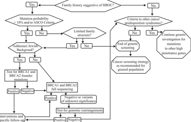
mutations in certain populations. Consequently, in HBOC families that test negative for BRCA point mutations by conventional approaches, screening for large gene rear-rangements inBRCA1and probably alsoBRCA2should be strongly considered. A suggested flowchart for investiga-tion in these families is presented in Figure 2. The availabil-ity of relatively inexpensive and technically
ward screening methods has greatly simplified this process, but often more than one method must be used to fully char-acterize a deletion or duplication in a given patient. Several studies in different populations have proven the usefulness of screening forBRCA1rearrangements, however the prev-alence of such mutations in a given population should be known before definitive recommendations are made re-garding the routine testing for rearrangements. In popula-tions where there are highly prevalent founder rearrange-ments, preliminary screening for pathogenic BRCA gene mutations may be a cost-effective initial strategy.
Acknowledgments
This study was partly supported by grants from Con-selho Nacional de Desenvolvimento Científico e Tecnoló-gico (CNPq, grant no. 477990/2006-1), Fundação de Apoio à Pesquisa do Hospital de Clínicas de Porto Alegre (FIPE, grant no. 04-081) and Susan G Komen for the Cure (POP 0403033). IPE was supported by a grant from Coordenação de Aperfeiçoamento de Pessoal de Nível Superior (CAPES), EIP and SLC were supported by grants from CNPq and PILR was supported by a grant from Fundação de Amparo à Pesquisa do Estado do Rio Grande do Sul (FAPERGS).
References
Agata S, Dalla Palma M, Callegaro M, Scaini MC, Menin C, Ghiotto C, Nicoletto O, Zavagno G, Chieco-Bianchi L, D’Andrea E,et al.(2005) Large genomic deletions inacti-vate the BRCA2 gene in breast cancer families. J Med Genet 42:e64.
Armour JA, Barton DE, Cockburn DJ and Taylor GR (2002) The detection of large deletions or duplications in genomic DNA. Hum Mutat 20:325-337.
Barker DF, Liu X and Almeida ER (1996) The BRCA1 and 1A1.3B promoters are parallel elements of a genomic dupli-cation at 17q21. Genomics 38:215-222.
Barrois M, Bièche I, Mazoyer S, Champème MH, Bressac-de Paillerets B and Lidereau R (2004) Real-time PCR-based gene dosage assay for detecting BRCA1 rearrangements in breast-ovarian cancer families. Clin Genet 65:131-136. Bastard C, Raux G, Fruchart C, Parmentier F, Vaur D, Penther D,
Troussard X, Nagib D, Lepretre S, Tosi M,et al.(2007) Comparison of a quantitative PCR method with FISH for the assessment of the four aneuploidies commonly evaluated in CLL patients. Leukemia 21:1460-1463.
Brown T (2001) Southern blotting. Curr Protoc Immunol Chapter 10: Unit 10.6A.
Brown MA, Xu CF, Nicolai H, Griffiths B, Chambers JA, Black D and Solomon E (1996) The 5’ end of the BRCA1 gene lies within a duplicated region of human chromosome 17q21. Oncogene 12:2507-2513.
Bunyan DJ, Eccles DM, Sillibourne J, Wilkins E, Thomas NS, Shea-Simonds J, Duncan PJ, Curtis CE, Robinson DO, Har-vey JF,et al.(2004) Dosage analysis of cancer predisposi-tion genes by multiplex ligapredisposi-tion-dependent probe amplifica-tion. Br J Cancer 91:1155-1159.
Casilli F, Di Rocco ZC, Gad S, Tournier I, Stoppa-Lyonnet D, Frebourg T and Tosi M (2002) Rapid detection of novel BRCA1 rearrangements in high-risk breast-ovarian cancer families using multiplex PCR of short fluorescent frag-ments. Hum Mutat 20:218-226.
Chin SF, Wang Q, Puisieux A and Caldas C (2001) Absence of re-arrangements in the BRCA2 gene in human cancers. Br J Cancer 84:193-195.
De Lellis L, Curia MC, Aceto GM, Toracchio S, Colucci G, Russo A, Mariani-Costantini R and Cama A (2007) Analysis of ex-tended genomic rearrangements in oncological research. Ann Oncol 18(Suppl 6):173-178.
Dunnen den JT and White SJ (2006) MLPA and MAPH: Sensitive detection of deletions and duplications. Curr Protoc Hum Genet Chapter 7: Unit 7.14.
Ford D, Easton DF, Stratton M, Narod S, Goldgar D, Devilee P, Bishop DT, Weber B, Lenoir G, Chang-Claude J, et al. (1998) Genetic heterogeneity and penetrance analysis of the BRCA1 and BRCA2 genes in breast cancer families. The Breast Cancer Linkage Consortium. Am J Hum Genet 62:676-689.
Gad S, Scheuner MT, Pages-Berhouet S, Caux-Moncoutier V, Bensimon A, Aurias A, Pinto M and Stoppa-Lyonnet D (2001) Identification of a large rearrangement of the BRCA1 gene using colour bar code on combed DNA in an American breast/ovarian cancer family previously studied by direct sequencing. J Med Genet 38:388-392.
Gad S, Klinger M, Caux-Moncoutier V, Pages-Berhouet S, Gau-thier-Villars M, Coupier I, Bensimon A, Aurias A and Stop-pa-Lyonnet D (2002) Bar code screening on combed DNA for large rearrangements of the BRCA1 and BRCA2 genes in French breast cancer families. J Med Genet 39:817-821. Gomez LC, Marzese DM, Adi J, Bertani D, Ibarra J, Mol B, Vos
IJ, De Marchi G and Roqué M (2009) MLPA mutation de-tection in Argentine HNPCC and FAP families. Fam Cancer 8:67-73.
Gutierrez-Enriquez S, de la Hoya M, Martínez-Bouzas C, San-chez de Abajo A, Ramón y Cajal T, Llort G, Blanco I,
Beristain E, Díaz-Rubio E, Alonso C, et al. (2007)
Screening for large rearrangements of the BRCA2 gene in Spanish families with breast/ovarian cancer. Breast Cancer Res Treat 103:103-107.
Hansen TV, Jonson L, Albrechtsen A, Andersen MK, Ejlertsen B and Nielsen FC (2009) Large BRCA1 and BRCA2 genomic rearrangements in Danish high breast - Ovarian cancer fami-lies. Breast Cancer Res Treat 115:69-76.
Hartmann C, John AL, Klaes R, Hofmann W, Bielen R, Koehler R, Janssen B, Bartram CR, Arnold N and Zschocke J (2004) Large BRCA1 gene deletions are found in 3% of German high-risk breast cancer families. Hum Mutat 24:534. Hauss O and Müller O (2007) The protein truncation test in
muta-tion detecmuta-tion and molecular diagnosis. Methods Mol Biol 375:151-164.
Hendrickson BC, Judkins T, Ward BD, Eliason K, Deffenbaugh AE, Burbidge LA, Pyne K, Leclair B, Ward BE and Scholl T (2005) Prevalence of five previously reported and recurrent BRCA1 genetic rearrangement mutations in 20,000 patients from hereditary breast/ovarian cancer families. Genes Chro-mosomes Cancer 43:309-313.
Hofmann W, Gorgens H, John A, Horn D, Huttner C, Arnold N, Scherneck S and Schackert HK (2003) Screening for large
rearrangements of the BRCA1 gene in German breast or ovarian cancer families using semi-quantitative multiplex PCR method. Hum Mutat 22:103-104.
Hogervorst FB, Nederlof PM, Gille JJ, McElgunn CJ, Grippeling M, Pruntel R, Regnerus R, van Welsem T, van Spaendonk R, Menko FH,et al.(2003) Large genomic deletions and dupli-cations in the BRCA1 gene identified by a novel quantitative method. Cancer Res 63:1449-1453.
Kolomietz E, Meyn MS, Pandita A and Squire JA (2002) The role of Alu repeat clusters as mediators of recurrent chromo-somal aberrations in tumors. Genes Chromosomes Cancer 35:97-112.
Lahti-Domenici J, Rapakko K, Paakkonen K, Allinen M, Nevan-linna H, Kujala M, Huusko P and Winqvist R (2001) Exclu-sion of large deletions and other rearrangements in BRCA1 and BRCA2 in Finnish breast and ovarian cancer families. Cancer Genet Cytogenet 129:120-123.
Lim YK, Lau PT, Ali AB, Lee SC, Wong JE, Putti TC and Sng JH (2007) Identification of novel BRCA large genomic rear-rangements in Singapore Asian breast and ovarian patients with cancer. Clin Genet 71:331-342.
Machado PM, Brandao RD, Cavaco BM, Eugenio J, Bento S, Nave M, Rodrigues P, Fernandes A and Vaz F (2007) Screening for a BRCA2 rearrangement in high-risk breast/ovarian cancer families: Evidence for a founder effect and analysis of the associated phenotypes. J Clin Oncol 25:2027-2034.
Mazoyer S (2005) Genomic rearrangements in the BRCA1 and BRCA2 genes. Hum Mutat 25:415-422.
Moisan AM, Fortin J, Dumont M, Samson C, Bessette P, Chi-quette J, Laframboise R, Lépine J, Lespérance B, Pichette R, et al.(2006) No evidence of BRCA1/2 genomic rearrange-ments in high-risk French-Canadian breast/ovarian cancer families. Genet Test 10:104-115.
Montagna M, Santacatterina M, Torri A, Menin C, Zullato D, Chieco-Bianchi L and D’Andrea E (1999) Identification of a 3 kb Alu-mediated BRCA1 gene rearrangement in two breast/ovarian cancer families. Oncogene 18:4160-4165. Montagna M, Dalla PM, Menin C, Agata S, De NA,
Chieco-Bianchi L and D’Andrea E (2003) Genomic rearrangements account for more than one-third of the BRCA1 mutations in northern Italian breast/ovarian cancer families. Hum Mol Genet 12:1055-1061.
Morozova O and Marra MA (2008) From cytogenetics to next-generation sequencing technologies: Advances in the detec-tion of genome rearrangements in tumors. Biochem Cell Biol 86:81-91.
Narod SA and Foulkes WD (2004) BRCA1 and BRCA2: 1994 and beyond. Nat Rev Cancer 4:665-676.
Nevanlinna H and Bartek J (2006) The CHEK2 gene and inherited breast cancer susceptibility. Oncogene 25:5912-5919. Pavlicek A, Noskov VN, Kouprina N, Barrett JC, Jurka J and
Larionov V (2004) Evolution of the tumor suppressor BRCA1 locus in primates: Implications for cancer predispo-sition. Hum Mol Genet 13:2737-2751.
Peelen T, van Vliet M, Bosch A, Bignell G, Vasen HF, Klijn JG, Meijers-Heijboer H, Stratton M, van Ommen GJ, Cornelisse CJ, et al.(2000) Screening for BRCA2 mutations in 81 Dutch breast-ovarian cancer families. Br J Cancer 82:151-156.
Peixoto A, Salgueiro N, Santos C, Varzim G, Rocha P, Soares MJ, Pereira D, Rodrigues H, Bento MJ, Fráguas A,et al.(2006) BRCA1 and BRCA2 germline mutational spectrum and evi-dence for genetic anticipation in Portuguese breast/ovarian cancer families. Fam Cancer 5:379-387.
Perrin-Vidoz L, Sinilnikova OM, Stoppa-Lyonnet D, Lenoir Gil-bert M and Mazoyer S (2002) The nonsense-mediated mRNA decay pathway triggers degradation of most BRCA1 mRNA bearing premature termination codons. Human Mol Genet 11:2805-2814.
Petrij-Bosch A, Peelen T, van Vliet M, van Eijk R, Olmer R, Drüsedau M, Hogervorst FB, Hageman S, Arts PJ, Ligten-berg MJ,et al.(1997) BRCA1 genomic deletions are major founder mutations in Dutch breast cancer patients. Nat Genet 17:341-345.
Preisler-Adams S, Schonbuchner I, Fiebig B, Welling B, Dworniczak B and Weber BH (2006) Gross rearrangements in BRCA1 but not BRCA2 play a notable role in predisposi-tion to breast and ovarian cancer in high-risk families of German origin. Cancer Genet Cytogenet 168:44-49. Puget N, Stoppa-Lyonnet D, Sinilnikova OM, Pages S, Lynch
HT, Lenoir GM and Mazoyer S (1999) Screening for germ-line rearrangements and regulatory mutations in BRCA1 led to the identification of four new deletions. Cancer Res 59:455-461.
Puget N, Gad S, Perrin-Vidoz L, Sinilnikova OM, Stoppa-Lyonnet D, Lenoir GM and Mazoyer S (2002) Distinct BRCA1 rearrangements involving the BRCA1 pseudogene suggest the existence of a recombination hot spot. Am J Hum Genet 70:858-865.
Ratajska M, Brozek I, Senkus-Konefka E, Jassem J, Stepnowska M, Palomba G, Pisano M, Casula M, Palmieri G, Borg A,et al.(2008) BRCA1 and BRCA2 point mutations and large re-arrangements in breast and ovarian cancer families in North-ern Poland. Oncol Rep 19:263-268.
Rouleau E, Lefol C, Tozlu S, Andrieu C, Guy C, Copigny F, Nogues C, Bieche I and Lidereau R (2007) High-resolution oligonucleotide array-CGH applied to the detection and characterization of large rearrangements in the hereditary breast cancer gene BRCA1. Clin Genet 72:199-207. Schouten JP, McElgunn CJ, Waaijer R, Zwijnenburg D, Diepvens
F and Pals G (2002) Relative quantification of 40 nucleic acid sequences by multiplex ligation-dependent probe am-plification. Nucleic Acids Res 30:e57
Smith TM, Lee MK, Szabo CI, Jerome N, McEuen M, Taylor M, Hood L and King MC (1996) Complete genomic sequence and analysis of 117 kb of human DNA containing the gene BRCA1. Genome Res 6:1029-1049.
Staaf J, Törngren T, Rambech E, Johansson U, Persson C, Sellberg G, Tellhed L, Nilbert M and Borg A (2008) Detec-tion and precise mapping of germline rearrangements in BRCA1, BRCA2, MSH2, and MLH1 using zoom-in array comparative genomic hybridization (aCGH). Hum Mutat 29:555-564.
Tancredi M, Sensi E, Cipollini G, Aretini P, Lombardi G, Di CC, Presciuttini S, Bevilacqua G and Caligo MA (2004) Haplo-type analysis of BRCA1 gene reveals a new gene rearrange-ment: Characterization of a 19.9 kbp deletion. Eur J Hum Genet 12:775-777.
BRCA1 and BRCA2 in western Denmark. Cancer Genet Cytogenet 168:168-171.
Tournier I, Paillerets BB, Sobol H, Stoppa-Lyonnet D, Lidereau R, Barrois M, Mazoyer S, Coulet F, Hardouin A, Chompret A,et al.(2004) Significant contribution of germline BRCA2 rearrangements in male breast cancer families. Cancer Res 64:8143-8147.
Unger MA, Nathanson KL, Calzone K, Antin-Ozerkis D, Shih HA, Martin AM, Lenoir GM, Mazoyer S and Weber BL (2000) Screening for genomic rearrangements in families with breast and ovarian cancer identifies BRCA1 mutations previously missed by conformation-sensitive gel electro-phoresis or sequencing. Am J Hum Genet 67:841-850.
Vanin EF (1985) Processed pseudogenes: Characteristics and evolution. Annu Rev Genet 19:253-272.
Vasickova P, Machackova E, Lukesova M, Damborsky J, Horky O, Pavlu H, Kuklova J, Kosinova V, Navratilova M and Foretova L (2007) High occurrence of BRCA1 intragenic rearrangements in hereditary breast and ovarian cancer syn-drome in the Czech Republic. BMC Med Genet 8:32.
Walsh T and King MC (2007) Ten genes for inherited breast can-cer. Cancer Cell 11:103-105.
Walsh T, Casadei S, Coats KH, Swisher E, Stray SM, Higgins J, Roach KC, Mandell J, Lee MK, Ciernikova S,et al.(2006) Spectrum of mutations in BRCA1, BRCA2, CHEK2, and
TP53 in families at high risk of breast cancer. JAMA 295:1379-1388.
Wang T, Lerer I, Gueta Z, Sagi M, Kadouri L, Peretz T and Abeliovich D (2001) A deletion/insertion mutation in the BRCA2 gene in a breast cancer family: A possible role of the Alu-polyA tail in the evolution of the deletion. Genes Chro-mosomes Cancer 31:91-95.
Weitzel JN, Lagos VI, Herzog JS, Judkins T, Hendrickson B, Ho JS, Ricker CN, Lowstuter KJ, Blazer KR, Tomlinson G,et al.(2007) Evidence for common ancestral origin of a recur-ring BRCA1 genomic rearrangement identified in high-risk Hispanic families. Cancer Epidemiol Biomarkers Prev 16:1615-1620.
Woodward AM, Davis TA, Silva AG, Kirk JA and Leary JA (2005) Large genomic rearrangements of both BRCA2 and BRCA1 are a feature of the inherited breast/ovarian cancer phenotype in selected families. J Med Genet 42:e31.
Internet Resources
Multiplex ligation-dependent probe amplification:
http://www.mrc-holland.com/pages/p002pag.html (July 19, 2008).
Associate Editor: Emmanuel Dias Neto
License information: This is an open-access article distributed under the terms of the Creative Commons Attribution License, which permits unrestricted use, distribution, and reproduction in any medium, provided the original work is properly cited.