REVIEW
Do hereditary syndrome-related gynecologic cancers have
any specific features?
Nelson Neto
1&Teresa Margarida Cunha
2Received: 29 May 2015 / Revised: 20 July 2015 / Accepted: 29 July 2015 / Published online: 4 September 2015 #The Author(s) 2015. This article is published with open access at Springerlink.com
Abstract
Hereditary syndromes are responsible for 10 % of
gynaecologic cancers, among which hereditary
breast-ovarian cancer and hereditary non-polyposis colon cancer
syndromes, known as HBOC and Lynch syndromes
re-spectively, present the highest relative risk. The latter
pre-disposes to endometrial cancer and both contribute to
ovarian cancer. Cowden syndrome-related endometrial
can-cer and the increased risk of ovarian, uterine and can-cervical
cancers associated with Peutz-Jeghers syndrome, are also
demonstrated, while Li-Fraumeni syndrome patients are
prone to develop ovarian and endometrial cancers. Despite
these syndromes
’
susceptibility to gynaecologic cancers
being consensual, it is still not clear whether these
tu-mours have any epidemiologic, clinical, pathologic or
im-aging specific features that could allow any of the
inter-vening physicians to raise suspicion of a hereditary
syn-drome in patients without known genetic risk. Moreover,
controversy exists regarding both screening and
surveil-lance schemes. Our literature review provides an updated
perspective on the evidence-based specific features of
tu-mours related to each of these syndromes as well as on
the most accepted screening and surveillance guidelines. In
addition, some illustrative cases are presented.
Teaching Points
•
HBOC syndrome is mainly associated with ovarian HGSC,
which arises in fallopian fimbriae.
•
LS-related endometrial tumours show histological diversity
and predilection for lower uterine segment.
•
LS and CS-related ovarian cancers are mostly of non-serous
type, usually endometrioid.
•
Ovarian SCTAT and cervical adenoma malignum are
strong-ly associated with PJS.
•
Unfortunately, hereditary gynaecologic cancers do not seem
to have distinctive imaging features.
Keywords
Gynaecologic neoplasms . Hereditary cancer
syndromes . Neoplasms by histological type . Diagnostic
imaging . Practice guideline
Introduction
Recently, there have been significant advances in the
knowl-edge of female genital tract malignancies related to hereditary
cancer susceptibility syndromes. According to the American
Society of Clinical Oncology, hereditary syndromes are
re-sponsible for about 10 % of gynaecologic cancers [
1
].
One striking example is the discovery of the association
between germline mutations in breast cancer (BRCA) 1 and
2 genes and ovarian cancers in hereditary breast-ovarian
can-cer (HBOC) syndrome. Another one is the role of germline
mutations in DNA mismatch repair (MMR) genes in
endome-trial and ovarian carcinogenesis related to hereditary
non-polyposis colon cancer, also known as Lynch syndrome (LS).
Increased risk of endometrial cancer caused by mutation in
the phosphatase and tensin homolog (PTEN) gene in Cowden
syndrome (CS) is also demonstrated, as well as ovarian,
uter-ine and cervical cancers related to Peutz-Jeghers syndrome
* Nelson Neto
1
Radiology Department, Centro Hospitalar de Lisboa Ocidental, Estrada do Forte do Alto do Duque, 1449-005 Lisboa, Portugal 2
Radiology Department, Instituto Português de Oncologia de Lisboa Francisco Gentil, Rua Professor Lima Basto,
(PJS), due to liver kinase b1 (LKB1/STK11) gene mutation.
Ovarian and endometrial cancers also occur excessively in
patients with Li-Fraumeni syndrome (LFS), although the
un-derstanding of the contribution of this inherited germline
mu-tation in p53 is less established.
Despite the clear evidence of these inherited disorders
’
sus-ceptibility to gynaecologic cancers, it is still not generically
clear whether these tumours have any epidemiologic, clinical,
pathologic or imaging specific features that could allow any of
the intervening physicians to raise suspicion of a hereditary
syndrome in patients without known genetic risk. Moreover,
thei r screeni ng and sur veillance schemes remain
controversial.
Our literature review provides an updated perspective on
the evidence-based specific features of tumours related to each
of these syndromes, as well as on the most accepted screening
and surveillance guidelines. In addition, some illustrative
cases are presented.
Hereditary breast-ovarian cancer syndrome
Ovarian cancer is the most lethal gynaecologic cancer, 70 %
being detected with advanced disease and therefore having
poor prognosis, with a 5-year survival rate of only 15 to
25 % for stage IV [
2
,
3
].
90 % have epithelial origin [
4
], hereditary ones accounting
for at least 10 % of cases [
2
,
5
,
6
] and the majority being due to
mutations in BRCA1 gene [
5
,
7
]. Lifetime risk for ovarian
cancers is 40
–
66 % and 10
–
20 % in BRCA1 and BRCA2
germline mutation carriers, respectively [
8
–
11
], in contrast
to 1.8 % in the general population [
12
].
BRCA1 locus on chromosome 17q and BRCA2 on
chro-mosome 13q both function as tumour suppressor genes
[
13
–
15
]. These mutations, also associated with increased risk
of breast cancer, are both on the basis of HBOC and
site-specific ovarian cancer syndromes. Ethnic background
signif-icantly influences the mutation rates, which are particularly
high among Ashkenazi Jews compared to other populations
[
16
,
17
].
Mean age of presentation of ovarian cancer in HBOC
syn-drome is 51
–
53 years, about 10 years earlier than in
non-BRCA mutations carriers [
5
,
12
].
Concerning its pathology, the great majority are high-grade
serous carcinomas (HGSC) of papillary type, diagnosed at
advanced stage [
2
,
7
,
12
,
18
]. Despite this, survival of ovarian
cancer seems to be surprisingly better in these women than in
sporadic ones [
7
], for unknown reasons.
Both cases of BRCA1 mutation-related ovarian cancer
pre-sented (Figs.
1
and
2
) were high-grade tumours with no tubal
involvement, diagnosed at younger ages than is typical for the
general population, which metastasized to lymph nodes. On
the other hand, a localized ovarian tumour in a BRCA2
mu-tation carrier is presented in Fig.
3
.
A curious detail regarding the origin of most ovarian
HGSC in patients with HBOC, suggested by recent studies
of prophylactically removed ovaries, is that instead of arising
primarily from the ovary as originally assumed, these tumours
actually seem to arise from the fallopian tube fimbriae and are
characterized by p53 signature mutations, typical of tubal
Fig. 1 a-d. High-grade serous carcinoma of the right ovary in a 56-year-old BRCA1-mutation carrier woman. Magnetic resonance imaging (MRI) scan with T2-weighted (a), post-gadolinium fat-suppressed (FS) T1-weighted (b) and diffusion-weighted (c) images, showing a complex cystic-solid
intraepithelial carcinomas. In addition, these studies failed to
identify reproducible precursor lesions in the ovaries [
2
,
12
].
For these reasons, some experts consider ovarian and fallopian
tube cancer in BRCA-mutations carriers to be more properly
termed as adnexal carcinomas [
19
]. However, interpretation of
p53 signatures, defined as p53 positivity by
immunohisto-chemical staining in at least 12 secretory cells with a low
proliferative index, must be done cautiously, as it does not
mean a neoplastic lesion is present and is a quite common
occurrence in fallopian tubes, regardless of BRCA status [
12
].
Another cancer type, in whose progression the loss of
BRCA1 function with concurrent deletion of p53 may be an
important step, is uterine leiomyosarcoma, a rare
gynaecologic malignancies with a low survival rate [
20
].
As genetic testing for BRCA genes is not cost-effective for
the general population, thus far the selection of candidates still
relies on family history [
1
]. Once a genetic risk is confirmed,
management is not consensual, but usually includes serial
serum determinations of multiple tumour markers together
with transvaginal ultrasound, another option being
prophylac-tic oophorectomy after planned childbearing is completed.
Although a protective effect of oral contraceptives against
ovarian cancer is nearly proven, their routine prophylactic
use is no longer recommended due to the possible increased
relative risk of breast cancer in patients with HBOC syndrome
[
5
].
Lynch syndrome
Lynch syndrome (LS), caused by an autosomal-dominant
he-reditary germline mutation in one of the MMR genes
—
MSH2, MLH1 and MSH6, in decreasing order [
21
]
—
predis-poses to early onset of multiple cancer types, including colon,
endometrial and ovarian ones, sometimes with synchronous
presentation [
22
,
23
]. MMR maintain genomic stability by
correcting mismatches generated during DNA replication,
their malfunction promoting cancer due to microsatellite
in-stability [
24
]. However, microsatellite instability is also
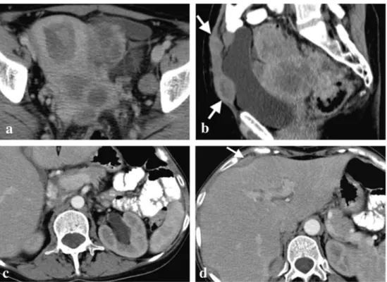
Fig. 2 a-d. Bilateral undifferentiated ovarian carcinomas in a 34-year-old BRCA1-mutation carrier woman. CT scan shows a large
multiloculated cystic left adnexal mass with thick septa (a). A right adnexal mass, similar to that one described in imagea, but with a more prominent solid component, was also present (b). Left para-aortic lymph node metastases are also present (c). Moderate ascites is evident in all images but there was no pleural effusion (d). Pathologic examination ruled out ovarian capsule and tubal invasion
present in 15
–
20 % of corresponding sporadic cancers,
usual-ly due to MLH1 methylation [
25
].
Traditionally associated with colorectal cancer, it is
nowa-days consensual that women with LS are at equal or even
higher risk for development of gynaecologic malignancy
[
26
–
29
], as their sentinel cancer in more than half of cases
[
30
,
31
]. Their lifetime cumulative risk of endometrial cancer
is 40
–
60 % [
26
,
28
,
29
,
31
] and that of ovarian cancer is 10
–
12 % [
18
,
21
,
27
,
32
–
34
], appearing to be particularly high for
MSH2-mutation carriers [
35
–
37
] and accounting for 2 % of all
ovarian cancers [
34
].
Although it has long been thought that the average age of
onset of LS-related endometrial cancer is 45
–
50 years, in
con-trast to 65 years in sporadic cases [
5
,
38
,
39
], other studies
have suggested that a cutoff age of 50 years old for screening
could lead to 25
–
60 % of LS missing cases [
40
,
41
], therefore
a cutoff age of 60 has already been advocated [
22
]. Regardless
of this controversy, a diagnosis of endometrial cancer at a
young age should raise suspicion for LS, especially if the most
typical constitutional factor of sporadic endometrial cancer
—
obesity
—
is absent and family history is positive [
42
,
43
].
Mean age for developing ovarian cancer is 40
–
48 years
among patients with LS [
5
,
36
,
39
].
While sporadic endometrial cancers that have
microsatel-lite instability are almost exclusively of endometrioid type,
usually well to moderately differentiated, those related to LS
tend to be histologically more diverse also occurring
non-endometrioid carcinomas, including serous, clear cell and
un-differentiated ones, although the majority is still of
endometrioid type [
38
,
43
]. LS-related endometrial cancers
also show a predilection for the lower uterine segment, with
up to one-third of these tumours arising in this location
[
44
–
46
].
Although certain distinctive microscopic features of
endometrioid carcinomas, like poor differentiation, tumour
heterogeneity and increased tumour-infiltrating lymphocytes,
have been shown to be suggestive of the presence of high
levels of microsatellite instability [
40
,
47
,
48
], there are
con-flicting data regarding their utility [
46
].
Contrary to ovarian cancers of the general population or
that are HBOC syndrome-related, those related to LS are
mostly of the non-serous type, including endometrioid, clear
cell and undifferentiated carcinomas [
36
,
49
]. Endometrioid is
most commonly associated with LS [
36
] and the second most
frequent histological subtype in the general population [
50
],
the majority being well to moderately differentiated and
pur-suing favourable clinical outcomes [
36
,
51
]. There may be
synchronous endometrial thickening, representing either
hy-perplasia or carcinoma [
52
]. Among ovarian carcinomas
as-sociated with MMR defects, including LS-related ones, clear
cell subtype represents the majority [
35
].
Prognostic impact of MMR status is not clear for either
endometrial or ovarian carcinomas [
12
].
LS cases illustrated in Figs.
4
and
5
correspond to patients
whose sentinel cancers were gynaecologic, the latter with
syn-chronous endometrial and ovarian tumours.
Current gynaecologic cancer screening guidelines for
women with LS, which include annual endometrial
sampling and transvaginal ultrasound beginning at 30
–
35 years, are not considered to be effective, but are still
a reasonable option [
12
,
53
].
Cowden syndrome
Cowden syndrome (CS), part of a broader category termed
PTEN hamartoma tumour syndrome, is characterized by a
mutation in the PTEN tumour-suppressor gene, which
leads to uncontrolled cell division and the formation
of hamartomatous neoplasms and certain cancers,
representing an increased lifetime risk of endometrial
carcinomas of 13
–
19 % [
54
].
Histologically, reported cases of CS-related endometrial
carcinomas [
54
–
57
] and the one illustrated in Fig.
6
are of
endometrioid type.
Although still not validated, the adoption of the screening
guidelines for LS, including annual endometrial biopsies
be-ginning at age 30 to 35, or 5 years before the earliest family
diagnosis of endometrial cancer and annual ultrasound
exam-ination with biopsy of suspicious areas for postmenopausal
women, has already been proposed [
54
]. Formal evaluation
for CS when endometrial cancer is diagnosed in adolescence
has also been suggested [
55
].
Li-fraumeni syndrome
Li-fraumeni syndrome (LFS) is an extremely rare
autosomal-dominant hereditary disorder characterized by a germline
mu-tation in the tumour-suppression gene p53, which leads to an
estimated 50-fold risk over the general population of
develop-ing several types of cancer [
58
], more than half occurring
before age 30 [
59
]. Although endometrial and ovarian cancers
have been found to occur excessively in at least some families
who have met criteria for LFS, their link to the syndrome is
not definitely established, occurring at a much lower rate than
other cancer types, like breast cancer [
60
].
Actually, the fact that distal fallopian tubes of women with
LFS are exquisitely prone to developing p53 signatures,
iden-tical to those described in BRCA1/2-mutations carriers and
general population, does not mean an association with ovarian
cancer, as at least one more genotoxic event is needed to
produce the malignant phenotype [
61
].
There are no studies specifically examining the relation
between LFS and endometrial cancer.
Fig. 5 a–d. Poorly differentiated endometrioid carcinomas of the left ovary and of the endometrium in a 56-year-old woman with Lynch syndrome. CECT shows a large complex cystic-solid multiloculated left adnexal mass with no cleavage plane from the uterine corpus (a) in concordance with invasion of the myometrium, as pathologically demonstrated. The same image depicts the endometrial tumour, seen as a polypoid solid lesion within the uterine cavity surrounded by fluid,
Peutz-jeghers syndrome
Peutz-jeghers syndrome (PJS) is a rare autosomal
domi-nant disease due to mutations in the tumour-suppressor
gene STK11 [
62
], which predisposes not only to
hamartomatous gastrointestinal polyps and
mucocutane-ous pigmentation, but also to numermucocutane-ous malignancies,
in-cluding gynaecologic ones, the latter having a reported
relative risk of 27.7 % in comparison to the general
pop-ulation [
63
].
Risk of ovarian, cervical and uterine cancers
associ-ated with PJS is 18
–
21 %, 10 % and 9 %, respectively
[
64
].
PJS-related gynaecological cancers are of some
character-istic histological types, particularly the sex cord tumours with
annular tubules (SCTAT) of the ovary [
63
,
65
–
67
], 36 %
oc-curring in association with this syndrome [
65
], although with
lower risk of malignant transformation than in the general
population [
63
]. This distinctive ovarian neoplasm, whose
predominant component has morphologic features
intermedi-ate between those of granulosa cell and Sertoli cell tumours,
may produce both oestrogen and progesterone [
68
]. This
his-tological type is followed by Sertoli cell, mucinous, serous
and mature teratoma [
67
].
There is also evidence that patients with PJS are prone to
develop endometrial adenocarcinomas [
69
,
70
], especially
highly invasive ones [
70
].
Among cervical tumours, there is an important association
with minimal deviation adenocarcinoma [
65
,
66
,
71
], known
as adenoma malignum, with 10 % of all cases occurring in PJS
patients [
66
]. This is a well-differentiated mucinous
adenocar-cinoma with highly aggressive behaviour, despite its
decep-tively benign appearance and very scarce cytological features
of malignancy within the tumour [
71
].
Gynaecologic cancer screening surveillance
recommenda-tions for patients with PJS include annual Papanicolaou test by
age 18 and annual pelvic examination and ultrasound by age
20 [
72
], which should also target the potential malignant
change of ovarian SCTAT [
63
].
Conclusions
Regardless of some genetic specifications, the following
par-ticularities concerning each of the syndromes discussed above
are consensual nowadays:
–
HBOC syndrome is mainly associated with ovarian
HGSC, which seem to arise in the fallopian fimbriae
and have better prognosis than sporadic cancers.
–
LS predisposes to endometrial cancer, at a lower rate than
ovarian cancers. Endometrial cancers show a predilection
for the lower uterine segment and tend to be histologically
more diverse in contrast to their sporadic counterparts,
including non-endometrioid carcinomas. Both LS and
CS-related ovarian cancers are mostly of non-serous type,
usually endometrioid.
–
LFS only slightly increases the risk of endometrial and
ovarian cancers.
–
PJS increases the risk of ovarian, cervical and uterine
cancers, in decreasing order, ovarian SCTAT and cervical
adenoma malignum being strongly associated.
–
Unfortunately, hereditary gynaecologic cancers do not
seem to have any imaging characteristics that might be
reliably used to distinguish them from sporadic cancers.
–
In order to ensure early identification of high-risk
pa-tients, every time a gynaecologic malignancy is
diag-nosed under the expected age in the general population,
a careful anamnesis including familial history of cancer
and genetic confirmation when indicated is required.
–
Screening and surveillance schemes usually consist of an
annual pelvic examination with endometrial sampling
and ultrasound beginning in young adulthood.
Fig. 6 a,b. High-grade endometrioid carcinoma within an endometrial hyperplastic polyp in a 62-year-old woman with Cowden syndrome. T2-weighted image (a) shows distension of the uterine cavity due to a polyp, more evident on post-gadolinium FS T1-weighted image (b), which is
Open AccessThis article is distributed under the terms of the Creative C o m m o n s A t t r i b u t i o n 4 . 0 I n t e r n a t i o n a l L i c e n s e ( h t t p : / / creativecommons.org/licenses/by/4.0/), which permits unrestricted use, distribution, and reproduction in any medium, provided you give appro-priate credit to the original author(s) and the source, provide a link to the Creative Commons license, and indicate if changes were made.
References
1. ASCO/Society of Gynecologic Oncology Special Session (2004) Clinical management of patients with hereditary predisposition to gynecologic (ovarian and endometrial) cancers. Program and ab-stracts of the 40th Annual Meeting of the American Society of Clinical Oncology. New Orleans, Louisiana
2. Lalwani N, Prasad SR, Vikram R et al (2011) Histologic, molecular, and cytogenetic features of ovarian cancers: implications for diag-nosis and treatment. Radiographics 31(3):625–646
3. Landis SH, Murray T, Bolden S et al (1998) Cancer statistics. CA Cancer J Clin 48:6–29
4. Feeley KM, Wells M (2001) Precursor lesions of ovarian epithelial malignancy. Histopathology 38(2):87–95
5. Lynch HT, Casey MJ, Shaw TG et al (1998) Hereditary Factors in Gynecologic Cancer. Oncologist 3(5):319–338
6. Lynch HT, Lynch JF, Conway TA (1993) Hereditary ovarian can-cer. In: Rubin SC, Sutton GP (eds) Ovarian cancan-cer. McGraw-Hill, New York, pp 189–217
7. Rubin SC, Benjamin I, Behbakht K et al (1996) Clinical and path-ological features of ovarian cancer in women with germ-line muta-tions of BRCA1. N Engl J Med 335(19):1413–1416
8. Easton DF, Bishop DT, Ford D et al (1993) Genetic linkage analysis in familial breast and ovarian cancer: results from 214 families. Am J Hum Genet 52:678–701
9. Easton DF, Ford D, Bishop DT et al (1995) Breast and ovarian cancer incidence in BRCA1 mutation carriers. Am J Hum Genet 56:265–271 10. Domanska K, Malander S, Masback A et al (2007) Ovarian cancer at young age: the contribution of mismatch-repair defects in a population-based series of epithelial ovarian cancer before age 40. Int J Gynecol Cancer 17:789–793
11. Schmeler KM, Lynch HT, Chen LM et al (2006) Prophylactic sur-gery to reduce the risk of gynecologic cancers in the Lynch syn-drome. N Engl J Med 354:261–269
12. Longacre T, Folkins A (2011) 217 Gynecologic familial cancer syn-dromes: what does the practicing pathologist need to know? American society for clinical pathology. Annual Meeting. Las Vegas, Nevada 13. Liu FS, Ho ESC, Shih A (1997) Mutational analysis of the BRCA1
tumor suppressor gene in endometrial carcinoma. Gynecol Oncol 66:449–453
14. Haber D, Harlow E (1997) Tumor-suppressor genes: evolving def-initions in the genomic age. Nat Genet 16:320–322
15. Merajver SD, Frank TS, Xu J et al (1995) Germline BRCA1 mu-tations and loss of the wild-type allele in tumors from families with early onset breast and ovarian cancer. Clin Cancer Res 1:539–544 16. Struewing JP, Hartge P, Wacholder S et al (1997) The risk of cancer
associated with specific mutations of BRCA1 and BRCA2 among Ashkenazi Jews. N Engl J Med 336:1401–1408
17. Abeliovich D, Kaduri L, Lerer I et al (1997) The founder mutations 185delAG and 5382insC in BRCA1 and 6174delT in BRCA2 ap-pear in 60 % of ovarian cancer and 30% of early-onset breast cancer patients among Ashkenazi women. Am J Hum Genet 60:505–514 18. Bewtra C, Watson P, Conway T et al (1992) Hereditary ovarian cancer: a clinicopathological study. Int J Gynecol Pathol 11:180–187
19. Piek JM, Dorsman JC, Zweemer RP et al (2003) Women harboring BRCA1/2 germline mutations are at risk for breast and female ad-nexal carcinoma. Int J Gynecol Pathol 22(3):315–316
20. Xing D, Scangas G, Nitta M et al (2009) A role for BRCA1 in uterine leiomyosarcoma. Cancer Res 69(21):8231–8235
21. Bonadona V, Bonaïti B, Olschwang S et al (2011) Cancer risks associated with germline mutations in MLH1, MSH2, and MSH6 genes in Lynch syndrome. JAMA 305:2304–2310
22. Wang Y, Li J, Cragun J et al (2013) Lynch syndrome related endo-metrial cancer: clinical significance beyond the endometrium. J Hematol Oncol 6:22
23. Soliman PT, Broaddus RR, Schmeler KM et al (2005) Women With Synchronous Primary Cancers of the Endometrium and Ovary: Do They Have Lynch Syndrome? J Clin Oncol 23(36):9344–9350 24. Jass JR, Cottier DS, Jeevaratnam P et al (1995) Diagnostic use of
microsatellite instability in hereditary non-polyposis colorectal can-cer. Lancet 346:1200–1201
25. Peltomaki P (2003) Role of DNA mismatch repair defects in the pathogenesis of human cancer. J Clin Oncol 21:1174–1179 26. Aarnio M, Sankila R, Pukkala E et al (1999) Cancer risk in mutation
carriers of DNA-mismatch-repair genes. Int J Cancer 81(2):214–218 27. Dunlop MG, Farrington SM, Carothers AD et al (1997) Cancer risk associated with germline DNA mismatch repair gene mutations. Hum Mol Genet 6(1):105–110
28. Quehenberger F, Vasen HF, van Houwelingen HC (2005) Risk of colorectal and endometrial cancer for carriers of mutations of the hMLH1 and hMSH2 gene: correction for ascertainment. J Med Genet 42:491–496
29. Vasen HF, Stormorken A, Menko FH et al (2001) MSH2 mutation carriers are at higher risk of cancer than MLH1 mutation carriers: a study of hereditary nonpolyposis colorectal cancer families. J Clin Oncol 19:4074–4080
30. Lu KH, Dinh M, Kohlmann W et al (2005) Gynecologic cancer as a "sentinel cancer" for women with hereditary nonpolyposis colorec-tal cancer syndrome. Obstet Gynecol 105:569–574
31. Vasen HF, Offerhaus GJ, den Hartog Jager FC et al (1990) The tumor spectrum in hereditary non-polyposis colorectal cancer: a study of 24 kindreds in the Netherlands. Int J Cancer 46:31–34 32. Prat J, Ribé A, Gallardo A (2005) Hereditary ovarian cancer. Hum
Pathol 36(8):861–870
33. Grindedal EM, Renkonen-Sinisalo L, Vasen H et al (2010) Survival in women with MMR mutations and ovarian cancer: a multicentre study in Lynch syndrome kindreds. J Med Genet 47:99–102 34. Malander S, Rambech E, Kristoffersson U et al (2006) The
contri-bution of the hereditary nonpolyposis colorectal cancer syndrome to the development of ovarian cancer. Gynecol Oncol 101:238–243 35. Jensen KC, Mariappan MR, Putcha GV et al (2008) Microsatellite instability and mismatch repair protein defects in ovarian epithelial neoplasms in patients 50 years of age and younger. Am J Surg Pathol 32:1029–1037
36. Ketabi Z, Bartuma K, Bernstein I et al (2011) Ovarian cancer linked to Lynch syndrome typically presents as early-onset, non-serous epithelial tumors. Gynecol Oncol 121:462–465
37. Vasen HF, Wijnen JT, Menko FH et al (1996) Cancer risk in fam-ilies with hereditary nonpolyposis colorectal cancer diagnosed by mutation analysis. Gastroenterology 110:1020–1027
38. Broaddus RR, Lynch HT, Chen LM et al (2006) Pathologic features of endometrial carcinoma associated with HNPCC: a comparison with sporadic endometrial carcinoma. Cancer 106(1):87–94 39. Aarnio M, Mecklin JP, Aaltonen LA et al (1995) Life-time risk of
different cancers in hereditary non-polyposis colorectal cancer (HNPCC) syndrome. Int J Cancer 64:430–433
Colorectal Cancer) among Endometrial Cancer Patients. Cancer Res 67:9603
41. Ryan P, Mulligan AM, Aronson M et al (2012) Comparison of clinical schemas and morphologic features in predicting Lynch syn-drome in mutation-positive patients with endometrial cancer en-countered in the context of familial gastrointestinal cancer regis-tries. Cancer 118:681–688
42. Chen S, Wang W, Lee S et al (2006) Prediction of germline muta-tions and cancer risk in the Lynch syndrome. JAMA 296:1479–
1487
43. Carcangiu ML, Radice P, Casalini P et al (2010) Lynch syndrome–
related endometrial carcinomas show a high frequency of nonendometrioid types and of high FIGO grade endometrioid types. Int J Surg Pathol 18:21–26
44. Westin SN, Lacour RA, Urbauer DL et al (2008) Carcinoma of the lower uterine segment: a newly described association with Lynch syndrome. J Clin Oncol 26:5965–5971
45. Goodfellow PJ, Buttin BM, Herzog TJ et al (2003) Prevalence of defective DNA mismatch repair and MSH6 mutation in an unse-lected series of endometrial cancers. Proc Natl Acad Sci U S A 100(10):5908–5913
46. Honore LH, Hanson J, Andrew SE (2006) Microsatellite instability in endometrioid endometrial carcinoma: correlation with clinically relevant pathologic variables. Int J Gynecol Cancer 16:1386–1392 47. Garg K, Leitao MM Jr, Kauff ND et al (2009) Selection of endo-metrial carcinomas for DNA mismatch repair protein immunohis-tochemistry using patient age and tumor morphology enhances de-tection of mismatch repair abnormalities. Am J Surg Pathol 33: 925–933
48. Shia J, Black D, Hummer AJ et al (2008) Routinely assessed mor-phological features correlate with microsatellite instability status in endometrial cancer. Hum Pathol 39:116–125
49. Lu FI, Gilks CB, Mulligan AM et al (2012) Prevalence of loss of expression of DNA mismatch repair proteins in primary epithelial ovarian tumors. Int J Gynecol Pathol 31(6):524–531
50. Gilks CB, Prat J (2009) Ovarian carcinoma pathology and genetics: recent advances. Hum Pathol 40(9):1213–1223
51. Watson P, Butzow R, Lynch HT et al (2001) The clinical features of ovarian cancer in hereditary nonpolyposis colorectal cancer. Gynecol Oncol 82:223–228
52. Jung SE, Lee JM, Rha SE et al (2002) CT and MR imaging of ovarian tumors with emphasis on differential diagnosis. RadioGraphics 22(6):1305–1325
5 3 . L i n d o r N M , P e t e r s e n G M , H a d l e y D W e t a l ( 20 0 6 ) Recommendations for the care of individuals with an inherited predisposition to Lynch syndrome: a systematic review. JAMA 296(12):1507–1517
54. ElNaggar AC, Spunt SL, Smith W et al (2013) Endometrial cancer in a 15-year-old girl: A complication of Cowden Syndrome. Gynecol Oncol Case Rep 3:18–19
55. Baker WD, Soisson AP, Dodson MK (2013) Endometrial cancer in a 14-year-old girl with Cowden syndrome: a case report. J Obstet Gynaecol Res 39(4):876–878
56. Edwards JM, Alsop S, Modesitt SC (2012) Coexisting atypical polypoid adenomyoma and endometrioid endometrial carcinoma in a young woman with Cowden Syndrome: Case report and
implications for screening and prevention. Gynecol Oncol Case Rep 2(2):29–31
57. Schmeler KM, Daniels MS, Brandt AC et al (2009) Endometrial cancer in an adolescent: a possible manifestation of Cowden syn-drome. Obstet Gynecol 114(2 Pt 2):477–479
58. Rédei G (2008) Encyclopedia of Genetics, Genomics, Proteomics and Informatics, vol 1, 3rd edn. Springer, p 1102
59. Le Bihan C, Moutou C, Brugieres L et al (1995) ARCAD: a method for estimating age-dependent disease risk associated with mutation carrier status from family data. Genet Epidemiol 12(1):13–25 60. Schneider K, Zelley K, Nichols KE et al (1999, Updated 2013)
Li-Fraumeni Syndrome. In: Pagon RA, Adam MP, Ardinger HH et al (eds) GeneReviews®, University of Washington, Seattle. Available viahttp://www.ncbi.nlm.nih.gov/books/NBK1311/
61. Xian W, Miron A, Roh M et al (2010) The Li Fraumeni Syndrome (LFS): a model for the initiation of p53 signatures in the distal fallopian tube. J Pathol 220(1):17–23
62. Jenne DE, Reimann H, Nezu J et al (1998) Peutz–Jeghers syndrome is caused by mutations in a novel serine threonine kinase. Nat Genet 18:38–43
63. Resta N, Pierannunzio D, Lenato GM et al (2013) Cancer risk associated with STK11/LKB1 germline mutations in Peutz–
Jeghers syndrome patients: Results of an Italian multicenter study. Dig Liver Dis 45(7):606–611
64. Gammon A, Jasperson K, Kohlmann W et al (2009) Hamartomatous polyposis syndromes. Best Pract Res Clin Gastroenterol 23(2):219–231
65. Young R, Welch W, Dickersin GR et al (1982) Ovarian sex cord tumor with annular tubules: review of 74 cases including 27 with Peutz-Jeghers syndrome and four with adenoma malignum of the cervix. Cancer 50(7):1384–1402
66. Srivatsa PJ, Keeney GL, Podratz KC (1994) Disseminated cervical adenoma malignum and bilateral ovarian sex cord tumors with an-nular tubules associated with Peutz-Jeghers syndrome. Gynecol Oncol 53:256
67. Papageorgiou T, Stratakis CA (2002) Ovarian tumors associated with multiple endocrine neoplasias and related syndromes (Carney complex, Peutz-Jeghers Syndrome, von Hippel Lindau disease, Cowden's disease). Int J Gynecol Cancer 12:337–347 68. Taheri D, Afshar-Moghadam N, Mahzoni P et al (2013) Cancer
problem in Peutz–Jeghers syndrome. Adv Biomed Res 2:35 69. Tanwar PS, Kaneko-Tarui T, Zhang L et al (2012) Stromal liver
kinase B1 [STK11] signaling loss induces oviductal adenomas and endometrial cancer by activating mammalian Target of Rapamycin Complex 1. PLoS Genet 8(8):e1002906
70. Contreras CM, Gurumurthy S, Haynie JM et al (2008) Loss of Lkb1 provokes highly invasive endometrial adenocarcinomas. Cancer Res 68(3):759–766
71. Connolly D, Katabuchi H, Cliby W et al (2000) Somatic mutations in the STK11/LKB1 gene are uncommon in rare gynecological tumor types associated with Peutz-Jeghers syndrome. Am J Pathol 1565(1):339–345