Ana Lopes Tavares
Dissertation presented to obtain the Ph.D degree in Biology
Instituto de Tecnologia Química e Biológica | Universidade Nova de Lisboa
Oeiras,
Staphylococcus aureus (CA-MRSA) in Portugal:
Ana Lopes Tavares
Dissertation presented to obtain the Ph.D degree in Biology
Instituto de Tecnologia Química e Biológica | Universidade Nova de Lisboa
Oeiras, December, 2014
Staphylococcus aureus
(CA-MRSA) in Portugal:
Second edition, January 2015
© Ana Tavares
!
!
!
!
!
!
Supervisors:
Dr. Hermínia de Lencastre
Dr. Maria Miragaia
Chairman of Examiners:
Dr. Cecília Arraiano, Full Professor at ITQB
Examiners:
Dr. Constança Pomba
Dr. João Paulo Gomes
Dr. José Melo Cristino
Dr. Isabel Couto
Dissertation presented to obtain the PhD degree in
Biology by Universidade Nova de Lisboa, Instituto de
To Dr. Hermínia de Lencastre, Professor and Head of the Laboratory of Molecular Genetics
(Microbiology of Human Pathogens Unit) at Instituto de Tecnologia Química e Biológica António
Xavier (ITQB), Universidade Nova de Lisboa (UNL), my supervisor, for accepting me in her
Laboratory and welcoming me into her group. For her vast scientific knowledge and fascinating
carrier in the field of staphylococci; she is a worldwide reference concerning the molecular
epidemiology and resistance mechanisms of
Staphylcoccus aureus
.
To Dr. Maria Miragaia, Auxiliary Investigator and Head of Laboratory of Bacterial Evolution and
Molecular Epidemiology (Microbiology of Human Pathogens Unit) at ITQB/UNL, my co-supervisor,
for guiding me trough the first steps in the molecular characterization of the staphylococci, for
teaching me how to be critical in science, for her incentive during the not so good moments, and for
her patience and valuable critical points of view throughout this work and along the years. Thanks
for her friendship.
To Dr. Henrik Westh, Associate Professor, Senior Consultant Microbiologist, in the Department of
Clinical Microbiology, Hvidovre Hospital, Copenhagen University Hospital, my co-supervisor for his
generosity and for welcoming me at the Department of Clinical Microbiology, where part of the work
included in this Thesis, regarding alpha-hemolysin studies, was performed. Thanks for providing me
all the resources that I needed for the development of the work, and also for making me feel at
home.
To Dr. Isabel Couto, Assistant Professor at Instituto de Higiene e Medicina Tropical (IHMT) at UNL,
who guide me through the first steps of the unbelievable world of
S. aureus
. For the person she is,
her scientific knowledge and for the unconditional dedication and recognition of her students.
Thanks for the friendship and confidence deposited in me, for the interesting discussions we had
and for the encouraging words and help throughout my master Thesis.
(CONCORD-HEALTH-F3-2008/ Project Number 222718/ European Commision) and to Fundação
Calouste Gulbenkian (037/BI-BI/2012) for the financial support during this PhD.
To all my colleagues and friends at the LGM along all these years, for their friendship and for
sharing with me the passion for science and, in some cases, the enthusiasm for such peculiar “bug”
as
Staph aureus
: Alexandra Simões, Ana Gomes, Bruno Guerra, Céline Coelho, Diana Espadinha,
Carina Valente, Débora Tavares, Inês Crisóstomo, Inês Grilo, Liliana Curto, Juliana Lamaro, Maria
Luís Amorim, Nuno Faria, Ons Bouchami, Pedro Arede, Raquel Portela, Rita Sobral, Sonia Nunes,
Sónia Almeida, Sofia Felix, Susana Gardete, Teresa Conceição, Teresa Figueiredo and Indiara
Sales.
To my friends Joana Rolo, Catarina Milheiriço and Nelson Frazão for sharing with me so many good
moments, for the enthusiastic discussions we had and, above all, for their friendship.
To Ana Cristina Paulo, very special thanks, not only for her precious help in finding consistency in
the alpha-hemolysin data, but also for her friendship and persistence in helping me during the very
difficult personal moment she was living.
To Cândida Delgado and Helen Accuri for their prompt and unconditional help in the Herculean task
of searching and rescuing data from the S3DB.
To “Dona Isilda”, for being as a “mother” to all of us, always available to help along all these years.
For sharing with me the happiness and sadness of life, and for her kindness and friendship.
To “Dona Manuela”, for always taking care of the hardest part of our work (bureaucracy), for her
help and precious advices through all these years.
very early times at Laboratory of Molecular Genetics, and also for their kindness. Special thanks to
Raquel Sá-Leão for her continuous attention to my carrier and personal life.
To Sissel Skovgaard, Department of Clinical Microbiology, Hvidovre Hospital, Copenhagen
University, the “special” person I was very lucky to meet in Copenhagen. For receiving me and
providing all the help I needed in the development of the part of the work, regarding growth curves,
performed at Life University. Thanks for making this part of the work possible; she was always there
for helping me, with her kindness and smile. Thanks for hosting me, but mostly thanks for the long
life friendship.
To Kristian Schønning and Jesper Nielsen, Department of Clinical Microbiology, Hvidovre Hospital,
Copenhagen University, for their enthusiastic and productive discussions about part of Thesis
studies related with the alpha-hemolysin (
hla
) expression. For their friendship and persistent
support. Special thanks to Jesper, a precious help in the bench work, who was always there when I
needed.
To Kit Boye and Susanne Rohde, Department of Clinical Microbiology, Hvidovre Hospital,
Copenhagen University, for their friendship and kindness. For their help in particular in the
hla
sequencing. I would have never finished the work without them.
To Mette Hogh, Department of Clinical Microbiology, Hvidovre Hospital, Copenhagen University, for
her “remote” help in the optimization of RNA extraction and probe design.
To my “old” friends Lidia, Daniela and Sofia for sharing so many good moments, while growing
together with me.
we have had and for always being ready to help.
To my small and special family for their unconditional support and love, my parents Terezinha and
Zé, to my sister Cristina and brother in law Ricardo and my nephews Francisco, Gaspar e Emilia.
To all of them, my apologies for having declined some of our family meetings because of the work
of the Thesis. “The gift is ready”.
Methicillin-resistant
Staphylococcus aureus
(MRSA), a human pathogen confined to hospitals
(HA-MRSA) for over 30 years have been emerging worldwide in the last two decades as a leading cause
of severe infections in healthy individuals in the community (CA-MRSA). Despite its clinical
significance, in the beginning of our studies no information existed on the prevalence, and
population structure of CA-MRSA in Portugal. Moreover, it remained to be clarified how CA-MRSA
emerged in our country. In particular, it was not known if CA-MRSA emerged locally by acquisition
of the staphylococcal cassette chromosome
mec
(SCC
mec
) by established methicillin-susceptible
S. aureus
(MSSA) in the community, if they were imported from abroad or have escaped from the
hospital.
CA-MRSA have specific genetic backgrounds, different from their hospital counterparts, and its
success as a pathogen in the community has been related with an enhanced virulence potential
believed to be associated to the presence and differential expression of specific virulence factors
like Panton-Valentine leukocidin (PVL), alpha-hemolysin (Hla), arginine catabolic mobile element
(ACME) and phenol soluble modulins (PSMs). Notwithstanding, no extensive study was ever
performed where expression of virulence genes was compared between CA-MRSA and HA-MRSA
genetic backgrounds.
In this Thesis we described for the first time the prevalence and population structure of CA-MRSA in
Portugal, provided clues on the origin of CA-MRSA in our country and produced new data that
contribute to a better understanding of CA-MRSA pathogenic potential.
small proportion was represented by typical CA-MRSA clones (11.4%). Surprisingly, the vast
majority of the MRSA in the community were HA-MRSA clones (88.6%).
A high genetic diversity among the CA-MRSA was identified, represented by more epidemic
(USA300, USA400, USA700, Southwest Pacific (SWP), European, and ST398) and less
disseminated (ST1810) clones. In contrast, among the HA-MRSA mostly only two epidemic clones
were found, the EMRSA-15 clone (77.2%) and the New York/Japan (NY/JP) (14.9%) – coincidently,
the two most prevalent clones in Portuguese hospitals. Altogether, these findings indicate that the
high prevalence of MRSA in the community in Portugal seem to result mainly from dissemination of
HA-MRSA clones from hospitals.
As expected, the MSSA population in the community was more prevalent (78.4%) and a higher
genetic diversity was observed when compared to the MRSA. Likewise, we found MSSA related to
both CA-MRSA (50.8%) and HA-MRSA (49.1%) genetic backgrounds. A considerable proportion of
MSSA (46.7%) were related with the CA-MRSA epidemic clones USA700 (30.3%), USA400
(26.3%), USA300 (18.2%), ST398 (14.1%), SWP (8.1%) and Taiwan (3%). Concerning the MSSA
related with HA-MRSA epidemic clones, the great majority (92.6%) were related to EMRSA-16
(38.4%), NY/JP (27.6%), Berlin (15.1%), EMRSA-15 (9.7%), and the Pediatric clone (9.2%). These
results suggest that in the community the MSSA population is highly related to the MRSA
population, and that in this environment SCC
mec
can be frequently acquired by MSSA and/or lost
by MRSA.
To evaluate the variation in virulence gene expression in CA and HA genetic backgrounds, we
analyzed one of the most common and clinically relevant virulence factors in
S. aureus
, the
alpha-hemolysin (Hla). The nucleotide sequences of the
hla
gene and its promotor and the relative
expression in a large and representative collection of epidemic and minor MRSA/MSSA of both
community and hospital origin were analyzed by DNA sequencing and Real-Time PCR (RT-PCR),
respectively. The results indicated that the
hla
gene has evolved together with the genetic
background, but the same could not be observed for the
hla
promotor region. Although no
correlations could be established between allotypes and expression profiles, the data obtained
suggest that CA backgrounds demonstrated, in general, a higher
hla
expression than HA
backgrounds. Notwithstanding, a high isolate-to-isolate variation in the level of gene expression was
detected in highly related isolates. Moreover, a high
hla
expression was observed in two isolates
belonging to the EMRSA-15 and NY/JP clones, both nosocomial MRSA clones found to be
predominant in Portuguese hospitals. These observations highlighted the need of including a
diverse and representative isolate collection, while evaluating the CA-MRSA pathogenesis, in future
studies. Moreover, we found that the two most predominant HA-MRSA clones in the hospitals and
community in Portugal have a high pathogenic potential.
!
!
!
!
!
Os
Staphylococcus aureus
resistentes à meticilina (MRSA), um agente patogénico humano
confinado aos hospitais (HA-MRSA) durante mais de 30 anos, emergiu nas últimas duas décadas
em todo o mundo como uma das principais causas de infecções graves em indivíduos saudáveis
na comunidade (CA-MRSA). Apesar da sua importância clínica, no início dos nossos estudos a
informação sobre a prevalência e estrutura populacional dos CA-MRSA em Portugal era quase
inexistente. Adicionalmente, não se conhecia a forma como os CA-MRSA emergiram no nosso
país. Em particular, desconhecia-se se os CA-MRSA surgiram localmente pela aquisição do
elemento genético móvel SCC
mec
(de
staphylococcal chromosome cassette mec
) que transporta o
gene
mecA
, por isolados
S. aureus
susceptíveis à meticilina (MSSA), ou se, alternativamente,
foram importados do exterior ou tiveram origem em estirpes disseminados do hospital.
Os isolados de CA-MRSA pertencem a tipos clonais específicos, que são diferentes daqueles
encontrados no ambiente hospitalar, e o seu sucesso como agentes patogénicos na comunidade
tem sido atribuído ao facto destas estirpes terem um maior potencial patogénico. Foi demonstrado
que a virulência dos CA-MRSA está associada à presença e à expressão diferencial de
determinados factores de virulência, nomeadamente a leucocidina Panton-Valentine (PVL), a
alfa-hemolisina (HLA), o elemento ACME (de
arginine catabolic mobile element
) e as PSMs (de
phenol
soluble modulins
). Não obstante, ainda não foi realizado nenhum estudo alargado, onde se tivesse
comparado a expressão de genes de virulência entre os diferentes tipos clonais de CA-MRSA e
HA-MRSA.
Neste estudo, descrevemos pela primeira vez a prevalência e a estrutura da população dos
CA-MRSA em Portugal, sugerimos as possíveis origens dos CA-CA-MRSA no nosso país e obtivemos
novos dados que contribuem para uma melhor compreensão do potencial patogénico dos
CA-MRSA.
fragmento interno do gene
spa
e de sete genes nativos (MLST, de
multilocus sequence typing
).
Os resultados obtidos mostraram uma elevada frequência de MRSA na comunidade (21,6%). No
entanto, verificámos que apenas uma pequena proporção dizia respeito a tipos clonais
característicos da comunidade ou CA-MRSA (11,4%). A vasta maioria dos isolados de MRSA
existentes na comunidade em Portugal pertenciam a tipos clonais característicos do hospital ou
HA-MRSA (88,6%).
Identificámos uma elevada diversidade genética nos isolados pertencentes a tipos clonais
associados à comunidade (CA-MRSA), incluindo clones considerados epidémicos (USA300,
USA400, USA700, Sudoeste do Pacífico (SWP), Europeu e ST398) e clones menos epidémicos
(ST1810). Em contraste, apenas dois clones epidémicos foram encontrados entre os MRSA
pertencentes a tipos clonais associados ao hospitais (HA-MRSA), em especial o tipo clonal
EMRSA-15 (77,2%), mas também o tipo clonal Nova Iorque/Japão (NY/JP) (14,9%) -
coincidentemente, os dois clones com maior prevalência nos hospitais portugueses.
Em resumo, estes resultados indicam que a elevada prevalência de MRSA na comunidade em
Portugal parece resultar, principalmente, da disseminação de isolados pertencente a tipos clonais
tipicamente hospitalares.
Apesar da elevada frequência de MRSA na comunidade, a grande maioria da população de
S.
Portugal poderá ter tido na estrutura da população de MSSA, analisámos a dinâmica populacional
dos MSSA e a distribuição geográfica ao longo de quase duas décadas em Portugal, na
comunidade e no hospital.
A caracterização molecular dos isolados de MSSA, com origem na comunidade e hospitalar,
recolhidos ao longo de um período de 19 anos (1992-2011), pelas técnicas de tipagem actualmente
usadas para
S. aureus
, mostraram que os MSSA são genéticamente diversos. No entanto,
verificou-se haver um clone maioritário (ST30-t012) que esteve presente ao longo de todo o
período de estudo e em todo o país, em contraste com outros clones (ST5-t002, ST8-t008,
ST15-t084, ST34-t166, ST72-t148, ST1-t127, ST7-t091 e ST398-t571) que foram detectados de forma
intermitente ao longo do tempo. Adicionalmente, verificámos que os isolados de MSSA
genéticamente relacionadas com tipos clonais característicos da comunidade (ST8-t008, ST72-t148
e ST1-t127) apareceram somente depois das primeiras descrições de CA-MRSA no mundo (1989).
Esses resultados sugerem que as mudanças que ocorrem na epidemiologia de MRSA poderão ter
impacto sobre a epidemiologia de MSSA.
Para avaliar a variação na expressão dos genes de virulência em tipos clonais característicos da
comunidade e hospital, analisámos a alfa-hemolisina (Hla), um dos factores de virulência mais
comuns e clinicamente mais relevantes em
S. aureus.
As sequências nucleotídicas do gene
hla
e
do seu promotor assim como a expressão relativa do gene foi determinada por sequênciação do
DNA e por PCR em tempo real (RT-PCR), respectivamente, numa colecção vasta e representativa
de MRSA/MSSA epidémicos e não epidémicos, com origem na comunidade e no hospital.
Os nossos resultados mostraram que o gene
hla
evoluiu em paralelo com as linhagens genéticas,
no entanto, o mesmo não se observou para a região do promotor do
hla
. Apesar de não se poder
estabelecer qualquer tipo de correlação entre os diferentes alelos do gene
hla
e do seu promotor
com os perfis de expressão, os dados obtidos sugerem que os isolados pertencentes a tipos
clonais epidémicos típicos da comunidade demonstraram, em geral, uma maior expressão de
hla
In Chapter I,
General Introduction
, a general overview is provided on the current knowledge of the
S. aureus
epidemiology, with particular emphasis on community-associated
Staphylococcus aureus
(CA-MRSA); the genetic basis of methicillin resistance and virulence in
S. aureus
are also
introduced.
Chapters II, III and IV include results from three different studies that analyzed the prevalence,
population structure and origin of CA-MRSA in Portugal, the relatedness of methicillin-resistant and
methicillin-susceptible
S. aureus
(MRSA and MSSA) populations, the dynamics of MSSA population
overtime and the variation in gene expression of one of the most important
S. aureus
virulence
factor (alpha-hemolysin).
In Chapter II, the study
“High prevalence of hospital-associated methicillin-resistant Staphylococcus
aureus in the community in Portugal: evidence for the blurring of community–hospital boundaries”
,
was focused on the molecular epidemiology and relatedness of MSSA/MRSA in the community in
Portugal.
S. aureus
isolates and epidemiological data were collected from representative hospitals
in Portugal and analyzed by state-of-the-art typing techniques. We described the prevalence and
population structure of MSSA and MRSA isolates
causing infections in the healthy individuals in the
community in Portugal. This study reveals a considerable frequency of MRSA in the Portuguese
community, although the majority of population was composed by MSSA. The data obtained
showed that the main MRSA clones circulating in the community have a hospital origin (HA-MRSA),
and only a small proportion was represented by CA-MRSA clones. Moreover, almost all epidemic
MRSA present in the community were related with contemporary MSSA.
In
Chapter III,
the study “
Population structure of methicillin-susceptible Staphylococcus aureus
emergence of CA-MRSA clones in Portugal.
In Chapter IV, the study “
Insights into the evolution and gene expression of alpha-hemolysin (hla)
among Staphylococcus aureus with hospital and community origin”
, aimed to compare the genetic
diversity and gene expression of alpha-hemolysin, one of the most important virulence factors in
S.
aureus
, in different genetic backgrounds of CA-MRSA/MSSA and HA-MRSA/MSSA. A collection of
well-characterized and representative MRSA isolates with community and hospital origins with
diverse geographical distribution and clinical origin was gathered. Isolates were analyzed for
nucleotide diversity
of
hla
gene and promoter gene by sequencing and for gene expression by
RT-PCR. This study demonstrated that the
hla
genetic diversity is evolving with the species, although
the promoter gene showed some variation to this pattern. Moreover, we confirmed that, in general,
isolates belonging to CA clones showed higher levels of
hla
expression than isolates belonging to
HA clones. However, we observed high isolate-to-isolate variation in the level of gene expression,
which was independent of the genetic background.
ABBREVIATIONS
A
ACME – arginine catabolic mobile element
ADAM10 – disintegrin and metalloprotease domain-containing protein 10
Agr – accessory gene regulator
AFLP – amplified fragment length polymorphism
arcC - carbamate kinase
aroE - shikimate dehydrogenase
B
BURP – based upon repeat pattern
BURST – based upon related sequence types
C
CA – community-associated
CC – clonal complex
ccr – cassette chromosome recombinase
CDC – Centers for Disease Control and Prevention
Clf – clumping factors
Coa – collagen-binding protein
CV – core variable
D
D-Ala-D-Ala– D-alanyl-D-alanine
DDD – defined daily dose
DGS – Direcção Geral de Saúde
DLV – double locus variant DNA – deoxyribonucleic acid
E
EARS-Net – European Antimicrobial Resistance Surveillance Network
EARSS – European Antimicrobial Resistance Surveillance System
EbpS –elastin-binding protein S
ECDC –European Centre for Disease Prevention and Control
EMRSA –epidemic methicillin-resistant Staphylococcus aureus
ESAC-Net - European Surveillance of Antimicrobial
F
FnBP– fibronectin-binding protein
G
G – guanidine
Gb – giga base pairs
GEN – gentamicin
goeBURST – global optimal eBURST
glpF - glycerol kinase
gmK - guanylate kinase
H
h – heterogeneous
HA– hospital-associated
HCW – healthcare worker
HIV - human immunodeficiency virus
Hla – alpha-hemolysin
hVISA – heterogeneous vancomycin-intermediate S. aureus
I
ITQB –Instituto de Tecnologia Química e Biológica
IS – insertion sequence
IWG-SCC – International Working Group on the Staphylococcal Cassette Chromosome Elements
J
J – joining/junkyard regions
JP –Japan
M
m – messenger
MDR – multidrug resistant profile
MGE –mobile genetic element
MIC - minimum inhibitory concentration
MLS – macrolide, lincosamide and streptogramin
MLST – multilocus sequence typing
MLVA - multiple locus variable-number tandem repeat analysis
MSSA - methicillin-susceptible Staphylococcus aureus
recognizing adhesive matrix molecules
N
NGS – next generation sequencing
NICU - neonatal intensive care units
NY –New York
O
OXA – oxacillin
P
p – plasmidPBP –penicillin-binding protein
PCR – polymerase chain reaction
PFGE –pulsed-field gel electrophoresis
PSM – phenol soluble modulin
PVL –Panton-Valentine leukocidin
pta - phosphatase acetyltransferase
R
RAPD - Random Amplified Polymorphic DNA RIF –rifampicin
RNA – ribonucleic acid
RT-PCR – real-time reverse transcriptase PCR
S
SaeRS– S. aureus exoprotein regulator SAg –superantigen
SarA – staphylococcal accessory regulator
SaPIs – pathogenicity islands
SCCmec – staphylococcal cassette chromosome mec
SCV – small colony variants
SEs – staphylococcal enterotoxins
SLV – single locus variant
SNPs – single nucleotide polymorphisms
spa –Staphylococcus aureus protein A
SSSS – staphylococcal scalded skin syndrome SSTIs –skin and soft tissue infections
ST –sequence type
SWP –Southwest pacific
T
TCSs – two-component systems
Tn – transposon
TSS – toxic shock syndrome
tpi - triosesphonate isomerase
U
UK –United Kingdom
USA – United States of America
V
VISA –vancomycin-intermediate S. aureus
VRSA –vancomycin-resistant S. aureus
W
WGS –whole-genome sequencing
Y
TABLE OF CONTENTS
ACKNOWLEDGMENTS
vii
ABSTRACT
xi
RESUMO
xv
THESIS OUTLINE
xix
ABBREVIATIONS
xxi
CHAPTER I
GENERAL INTRODUCTION
7
1.
Staphylococcus aureus
7
General features 7
Genome 7
Opportunistic pathogen in the Hospital and Community 8
Reservoirs and transmission routes 10
Cost of infections and infection control 11
Antimicrobial consumption 12
Antimicrobial Resistance 14
Beta-lactam antibiotics: Methicillin 18
Non beta-lactam antibiotics: Vancomycin 20
New therapeutic strategies 21
2. Typing methods used to characterize
S. aureus
21Pulsed-Field Gel Electrophoresis (PFGE) 22
Staphylococcus aureus protein A (spa) typing 23
Multilocus Sequence Typing (MLST) 24
Staphylococcal cassette chromosome mec (SCCmec) typing 25
Clone Definition 26
Whole Genome Sequencing (WGS) 26
3. Molecular epidemiology of
S. aureus
27
3.1. Methicillin-susceptible
S. aureus
(MSSA)
27
4. Virulence factors in
S. aureus
45
4.1 Host-pathogen infection process
45
4.2 Global regulators
47
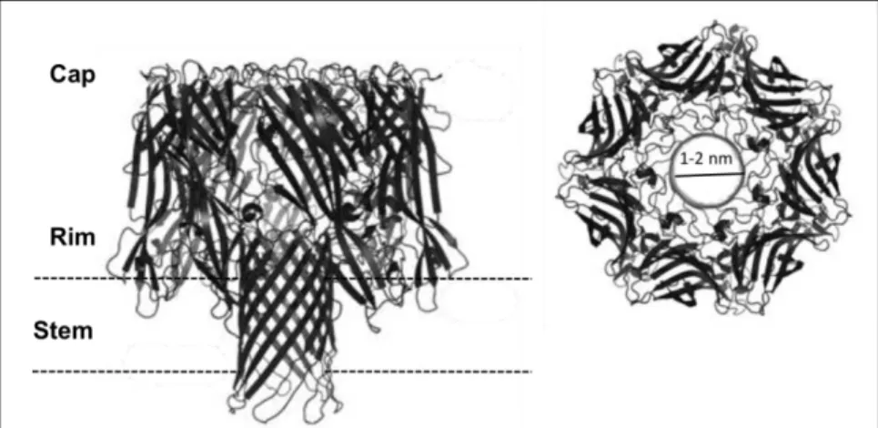
4.3 Alpha-hemolysin
47
5. References
52
CHAPTER II
High prevalence of hospital-associated methicillin-resistant
Staphylococcus aureus
(HA-MRSA) in the community in Portugal: evidence for the blurring of
community–hospital boundaries
79
ABSTRACT 81
INTRODUCTION 82
MATERIALS AND METHODS 84
RESULTS 88
DISCUSSION 97
ACKNOWLEDGEMENTS 102
REFERENCES 104
SUPPLEMENTARY MATERIAL 110
CHAPTER III
Population structure of methicillin-susceptible
Staphylococcus aureus
(MSSA) in Portugal over a 19-year period (1992–2011)
111
ABSTRACT 113
INTRODUCTION 114
MATERIALS AND METHODS 116
RESULTS 119
DISCUSSION 126
ACKNOWLEDGEMENTS 128
CHAPTER IV
Insights into alpha-hemolysin (Hla) evolution and expression among
Staphylococcus aureus
clones with hospital and community origin
133
ABSTRACT 135
INTRODUCTION 136
MATERIALS AND METHODS 138
RESULTS 142
DISCUSSION 152
ACKNOWLEDGEMENTS 155
REFERENCES 156
SUPPLEMENTARY MATERIAL 159
CHAPTER V
CONCLUDING REMARKS
167
CHAPTER I
General Introduction
CHAPTER I
General Introduction
1.
Staphylococcus aureus
GENERAL FEATURES
Staphyloccoccus aureus
was first discovered in 1880 in a human abscess pus by the surgeon Sir
Alexander Ogston, who after the observation of its characteristic grape-like clusters, named them
Staphylococcus
from the Greek expression “staphylé” (a bunch of grapes) (267). Four years later
(1884), Rosenbach was able to isolate and grow these bacteria also from abscesses and called
them
Staphyloccocus aureus
, because of the gold-like pigmentation appearance of the colonies
(“
aureus
” from Latin “golden”) (304) in opposition to
S. albus
(“albus” from Latin “white), nowadays
known as
S. epidermidis
.
S. aureus
belongs to a distinct monophyletic group within the
Firmicutes
Phylum, to the class
Bacilli
,
order
Bacillales,
family
Staphylococcaceae and
genus
Staphylococcus
(123, 281, 340).
Presently,
the
genus
Staphylococcus
contains
49
species
and
26
subspecies
(http://www.bacterio.net/s/staphylococcus.html accessed on September 2014).
S. aureus
are a
Gram-positive cocci, with 0.5 to 1.5
µ
m diameter, that can contain or not a polysaccharide capsule,
are non-motile and non-sporeforming, facultative anaerobes, which produce catalase and
coagulase (266, 393).
GENOME
The
S. aureus
genome consists of a circular chromosome of approximately 2,700 to 2,900 Mb, with
low G+C content (32.8%) (17). There are currently 56 annotated complete whole-genome
sequences
of
S.
aureus
available
in
the
public
domain
(http://www.ncbi.nlm.nih.gov/genome/genomes/154? assessed on September 2014). The genome
is composed of the core genome and the accessory genome (205).
larger regions of DNA diversity (from few nucleotides within a gene to insertion/deletion of several
kilobase pairs) (205). Core genes are located on the bacterial chromosome and, therefore, are
typically stable and transferred vertically.
The accessory genome represents up to 25% of the
S. aureus
genome and includes
bacteriophages, plasmids,
S. aureus
pathogenicity islands (SaPI), transposons and staphylococcal
cassette chromosome (SCC) elements (107, 204, 205). The accessory genome largely contributes
to the high genetic and phenotypic plasticity of
S. aureus
and mainly contains resistance and
virulence genes. The accessory genes can be transmitted by horizontal transfer to other strains and
species, but also vertically to the daughter cells (205).
OPPORTUNISTIC PATHOGEN IN THE HOSPITAL AND COMMUNITY
S. aureus
is a remarkable versatile bacterium with a two-faced lifestyle. On one hand,
S. aureus
behaves as a harmless colonizer, on the other hand is one of the most successful human
pathogens and a worldwide leading cause of infections.
S. aureus
can exist as a commensal (251) and colonizer of skin and mucous membranes of
humans and animals, including, pigs, cattle, rabbits, dogs and cats among others (212, 251).
Despite being found in multiple human body sites, colonization of the anterior nares is the
preferential ecological niche (79, 367, 385). Notwithstanding, some studies indicated that the rate of
throat colonization is higher than colonization of the anterior nares (221, 258).
children attending day-care centers (344). Moreover, the authors observed that the carriage rate
was associated with age, ranging from 6.3% in children less than two years old to up to 27.5%
among six years old children (344).
Frequent exposure to health care facilities and/or hospitalization are commonly associated with an
increased rate of
S. aureus
colonization, most of the time with
S. aureus
carrying antimicrobial
resistance determinants. Among hospitalized individuals in the USA, prevalence of
S. aureus
nasal
colonization was 28.6% in 2003-2004 (132). Kampf
et al.
reported that
S. aureus
carriage among
hospital staff was approximately 33.8% (174). Moreover, rates of colonization were described to be
higher among particular patient groups, e.g. HIV patients (257, 327), individuals with
S. aureus
skin
infections and skin diseases (153, 390), particular with insulin-dependent diabetes
(206), and
patients undergoing hemodialysis (183, 211, 401) reaching up to 100% in individuals with atopic
dermatitis (153, 245).
The carriage
status
has been associated to an increased risk for the development of staphylococcal
infection (148, 185, 367, 386). Hospitalized patients are particularly at risk of developing a
staphylococcal infection not only due to their increased
S. aureus
colonization rate, but also as a
consequence of their general compromised immunity, and the use of invasive clinical procedures,
such as surgery or the introduction of foreign indwelling medical devices, that serve as a port of
entry of
S. aureus
into presumably sterile body sites.
The severity of
S. aureus
infections can range from minor to life-threatening, local to systemic and
acute to chronic.
S. aureus
infections are categorized in three general types: 1. local infections -
superficial lesions such wound infections, skin and soft tissue infections (SSTIs); 2. systemic and
life-threatening - endocarditis, osteomyelitis, pneumonia, brain abscesses, meningitis, bacteremia
and septicemia; and 3. toxinoses- toxic shock syndrome (TSS), staphylococcal scalded-skin
syndrome and food poisoning (65).
patients with at least one HAI over the total number of patients, ranged from 2.3% in Latvia to the
highest prevalence registered in Portugal of 10.8% (98). As opposed to the majority of European
countries, in Portugal,
S. aureus
takes the lead over
E. coli
as the most frequent cause of
nosocomial infections (98).
Besides being one of the most important pathogens in healthcare settings,
S. aureus
is also an
important agent of infections in immunocompetent individuals outside hospitals.
S. aureus
in the
community have been described to cause mainly skin and soft tissue infections (SSTI), but also
invasive infections such as bacteremia, necrotizing pneumonia and necrotizing fasciitis (73, 148).
RESERVOIRS AND TRANSMISSION ROUTES
The Centers for Disease Control and Prevention (CDC) estimated that for every 20 people
hospitalized in the USA, one will develop a nosocomial infection (98). As the result of the high
burden caused by
S. aureus
in hospitals, staphylococcal infections are the principal focus of
infection control programs.
improving active surveillance, mainly by screening patients and HCW, by implementing appropriate
hand hygiene practices, using personal protective equipment and performing good environmental
desinfection. Moreover, decolonization of patients and health-care workers has been proved
efficient in reducing the risk of staphylococcal infection (29, 278, 339).
COST OF INFECTIONS AND INFECTION CONTROL
Hospital Infections have a substantial cost all over the world. The costs associated with treatment
and control of
S. aureus
infections are approximately $14.5 billion per year in the USA (260). In
2005, the healthcare system of the USA spent an estimated value of $830 million to $9.7 billion with
S. aureus
infections (229). The infections associated with drug resistant
S. aureus
have an
increased cost when compared to those susceptible, being a direct cost ($3,000 to $35,000) for the
treatment of a single infection episode (62).
The decreasing of
S. aureus
frequency in invasive disease in many European countries is
encouraging (97). Many of these countries adopted strict infection control measures, with good
results. In the UK, since 2001, specific measures were applied in hospitals at the country level with
an amazing success (89, 284). These included: 1) mandatory reporting of all
S. aureus
resistant
bacteraemia, 2) public standardization of incidence rates, 3) guidelines for preventing
hospital-associated infections, 4) establishment of a national hand hygiene campaign, 5) prudent use of
antibiotics, and 6) the implementation of the so called ’high impact interventions’, i.e. care “bundles”
focusing on key clinical procedures that, when not appropriately performed, can increase the risk of
infection. A decreasing trend of MRSA bacteraemia was clear from 2001 to 2009 in the UK, with a
62% reduction in the incidence of MRSA in blood cultures (158).
audited by the ECDC (European Centre for Disease Prevention and Control) (87). This program
aims to normalize the mission and structure of the commissions of infection of all hospitals in
Portugal in what regards to prevention and control of antimicrobial resistance. Moreover, the
program intends to standardize the procedures in clinical practice through the implementation of an
hospital “bundle” with the following rules: five moments of hand hygine accordingly to the World
Health Organization (WHO) guidelines (http://www.who.int/gpsc/tools/Five_moments/en/), correct
use of gloves, frequent cleaning of touch surfaces, and antimicrobial correct procedures. Finally,
finantial incentives for good pratices related with infection control, antimicrobials resistance
prevention and antibiotic consumption were implemented from 2013 on. No results on the impact of
these measures are available yet.
ANTIMICROBIAL CONSUMPTION
The excessive use of antimicrobials is one of the main forces driving the development and spread of
antimicrobial resistance. Antimicrobial resistance became a serious public health concern as the
result of the emergence and spread of highly resistant bacteria and also due to the limited choice of
antimicrobial agents to treat these pathogens.
Different actions to control the use of antimicrobials in the community and hospitals have been
followed in the different European countries, as demonstrated by the consumption of antimicrobials
report from the European Surveillance of Antimicrobial Consumption Network (ESAC-Net) published
by the European Centre for Disease Prevention and Control in 2010 (ECDC) (See Figure 1) (99).
The consumption of antimicrobial for systemic use (ATC (anatomical therapeutic chemical) group
J01: beta-lactams, penicillins; other beta-lactam antibacterials; tetracyclines; sulfonamides and
trimethoprim; macrolides, lincosamides and streptogramins; quinolones; and others) in both
hospitals and community is defined by number of DDD (defined daily dose) per 1 000 inhabitants
and per day.
beta-lactams, quinolones and macrolides.
Figure 1. Distribution by country of antimicrobial consumption for systemic use in the hospital, in 18 countries (2010). DDD: defined daily dose; (a) Finland: data include consumption in remote primary health care centres and nursing homes; (b) Portugal: data correspond to public hospitals only [adapted from (99)].
Figure 2. Distribution by country of antimicrobial consumption for systemic use in the community, in 26 countries (2010) [adapted from (99)].
The lowest consumption (< 16.7 DDD per 1 000 inhabitants and per day) was reported in the north
of Europe, e.g. Scandinavian and Baltic countries and the highest (
!
22.4 DDD per 1 000
inhabitants and per day) in the south of Europe e.g. Greece, Italy and also in Portugal with 22.4. In
Portugal, the most commonly used antimicrobials were the combinations of penicillins and
penicillins with extended-spectrum, followed by macrolides and tetracyclines. Portugal was the
second European countries with the highest consumption of macrolides, just preceded by Italy.
ANTIMICROBIAL RESISTANCE
introduction of different antibiotics into clinical practice.
In the pre-antibiotic era, the mortality rate associated to
S. aureus
infections was above 80% (328).
This scenario changed after the occasional discovery by Alexander Fleming in 1928 of penicillin
(109), later referred to as the first antibiotic of the antibiotic era. With the introdution of penicillin into
clinical practice, in the early 1940s, an effective treatment was provided in the treatment of bacterial
infections for the first time in medical history, leading to a dramatic decrease in the mortality and
morbility associated to
S. aureus
infections (41, 317). Despite its promising efficiency, ten years
later, more than 90% (or virtually all)
S. aureus
became resistant to penicillin (184). Similarly to what
was observed for penicillin, the development of new antibiotics and its application into clinical
practice was consecutively followed within just a few years, by the emergence of resistance (See
Figure 3).
Figure 3. Timeline of antibiotic introduction in the clinical practice and the emergence of antibiotic resistance [adapted from (53)].
Table 1. Antimicrobial agent classes, mechanisms of action and resistance [adapted from (8, 348, 395)].
Antibiotic classes (examples) Mechanism action Resistance type Mechanism of resistance Beta-lactams Cell wall synthesis: Altered target site Additional and altered PBPs (Penicillins, Cephalosporins,
Carbapenems, Monobactams)
Bind to penicillin-binding proteins (PBPs) and inhibit the transpeptidation step in the peptidoglycan synthesis stimulate autolysins
Enzymatic degradation Beta-lactamase
Glycopeptides Cell wall synthesis: Altered target site Altered peptidoglycan cross-link target (Vancomycin, Teicoplanin) Inhibit transglycosylation and
transpeptidation steps in peptidoglycan synthesis - bind to D-Ala-D-Ala
Excess of peptidoglycan Target overproduction
Protein synthesis: Enzymatic modification (AMEs) Phosphotransferase; Adenyltransferase; Acetyltransferase; Bifunctional enzyme
Inhibit 30S ribosomal subunits
Inactivation by aminoglycoside – modifying enzymes Decreased uptake Changes in outer membrane permeability Tetracyclines
(Tetracycline, Tigecycline)
Protein synthesis: Altered target Production of proteins that bind to the ribosome and alter the conformation of the active site
Inhibit 30S ribosomal subunits
Disrupt bacterial membrane Efflux New membrane transporters
Protein synthesis: Altered target Methylation of ribosomal active site with reduced binding Inhibit 50S ribosomal subunits
Efflux Mef type pump Protein synthesis: Enzymatic degradation CAT Inhibit 50S ribosomal subunits
Efflux New membrane transporters Mupirocin Protein synthesis:
(Mupirocin) Inhibit isoleucyl-tRNA synthetase
Fusidic acid Protein synthesis: Altered target Mutation leading to reduced binding to active site(s) (Fusidic Acid) Inhibit protein synthesis (elongation factor G)
Decreased permeability Chloramphenicol acetyltransferase
Quinolones Nucleic acid synthesis Altered target Mutation leading to reduced binding to active site(s) (Ciprofloxacin, Norfloxacin,
Levofloxacin)
Bind DNA gyrase
Efflux New membrane transporters Rifampin RNA synthesis
(Rifampicin) Bind to beta-subunit of bacterial RNA polymerase Sulfonamides/ Trimethoprim
(Trimethoprim,
Beta-lactam antibiotics: Methicillin
Among all classes of antibiotics, beta-lactams are considered the gold standard in the treatment of
S. aureus
infections. Beta-lactams act by acylation (inactivating) the active site of the
transpeptidase of the penicillin-binding proteins (PBPs), preventing cell wall synthesis.
Resistance to beta-lactam antibiotics was first described for penicillin and was found to be
associated to the presence of the
blaZ
gene. This gene can be transported in a plasmid or be
chromosomally encoded and mediates the production of a beta-lactamase that acts by hydrolyzing
the beta
!
lactam ring of penicillin.
In order to overcome the problem of the penicillin-resistant
S. aureus
infections, in 1959 a
semisynthetic antibiotic, resistant to beta-lactamases, was developed - methicillin (originally called
celbenine) (302). At that time methicillin was believed to be a definitive cure to
S. aureus
infections.
However, within only two years (1961), in the UK, the first treatment failure was reported, and
methicillin-resistant
S. aureus
(MRSA) emerged (170). Althought currently accumulating resistance
to other semisynthetic beta-lactams, the designation of MRSA is still extensively used in opposition
to MSSA (methicillin-susceptible
S. aureus
).
The main mechanism associated with methicillin resistance is related with the presence of an extra
penicillin-binding protein (PBP), named PBP2A (147) with reduced affinity to methicillin and most
beta-lactams antibiotics. Notwithstanding
,
other types of resistance to beta-lactams have been
described in
S. aureus
, namely
the overexpression of the beta-lactamase (118, 227), the presence
of chromosomal mutations (16, 141, 253) or the overexpression of PBPs (352).
Mechanism of methicillin-resistance
In the presence of beta-lactams, transpeptidase domain of the four native PBPs (PBP1 to 4) of
S.
aureus
is inactivated, and the extra PBP, PBP2A, a peptidoglycan transpeptidase, in cooperation
with the transglycosylase domain of PBP2, catalyzes the cell wall biosynthesis (147, 333, 357).
PBP2A is encoded by the
mecA
gene, which is carried by a heterologous mobile genetic element
called staphylococcal cassette chromosome
mec
(SCC
mec
) (177). More recently, a
mecA
homologue –
mecC
- was described in a
S. aureus
strain of animal origin (LGA251) (180); the
mecC
the PBP slide agglutination tests (119, 320). Since
mecC
first description, in 2011, several studies
have reported the presence of this gene in different
S. aureus
genetic backgrounds of both animal
and human origin (22, 82, 283), with limited geographic distribution in Europe (22).
SCC
mec
structure
SCC
mec
integrate into the staphylococcal chromosome at a specific site (
attB
or the integration site
sequence ISS), within the 3’ end of an open reading frame (
orfX
) coding for a methyltransferase of
RlmH type, located near the origin of replication (30, 76, 150).
SCC
mec
elements are composed by i)
mec
gene complex, ii)
ccr
gene complex and iii) three
flanking regions, the joining (J) regions. The
mec
gene complex, besides the
mecA
gene include
also intact or truncated
mecA
regulators,
mecI
(repressor) and
mecR1
(sensor inducer). More
recently,
mecR2
was also identified as a regulator of
mecA
through binding to the methicillin
repressor
mecI
(11). To date five different
mec
complex classes (A, B, C1, C2 and E) have been
described in
S. aureus
according to its structure
(See Table 2) (76, 144, 163, 165, 168). The
ccr
complex encodes for recombinases of the invertase resolvase family responsible for mediation of
site- and orientation-specific integration and excision of SCC
mec
from the chromosome (168, 177).
A total of five
ccr
allotypes have been described: four allotypes containing combinations of
ccrA
and
ccrB
genes (
ccrAB1
,
ccrAB2
,
ccrAB3
,
ccrAB4
); and another allotype containing a single gene,
ccrC
(See Table 2). The remaining part of the SCC
mec
is composed by the very heterogeneous J
regions (previously called “junkyard” regions) (J1, J2, J3), containing non-essential components and
genes conferring antibiotic and heavy metal resistance. The J1 region is located between the right
junction and the
ccr
complex, the J2 region located between the
ccr
complex and the
mec
complex
and J3 region extends from the
mec
complex to the
orfX
.
SCC
mec
is classified into types and subtypes. The SCC
mec
types results from the combination of
the class of the
mec
gene complex and the allotype of the
ccr
gene complex and the variations in
the J regions are used to define the SCC
mec
subtypes (237). Currently, eleven different types of
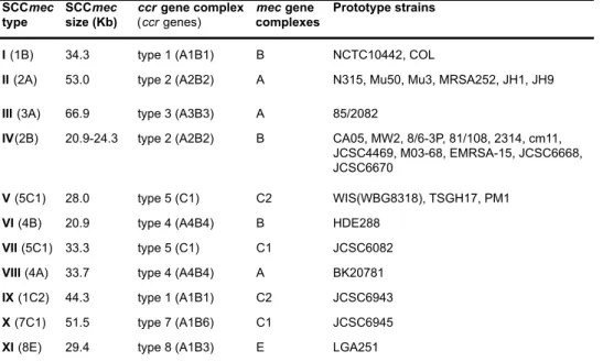
Table 2. SCCmec types identified in S. aureus (I to XI).
Adapted from http://www.sccmec.org/Pages/SCC_TypesEN.html
In contrast to the SCC
mec
types IV, V, VI and VII encoding resistance to beta-lactams only, the
SCC
mec
types I, II, III and VIII carry additional resistance determinants for other antibiotics and
heavy metal. This is conferred by the presence of integrating plasmids, like pUB110 encoding for
resistance to kanamycin, tobramycin and bleomycin, pT181 coding for tetracycline resistance and
pI258 coding for resistance to mercury; and transposons such as
Tn
554 carrying the
ermA
gene,
which is responsible for inducible macrolide, lincosamide and streptogramin (MLS) resistance (168).
In a single event the acquisition by
S. aureus
of these SCC
mec
types, gives rise to a multidrug
resistant (MDR) bacteria.
Non beta-lactam antibiotics: Vancomycin
Throughout the past years, antimicrobials commonly used to treat MRSA infections were
vancomycin and teicoplanin, tetracyclines, clindamycin, quinolones and fusidic acid, or the more
recently released antibiotics linezolid, daptomycin, tigecycline, telavancin and ceftaroline. However,
MRSA have progressively become resistant to a number of antimicrobials, including clindamycin
and tetracycline (143). Based on antimicrobial resistance surveillance in Europe, in 2012, that
included 29 countries, a high percentage of resistance to fluoroquinolone (81%) was registered
among MRSA in invasive disease (97). Resistance was also noticed, although in lower frequency,
SCCmec
type
SCCmec
size (Kb)
ccr gene complex
(ccr genes)
mec gene
complexes
Prototype strains
I (1B) 34.3 type 1 (A1B1) B NCTC10442, COL
II (2A) 53.0 type 2 (A2B2) A N315, Mu50, Mu3, MRSA252, JH1, JH9
III (3A) 66.9 type 3 (A3B3) A 85/2082
IV(2B) 20.9-24.3 type 2 (A2B2) B CA05, MW2, 8/6-3P, 81/108, 2314, cm11, JCSC4469, M03-68, EMRSA-15, JCSC6668, JCSC6670
V (5C1) 28.0 type 5 (C1) C2 WIS(WBG8318), TSGH17, PM1
VI (4B) 20.9 type 4 (A4B4) B HDE288
VII (5C1) 33.3 type 5 (C1) C1 JCSC6082
VIII (4A) 33.7 type 4 (A4B4) A BK20781
IX (1C2) 44.3 type 1 (A1B1) C2 JCSC6943
X (7C1) 51.5 type 7 (A1B6) C1 JCSC6945

![Figure 2. Distribution by country of antimicrobial consumption for systemic use in the community, in 26 countries (2010) [adapted from (99)].](https://thumb-eu.123doks.com/thumbv2/123dok_br/15764230.640109/36.748.84.678.80.602/figure-distribution-country-antimicrobial-consumption-systemic-community-countries.webp)
![Figure 3. Timeline of antibiotic introduction in the clinical practice and the emergence of antibiotic resistance [adapted from (53)]](https://thumb-eu.123doks.com/thumbv2/123dok_br/15764230.640109/37.748.78.678.446.757/timeline-antibiotic-introduction-clinical-practice-emergence-antibiotic-resistance.webp)
![Table 1. Antimicrobial agent classes, mechanisms of action and resistance [adapted from (8, 348, 395)]](https://thumb-eu.123doks.com/thumbv2/123dok_br/15764230.640109/39.1062.86.851.90.681/table-antimicrobial-agent-classes-mechanisms-action-resistance-adapted.webp)
![Table 3. Molecular characterization and geographical distribution of the major HA-MRSA [adapted from (38, 85, 86)]](https://thumb-eu.123doks.com/thumbv2/123dok_br/15764230.640109/57.1062.85.876.94.553/table-molecular-characterization-geographical-distribution-major-mrsa-adapted.webp)
![Table 4. Molecular characterization and geographical distribution of the major CA-MRSA and LA-MRSA clones [adapted from (38, 85, 86)]](https://thumb-eu.123doks.com/thumbv2/123dok_br/15764230.640109/63.1062.87.877.92.499/table-molecular-characterization-geographical-distribution-major-clones-adapted.webp)
![Table 5. S. aureus virulence factors involved in the host infection process [adapted from (402)]](https://thumb-eu.123doks.com/thumbv2/123dok_br/15764230.640109/68.748.84.696.107.779/table-aureus-virulence-factors-involved-infection-process-adapted.webp)