OCCURRENCE OF METHICILLIN RESISTANT STAPHYLOCOCCUS AUREUS (MRSA) AMONG CLINICAL SAMPLES IN TEHRAN-IRAN AND ITS CORRELATION WITH POLYMORPHISM OF SPECIFIC ACCESSORY
GENE REGULATOR (AGR) GROUPS
Amir Azimian1,Shahin Najar-pirayeh1, Siamak Mirab-Samiee 2,5, Mahmood Naderi3,4
1
Department of Medical Bacteriology, School of medical Science, Tarbiat Modares University, Tehran, P.O.BOX: 14115-331;
2
Food and Drug Laboratory Research Center, Ministry of Health and Medical Education, No. 408, Emam Khomeini Ave., Tehran
11136-15911, Iran; 3Department of Medical Biotechnology, School of Medical Science, Tarbiat Modares University, Tehran,
P.O.BOX: 14115-331; 4Molecular Biology and Genetic Engineering Department, Stem Cell Technology Research Center, Tehran,
Iran; 5Reference Health Laboratories, Ministry of Health and Medical Education, Tehran, Iran.
Submitted: March 31, 2011; Returned to authors for corrections: November 09, 2011; Approved: January 16, 2012.
ABSTRACT
Methicillin-Resistant Staphylococcus aureus (MRSA) is responsible for an increasing number of serious
hospital and community acquired infections. Virulence gene expression in Staphylococcus aureus is
orchestrated by regulators such as the accessory gene regulator (agr). Staphylococcal strains are divided into
four major agr groups (agrI-IV) on the basis of agrD and agrC polymorphisms. The purpose of this study
was to define the prevalence of MRSA strains in appointed Tehran’s hospitals and then to define and
compare the proportion of agr I, II, III, IV polymorphisms between MRSA and Methicillin Sensitive
Staphylococcus aureus (MSSA) strains. A total of 235 isolates were evaluated by conventional antibiotic
susceptibility tests and PCR for agr and mecA genes. 112 strains were MRSA (47.5%) and the most
prevalent agr specific group was agr I followed by agr III, agr II and agr IV, respectively. The prevalence
of agr groups amongst MRSA and MSSA strains was not statistically significant (P≥0.05). This study
suggests that agr I is not only the most prevalent agr type in MRSAs but also the most common one in
Methicillin Sensitive Staphylococcusaureus (MSSA) strains in Iran.
Key words: Methicillin Resistant Staphylococcus aureus, agr, PCR.
INTRODUCTION
Staphylococcus aureus is the major pathogen responsible
for both hospital and community acquired infections. Based on
numerous reports S. aureus has become resistant to most
available antibiotics (4, 1, 14). In the early 1950s acquisition
and spread of beta lactamase producing plasmids thwarted the
effectiveness of penicillin for treating S. aureus infections. In
1950 methicillin, a semisynthetic penicillin, was introduced,
even though in 1960 methicillin resistant Staphylococcus
aureus (MRSA) strains were identified. The mechanism by
which S. aureus acquires resistance to Methicillin is dependent
upon the production of an altered penicillin binding protein
(PBP2a) which is encoded by mecA gene. Increasing number of
isolated MRSA strains has led to complication in treatment of
staphylococcal diseases (7, 10, 24).
This pathogen causes a wide range of diseases including
septicemia, meningitis, endocarditis, osteomyelitis, septic
arthritis, toxic shock syndrome and food poisoning (4, 1, 14).
The accessory gene regulator (agr) locus was identified as the
regulator of virulence factors in S. aureus. It controls a large
set of genes, including most of those encoding cell wall
associated and extracellular proteins(2, 18). The agr locus is
composed of two divergent transcriptional units, RNAII and
RNAIII, driven by P2 and P3 promoters, respectively. The P2
operon encodes four proteins that generate the agr-sensing
mechanism and as a result of their activation, the effector
molecule (RNAIII) is produced and affects the expression of
virulence genes. The association between agr specific group,
the type of infection, and also antibiotic resistance has been
reported by many researchers (29, 30). In this study we
investigated the occurrence of the Methicillin Resistant S.
aureus (MRSA) among clinical samples while considering
their specific accessory gene regulator (agr) groups and the site
of infection.
MATERIALS AND METHODS
Bacterial isolates
A total of 235 S. aurous isolates were isolated from
patients and healthy individuals. Isolates were taken from
blood culture [60], urine [37], skin [43], respiratory tract
specimens [55] and miscellaneous specimens such as tissue
biopsies, exudates and bone marrow [9]. Also 31 nasal swabs
of S. aureus were taken from healthy volunteers.
Laboratory methods
S. aureus isolates were identified with the use of
conventional tests consisting of gram staining, catalase test,
growth in manitol salt agar media, DNase and coagulase test.
Antibiotic susceptibility test
Disk diffusion and MIC agar dilution were performed for
all isolates. Disk diffusion and MIC were accomplished
according to the guidelines of Clinical and Laboratory
Standards Institute (CLSI) (28). We used 1 μg Oxacillin disk
(HiMedia Code: SD088) for disk diffusion test. Oxacillin
powder (Sigma code: O1002) was utilized for MIC (Agar
dilution method) while Staphylococcus aureus ATCC25923
was used as the control.
Genomic DNA extraction
Bacterial DNA lysates were prepared from 1 ml of an
overnight Tripticase Soy Broth (TSB) culture. After
centrifugation at 6000 g for 5 min the bacterial pellet was
resuspended in 500µl of TE buffer [50mM Tris-Hcl (PH=8),
50mM disodium EDTA] containing 20 unit lysostaphin (Sigma
code: L7386 ) (25), and incubated at 37°C for 30-60 min and
then extracted by conventional Phenol-Chloroform method.
DNA amplification
Thermal cycling for amplification of mecA gene was
performed in an Eppendorf thermal cycler (Mastercycler®
gradient). Amplification protocol consisted of 5 min initial
denaturation at 94°C, followed by 30 cycles of denaturation
(94°C/30 seconds), annealing (55°C/30 seconds) and extension
(72°C/60 seconds), and an additional post-amplification
extension step at 72°C for 7 min.
The same device was used for agr group amplification.
Amplification protocol consisted of 5 min initial denaturation
at 94°C, followed by 30 cycles of denaturation (94°C/60
seconds), annealing (57°C/60 seconds) and extension (72°C/60
seconds), and a final post-amplification extension at 72°C for 7
min (23). The list of primers used for this experiment is
depicted in Table 1. The products of amplified samples were
with ethidium bromide. S. aureus strains RN6390 (agr groupI),
RN6607 (agr groupII), RN8465 (agr groupIII), RN4550 (agr
group IV) and RN6911 (agr negative) were included as run
controls for agr group identification.
Table 1. primers
Primer name sequence Product size
Forward mecA 5'- AAAATCGATGGTAAAGGTTGGC-3' Reverse mecA 5'-AGTTCTGCAGTACCGGATTTG-3'
533 bp
pan forward agr 5'-GTCACAAGTACTATAAGCTGCGAT-3' - Reverse agrI 5'-GTATTACTAATTGAAAAGTGCCATAGC-3' 440bp Reverse agrII 5'-GTATTACTAATTGAAAAGTGCCATAGC-3' 572bp Reverse agrIII 5'-CTGTTGAAAAAGTCAACTAAAAGCTC-3' 406 bp Reverse agrIV 5'-CGATAATGCCGTAATACCCG-3' 588 bp
Statistical analysis
Statistical significance of differences between groups was
analyzed by means of T-student or ANOVA test. Multivariate
analysis was performed to assess the independence of the
statistically significant variables in unvariate analysis. A ρ
-value < 0.05 was considered significant.
RESULTS
A total of 235 strains from patients and healthy
individuals (163 men and 72 women; 69% and 31%,
respectively) were evaluated. Among the 235 isolates tested by
disk diffusion method for detection of oxacillin resistance, 127
strains (54%) were susceptible and 108 strains (46%) showed
resistance. By MIC agar dilution method, 130 (55%) strains
were susceptible and 105 (45%) strains were resistant. Finally,
PCR for mecA gene showed that 110 strains (47%) had mecA
gene while 125 strains (53%) showed no amplification for this
target.
Our strains were isolated from blood, urine, coetaneous
samples, respiratory tract, nasal swabs and miscellaneous
samples. Prevalence of MRSA strains in different samples was
depicted in Table 2.
Our results showed good correlation between phenotypic
and genotypic methods for detection of antibiotic susceptibility
tests. According to Table 2, the highest percentage of MRSA
strains were isolated from respiratory tract specimens (49%)
followed by blood cultures (48%), miscellaneous specimens
such as tissue biopsies , exudates and bone marrow (45%),
urine (43%), cutaneous specimens (41%) and nasal swabs
(34%), respectively. The observed differences were not
statistically significant (p>0.05).
Table 2. Resistance against oxacillin in different specimens with various phenotypic and genotypic tests
mecA gene positive n(%) Disk diffusion
n(%) MIC*
n(%) resistant%
sample
30(50) 50(30)
29(48) Blood (n=60)
17(46) 16 (43)
16(43) Urine (n=37)
19 (44) 18(42)
18(42) Coetaneous (n=43)
28(51) 27(49)
27 (49) Respiratory tract (n=55)
5(46) 5(46)
4(45) Other (n=9)
11(35) 12(39)
11 (35) Nasal swab (n=31)
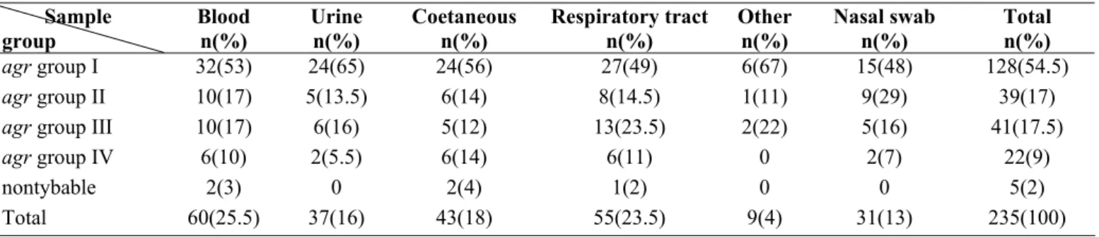
The majority of S. aureus strains isolated from clinical and
healthy cases belonged to agr group I (128 strains), followed by
agr group III (41 strains), agr group II (39 strains) and finally agr
group IV (22 strains). Five isolated strains were untypable by our
assay (Table 3).
There was a difference in prevalence of specific agr groups
between MRSA and MSSA isolates. In MRSA isolates, agr group
I had the highest prevalence (57%) followed by group III (19%),
group II (14%) and group IV (8%). Two percent of MRSA isolates
were untypable. In MSSA isolates, most of the strains belonged to
agr group I (52%) followed by group II (19%), group III (16%)
and group IV (11%). Two percent of MSSA isolates were
untypable. Results are depicted in Table 4. The differences were
not statistically significant (P>0.05).
Table 3. Genetic polymorphism of the agr locus in staphylococcus aureus isolates from different specimens
Total n(%) Nasal swab n(%) Other n(%) Respiratory tract n(%) Coetaneous n(%) Urine n(%) Blood n(%) Sample group 128(54.5) 15(48) 6(67) 27(49) 24(56) 24(65) 32(53)
agr group I
39(17) 9(29) 1(11) 8(14.5) 6(14) 5(13.5) 10(17)
agr group II
41(17.5) 5(16) 2(22) 13(23.5) 5(12) 6(16) 10(17)
agr group III
22(9) 2(7) 0 6(11) 6(14) 2(5.5) 6(10)
agr group IV
5(2) 0 0 1(2) 2(4) 0 2(3) nontybable 235(100) 31(13) 9(4) 55(23.5) 43(18) 37(16) 60(25.5) Total
Table 4. Genetic polymorphism of the agr locus in MRSA and MSSA strains
agr I n(%)
agr II n(%)
agr III n(%)
agr IV n(%)
N n(%)
Total n
MRSA 64(57) 16(14) 21(19) 9(8) 2(2) 112
MSSA 64(52) 23(19) 20(16) 13(11) 3(2) 123
Total 128(54.5) 39(16.5) 41(17.5) 22(9.5) 5(2) 235
DISCUSSION
Since the introduction of semisynthetic penicillins such as
methicillin and oxacillin for the therapy of infections caused by
S. aureus, the occurrence of resistantstrains to methicillin has
steadily increased and MRSA strains have become the major
nosocomial pathogens (19, 27). Infections with MRSA strains
require treatment with glycopeptide antibiotics which could be
nephro- and ototoxic (9). Staphylococcus aureus is the major
pathogen in both community and hospital acquired infections
(26). The ability of this organism to cause a multitude of
human diseases such as endocarditis, pneumonia, bacteremia
and Toxic Shock Syndrome (TSS) suggests that the
pathogenesis of Staphylococcus aureus infections is highly
complex. The growth phase is not only affected by many cell
surface proteins as well as exotoxins but also influenced by the
environmental and host signals which contribute to the
regulation of virulence factors (18).
The agr operon involves in the coordinated regulation of a
number of Staphylococcus aureus virulence factors.
Staphylococcus aureus strains exhibit well-defined genetic
polymorphisms within the agr locus. Four agr genotypes,
group I to IV, have been described to date (4, 6). Although
there is massive amounts of data relating agr type and specific
infections, Jarraud et al. have shown that specific agr genotype
strains may be associated with particular infectious syndromes,
with enterotoxin disease linked to agr group I, endocarditis
linked to agr groups I and II, toxic shock syndrome linked to
agr group III and exofoliative disease linked to agr group IV
(12). The agr group III has been overrepresented among strains
isolated from community-acquired MRSA infections, whereas
hospitals (17, 21).
In our study, resistance to oxacillin between four agr
groups was almost similar. S. aureus strains belonging to agr
groups II and IV were equally resistant to oxacillin (41%)
whereas strains carrying agr group I and agr group III were
more resistant with resistance rates of 50% and 51%,
respectively. However, the differences were not statistically
significant (P>0.05). Other studies showed a correlation
between induction of Glycopeptide Intermediate-resistant
Staphylococcus aureus (GISA) phenotype and autolytic
deficiency, especially in the context of agr genotype II (13).
Some reports stated that there are clinical trends according to
each agr group. For example, agr group I was prevalent in a
collection of 192 S. aureus strains in which 71% were
methicillin resistant (11, 26). Recently, Jarraud et al. reported
an overrepresentation of agr genotype II in S. aureus isolates
from patients with infective endocarditis (12). Pamela et al.
showed that agr group II polymorphism in MRSA predicts the
failure of vancomycin therapy (16). Moreover, it has been
reported that community-acquired MRSA, Methicillin
Sensitive S. aureus (MSSA) (3, 20) and Toxic Shock
Syndrome Toxin (TSST-1) producing isolates belong to agr
specificity group III (6).
In our study most of MRSA strains belonged to agr group
I and III, respectively, and most of MSSA strains belonged to
agr groups II and IV (%59). Van Leeuwen et al. screened a
collection of 55 MSSA isolates, mostly taken from healthy
nasal carriers, but did not find any agr III isolate (26). Most
exofoliatin producing strains responsible for Staphylococcal
Scalded Skin Syndrome (SSSS) belongs to group IV (11). The
agr group IV was absent in many previously reported articles
(23, 26, 15, 22), nevertheless, we detected agr group IV (9.5%)
in our experiments that was more likely due to ecological and
geographical differences. Goerke et al. reported that the
majority of S. aureus strains, taken from patients undergoing
intubations, belonged to group III (5). Manago et al. found that
most of agr I strains show poor biofilm formation, compared
with other agr groups. They also found a lower prevalence of
groupI strains and a higher prevalence of group II strains in the
nosocomial infections (15). Most of the agr group I clones
which had been previously reported by the Brazilian,
Portuguese, Hungarian and Berlin Research Groups. Group II
strains were mainly isolated in Japan and North America. On
the other hand, strains of group III were mainly isolated in
Europe (8). Recent data demonstrate that the vast majority of
MRSA in France and around the world belongs to agr group III
(20, 3). Our experience revealed that group I is the most
prevalent group in Iran, followed by groups III, II and IV. Iran
is one of the several countries with high antibiotic resistance
rate, including methicillin resistance. Therefore, it is important
to emphasize on the verification of characteristics of MRSA in
this country. This report has evaluated the correlation between
agr groups and antibiotic resistance in Iran population. This
result will be helpful to encourage verification of the
characteristics of MRSA in other Asian countries. In addition,
this study may also aid in evaluating the global spread of
MRSA strains based on agr locus polymorphisms.There seems
to be a geographic distribution difference between agr groups.
ACKNOWLEDGEMENTS
We thank Professor Richard P. Novick (Skirball Institute,
New York) for generously providing standard strains and Dr.
Patrice Francois for standard DNA controls. Moreover, we
thank Dr. Yousof Gheisari and Ehsan Arefian for their
assistance in statistical analyses.
REFERENCES
1. Arbuthnott, J.P.; Coleman, D.C.; de Avazedo, J.S. (1990). Staphylococcal toxins in human disease. Soc Appl Bacteriol Symp Ser.19:s101-7.
2. Balaban, N.; Novick, R.P. (1995). Autocrine regulation of toxin synthesis by Staphylococcus aureus. Proc. Natl. Acad Sci USA. 92: 1619-1623.
4. Fischetti, V.A.; Novick, R.P.; Ferretti, J.J.; Portnoy, D.A.; Rood, J.I.; editors (2000). Pathogenicity factors and their regulation. Gram-positive pathogens. Washington DC: ASM Press.392-407.
5. Georke, C.; Kymmel, M.; Dietz, K.; Wolz, C.; (2003). Evaluation of interspecies interference due to agr polymorphism in Staphylococcus aureus during infection and colonization. J Infect Dis. 188:250-256. 6. Gi, J.; Beavis, R.; Novick, R.P. (1997). Bacterial interference caused by
autoinducing peptide variants. Science. 276: 2027-2030.
7. Gold, H.S.; Moellering, R.C. (1996).Antimicrobial-drug resistance. Engl. J. Med. 335:1445-1453.
8. Gomes, A.R.; Vinga, S.; Zavolan, M.; de Lencastre, H. (2005). Analysis of the genetic variability of virulence-related loci in epidemic clones of Methicillin-Resistant Staphylococcus aureus. Antimicrob Agents Chemother. 49:366-379.
9. Grisold, A.J.; Leitner, E.; Muhlbauer, G.; Marth, E.; Kessler, H.H. (2002). Detection of Methicillin Resistant Staphylococcus Aureus and simultaneous confirmation by automated nucleic acid extraction and Real-Time PCR. J. Clin. Microbiol. 40: 2392-2397.
10. Hiramatsu, K.; Chui, L.; Kuroda, M.; Ito, T. (2000). The emergence and evolution of methicillin resistant Staphylococcus aureus. Trends. Microbiol. 9:486-493.
11. Jarraud, S.; Lyon, G.J.; Figueiredo, J.M.; Jerard, L.; Vandenesch, F.; Etienne, J. et al. (2000). Exofoliatin-producing strains define a forth agr specificity group in Staphylococcus aureus. J Bacteriol. 182:6517-6522. 12. Jarraud, S.; Mougel, C.; Thioulous, J.; Lina, G.; Meugnier, H.; Forey, F.;
Nesme, X.; Etienne, J.; Vandenesch, F. (2002). Relation between Staphylococcus aureus genetic background, virulence factors, agr groups (alleles) and human diseases. Infect. Immun. 70:631-641.
13. Kohel, J.L.; Muthaiyan, A.; Jayaswal, R.K.; Ehlert, K.; Labinschinski, H.; Wilkinson, B.J. (2004). Cell wall composition and decreased autolytic activity and lysostaphin susceptibility of glycopeptide-intermediate Staphylococcus aureus. Antimicrob. Agents Chemother. 48:3749-3757.
14. Lina, G.; Gillet, Y.; Vandenesch, F.; Jones, M.E.; Floret, D.; Etienne, J. (1997). Toxin involvement in staphylococcal scalded skin syndrome. Clin Infect Dis. 25:1369-73.
15. Manago, K.; Nishi, J.; Wakimoto, N.; Miyanohara, H.; Sarantuya, J.; Tokuda, K.; Iwashita, M.; Yamamato, K.; Yoshinaga, M.; Maroyama, I.; Kwano, Y. (2006). Biofilm formation by and accessory gene regulator typing of Methicillin-Resistant Staphylococcus aureus strains recovered from patients with nosocomial infections. Infect Control Hosp Epidemiol. 27:188-190.
16. Moise-broder, P.A.; Sakulas, J.; Eliopoulos, G.M.; Schentag, J.J.; Forrest, A.; Moellering, R.C. (2004). Accessory gene regulator group II polymorphism in Methicillin-Resistant Staphylococcus aureus is predictive of failure of vancomycin therapy. Clin Infect Dis. 38:1700-1705.
17. Naimi, T.S.; LeDell, K.H.; Como-Sabetti, K.; Borchardt, S.M.; Boxrud, D.J.; Etienne, J.; Johnson, S.K.; Vandenesch, F.; Fridkin, S.; O'Boyle, C.; Danila, R.N.; Lynnfield, R. (2003). Comparison of community and health care associated Methicillin Resistant Staphylococcus aureus infection. JAMA. 290:2976-2984.
18. Novick, R.P. (2003). Autoinduction and signal transduction in the regulation of Staphylococcal virulence. Mol Microbiol. 48:1429-1449. 19. Panlilio, N.L.; Culver, D.H.; Gaynes, R.P.; Banerjee, S.; Henderson,
T.S.; Tolson, J.S.; Martone, W.J. (1992). Methicillin Resistant Staphylococcus aureus in U.S hospitals ,1975-1991. Infect. Hosp Control. Epidemiol. 13:582-586.
20. Pearman, J.W. (2002). Community-acquired MRSA: the Australian experience [abstract 359]. In: program and abstracts of the 10th international symposium on Staphylococci and Staphylococcal diseases (Tsukuba, Japan). Japan: Japan Symposium on Staphylococci and Staphylococcal.Disease. p 18.
21. Sakoulas, G.; Eliopoulos, G.M.; Mollering, R.C.; Wennersten, C.; Venkataraman, L.; Novick, R.P.; Gold, H.R. (2002). Accessory gene regulator (agr) locus in geographically divers Staphylococcus aureus isolates with reduces susceptibility to vancomycin. Antimicrob Agents Chemother. 46:1492-1502.
22. Sakulas, G.; Eliopoulos, G.M.; Moellering, R.C.; Novick, R.P.; Venkataraman, L.; Wennersten, C.; DeGirolami, P.C.; Schwaber, M.J.; Gold, H.S. (2003). Staphylococcus aureus accessory gene regulator (agr) group II: is there a relationship to the development of intermediate-level glycopeptide resistance? J. Infect. Dis. 187:929-938.
23. Shopsin, B.; Mathema, B.; Alcabes, P.; Said-Salim, B.; Lina, G.; Matuska, A.; Matinez, J.; Kreiswirth, B. (2000). Prevalence of agr specificity groups among Staphylococcus aureus strains colonizing children and their guardian's Clin Microbil. 1:456-459.
24. Stefani, S.; Varaldo, P.E. (2003). Epidemiology of methicillin resistance staphylococci in Europe. Clin. Microbiol. Infect. 9: 1179-1186.
25. Takeushi, S.; Maeda, T.; Hashimoto, N.; Imaziumi, K.; Kaidoshi, T.; Hayakawa, Y. (2001). Variation of the agr locus in Staphylococcus aureus isolates from cows with mastitis. Vet Microbiol. 79:267-274. 26. Van Leeuwen, W.; Van Nieuwenhuizen, W.; Gijzan, C.; Vebrugh, H.;
Van Belkum, A. (2000). Population studies of Methicillin Resistant and Sensitive Staphylococcus aureus strains reveal a lack of variability in the agrD gene, encoding a Staphylococcal autoinducer peptide. J Bacteriol. 182: 5721-5729.
27. Voss, A.; Milatovic, D.; Wallarauch-Schwarz, C.; Rosdahl, V.T.; Braveny, I (1994). Methicillin Resistant Staphylococcus aureus in Europe. Eur. J. Clin. Microbiol. Infect. Dis. 13:50-55.
28. Wayane, P.A. (2000). National Committee for Clinical Laboratory Standards. Performance standards for antimicrobial disk susceptibility tests, 7th ed. Approved standard M2-A7.
Novick, R.P. (2005). The agr radiation: an early event in the evolution of staphylococci. J Bacteriol, 16: 5585-5594.
30. Yarwood, J.M.; Schielvert, P.M. (2003). Quorum sensing in Staphylococcus infections. J Clin Invest., 11: 1620-1625