Dengue in Nicaragua, 1994:
reintroduction of serotype 3 in the Americas
1
María G. Guzmán,
2Susana Vázquez,
2Eric Martínez,
3Mayling Álvarez,
2Rosmary Rodríguez,
2Gustavo Kourí,
2José de los Reyes,
4and Francisco Acevedo
4The principal aim of the report presented here is to describe the reappearance of dengue serotype 3 in the Americas, following a 17-year absence, through the recent experience of Nicaragua. In all, 356 serum samples obtained through Nicaragua’s dengue monitoring system in October 1994 during an epidemic were examined. Anti-dengue IgM antibodies were detected in 43% of these, with sera from 12 of the 18 areas covered by Nicaragua’s local integrated health care systems yielding positive results. In addition, dengue virus was isolated from 5 of 24 sera obtained from patients with hemorrhagic symptoms, dengue 3 being isolated from 3 of these samples and dengue 1 from the other 2. A diagnosis of dengue with hemorrhagic manifesta-tions or of hemorrhagic dengue was supported or confirmed by laboratory findings obtained from 26 of 39 patients hospitalized in León or Managua. The most frequent symptoms of 18 patients diagnosed as having dengue with hemorrhagic manifestations were fever, headache, vomiting, myalgia, arthralgia, and epistaxis. The remaining eight patients, diagnosed as hav-ing probable hemorrhagic dengue, exhibited fever, general malaise, hemorrhaghav-ing, thrombocy-topenia, hemoconcentration, and hemagglutination-inhibition antibody titers ranging from 640 to 20 480. Overall, the reappearance of dengue serotype 3 in the Region was confirmed, together with its ability to produce cases of hemorrhagic dengue. At least in Nicaragua, it is apparent that the introduction of dengue serotype 3 has prompted an increase in the number of classical dengue and hemorrhagic dengue cases, a scenario that might constitute the grim prelude to future developments in the Americas if urgent attention is not given to controlling the disease’s mosquito vector.
SUMMARY
In recent times a progressive in-crease has been noted in the numbers of dengue epidemics, dengue cases, and hemorrhagic dengue patients in
the Americas (1). Generally speaking, ever since the dengue 1 epidemic that struck the Caribbean in 1977 and the first reported epidemic of hemorrhagic dengue in Cuba in 1981—that coun-try’s largest and most serious dengue hemorrhagic fever (DHF) outbreak to date—the status of dengue in the Americas has become increasingly ominous and hard to resolve (2, 3). Events in Central America have reflected this continental trend. For example, while Costa Rica and Panama had no reported dengue until 1992,
epidemics occurred in both countries in 1993 (4), facilitating spread of the disease from Mexico in the north to Bolivia, Brazil, and Paraguay in the south. No cases from this epidemic were detected in Chile (5).
Nicaragua’s first recorded dengue epidemic occurred in 1985. During that event dengue serotypes 1 and 2 were isolated, over 17 000 cases oc-curred, and seven patients diagnosed with hemorrhagic dengue died (6). Sporadic cases were then reported until 1990, when a second outbreak 1 This article was published in Spanish in the Boletín
de la Oficina Sanitaria Panamericana,Vol. 121, No. 2, August 1996, pp. 102–110, with the title “Dengue en Nicaragua, 1994: reintroducción del serotipo 3 en las Américas.”
2 Pedro Kourí Institute of Tropical Medicine,
Havana, Cuba. Mailing address: Instituto de Me-dicina Tropical “Pedro Kourí,” Apartado 601, Ma-rianao 13, La Habana, Cuba.
3 Juan Manuel Márquez Pediatric Teaching
Hospi-tal, Havana, Cuba.
caused 4 137 recorded cases. This number rose in 1992 to 4 936, most of them in the city of León where dengue serotypes 2 and 4 were isolated. In 1993 a total of 8 938 cases was re-ported, the clinical symptoms were more serious, and consequently the number of hospitalized patients was greater (7).
In July 1994 a new increase in the number of dengue and DHF cases became discernible in León, a total of 1 680 dengue cases being reported that month in the city. The dengue case incidence then increased abruptly in September and October in Managua, forcing health authorities to declare a state of epidemiologic emergency and intensify control activities (7). By the end of 1994, a total of 20 469 dengue cases had been reported for the year, of which 1 247 exhibited hemorrhagic manifestations.
The main aim of this article is to report on the reappearance of dengue 3 in the Americas through the guan situation. (Prior to 1994 Nicara-gua had never reported dengue 3 virus.) It also describes the more fre-quently cited symptoms of classical dengue and hemorrhagic dengue patients in Nicaragua and reports on dengue-specific IgM antibodies de-tected in serum samples obtained from patients with clinical signs of the dis-ease by the country’s seroepidemio-logic surveillance system.
MATERIALS AND METHODS
In Nicaragua, seroepidemiologic monitoring of dengue is carried out through the remittance of blood sam-ples by the country’s 18 local inte-grated health care systems (sistemas locales de atención integral en salud— SILAIS), three of which are located in Managua. These samples, drawn from patients presenting a clinical picture of dengue, are tested for anti-dengue IgM antibodies at the National Diag-nostic and Reference Center (Centro Nacional de Diagnóstico y Referencia— CNDR) in Managua. As part of this surveillance, the CNDR’s virology lab-oratory received 356 serum samples
drawn from patients with a clinical diagnosis of dengue who had experi-enced the disease in early October 1994. A total of 41 samples (including 2 acute serum samples) were also obtained from 39 hospital patients diagnosed as having either clinical dengue with hemorrhagic manifesta-tions or hemorrhagic dengue.5 This article describes analyses conducted with the sera from these 395 patients.
All of the 39 patients with hemor-rhagic symptoms had been hospital-ized in the La Mascota or Manolo Morales Hospital in Managua or the Oscar Danilo Rosales Argüello Teach-ing Hospital (HEODRA) in León dur-ing the second half of October. In 14 cases it was possible to draw a second sample between 44 and 58 days after the onset of symptoms. In addition, the clinical history of each of the 39 patients was reviewed, and important data were noted on a form designed for the purpose.
The presence of IgM anti-dengue antibodies was detected with the MAC-ELISA technique, using antigens prepared from the four dengue virus serotypes and following the procedure described by Kuno et al. (8). Hemag-glutination inhibition (HI) tests were also performed using the technique described by Clarke and Casals (9), and a further search for total dengue virus antibodies was made using the ELISA inhibition method described by Fernández and Vázquez (10).
In addition, an attempt was made to isolate dengue virus from 24 serum samples obtained from the 39 patients hospitalized with clinically diagnosed dengue, with hemorrhagic symptoms, or dengue hemorrhagic fever. These samples were obtained during the first 5 days following the onset of symptoms.
The 24 blood samples were trans-ported in refrigerated thermoses at 4 °C to the CNDR virology laboratory in Managua, where the sera were
sep-arated under sterile conditions and stored at –20 °C pending shipment to the Pedro Kourí Institute of Tropical Medicine in Havana, Cuba, for further processing. There they were inocu-lated onto confluent monolayers of C636 and Ap61 cells at final dilutions of 1:50 and 1:20, respectively, using MEM medium as a maintenance medium with 2% fetal calf serum for the C636 cells and L15 medium with 2% fetal calf serum for the Ap61 cells.6 Following incubation for 10 days at 28 °C, viral antigens were sought by means of the indirect immunofluores-cence technique using polyclonal mouse ascitic fluids hyperimmune to dengue virus serotypes 1 and 2. When positive immunofluorescence was ob-served, the serotype responsible was determined with specific monoclonal antibodies for the four dengue virus serotypes, which were provided by the U.S. Centers for Disease Control and Prevention’s Division of Arthropod-transmitted Infectious Diseases lo-cated in Fort Collins, Colorado. As a positive control for immunofluores-cence, cells inoculated with the four serotypes were used, the strains in-volved being as follows: dengue 1, Cuban strain isolated during the 1977 epidemic; dengue 2, strain A15 iso-lated during the Cuban epidemic of 1981; dengue 3, strain H87; and dengue 4, strain H241. Two blind pas-sages in cell cultures were provided for those samples negative on the first passage; these samples were also tested again by the immunofluores-cence technique.
The criteria for confirming a case of dengue included the following: the presence of a clinical picture suggest-ing the disease, isolation of the virus, seroconversion of total immunoglobu-lins, and seroconversion of IgM anti-bodies or at least a fourfold increase in the IgG antibody titer of a second serum sample drawn during convales-cence as compared to the titer of a first
5 The former cases, which exhibited relatively mild
hemorrhagic manifestations, were not classified as hemorrhagic dengue cases because the patients did not also present the hemoconcentration and thrombocytopenia required for that diagnosis.
6 The cells, previously maintained by means of
serum sample drawn during the acute phase of the disease (11). Patients exhibiting dengue-like symptoms and those whose sera (a single sample) exhibited anti-dengue IgM antibodies or HI antibody titers of 1 280 or more were classified as probable cases (11). All cases with HI antibody titers of 2 560 or more were classified as proba-ble cases possibly involving secondary dengue infection (12). Patients with a clinical diagnosis of hemorrhagic dengue were classified according to the severity of the symptoms in accordance with PAHO’s Dengue and Dengue Hem-orrhagic Fever in the Americas: Guidelines for Prevention and Control(11).
RESULTS
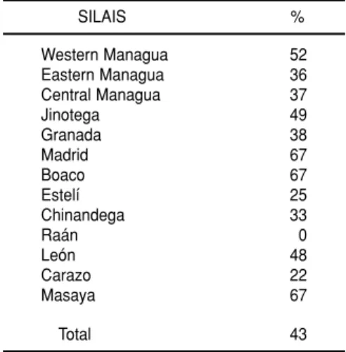
IgM antibodies were detected in 152 (43%) of the 356 samples tested (Table 1). More than 35% of the sera received from the three SILAIS located in Man-agua were positive. The groups of serum samples provided by the 13 SILAIS submitting samples yielded positive results in all but one instance (none of the samples from the Raán
SILAIS being positive). Table 2 shows the distribution by age and sex of the 143 patients for whom age and sex data had been recorded. Most of the positive sera were obtained from patients under age 30.
All serum samples from 39 patients hospitalized with a clinical diagnosis of dengue and exhibiting hemorrhagic manifestations (29) or hemorrhagic dengue (10) were studied for IgM anti-bodies and total antianti-bodies by HI and ELISA. Thirty of these patients were under 15 years old. In 24 of these 39 cases an attempt was made to isolate the virus.
In five of these 39 patients, paired acute and convalescent sera did not yield confirmation of dengue virus infection, and so the diagnosis was rejected. In another eight patients from whom only one serum sample was obtained (during the acute phase of the disease), no IgM antibodies or high total immunoglobulin titers affirming dengue infection were detected. De-spite the fact that no viruses were iso-lated from five of these eight samples, all eight patients were diagnosed as having probable dengue cases because their clinical symptoms were consis-tent with the disease and had evolved during a dengue epidemic. Specifi-cally, six were diagnosed as having hemorrhagic manifestations and two as having grade II hemorrhagic dengue. Even so, in the absence of a second serum sample, the serologic studies of these eight patients were inconclusive.
In the remaining 26 patients, it was possible to associate the clinical diag-nosis with a positive laboratory result
(virologic or serologic) that supported or confirmed the clinical diagnosis. Of these, 18 were clinically classified as probable cases of dengue with hemor-rhagic manifestations and eight as cases of hemorrhagic dengue. Table 3 shows the primary symptoms and clinical signs observed in these 18 patients, as well as the laboratory results. The clinical picture was charac-terized by fever, headache, and vomit-ing, as well as by myalgia and arthral-gia. With less frequency, retroocular pain, abdominal pain, and rash were reported. Eight (44%) of these patients had thrombocytopenia, and 10 (56%) yielded a positive tourniquet test. The most frequent hemorrhagic manifesta-tion, found in 12 cases (67%), was epis-taxis. In eight patients seroconversion was observed, and in five the dengue virus was isolated (three of the isolates were dengue serotype 3 and two were serotype 1). These latter findings made it possible to confirm the clinical diag-nosis in these eight patients. The other 10 patients were classified as having probable dengue cases.
The first two serotype 3 isolations were obtained from children with clin-ical pictures of fever, vomiting, head-ache, general symptoms, and some hemorrhagic symptoms. Both had been admitted to Managua’s La Mas-cota Hospital. In both, seroconversion to flavivirus and primary infection were detected by the HI and ELISA methods. The clinical evolution of their cases, classified as confirmed dengue with hemorrhagic manifesta-tions, was satisfactory. Repeated isola-tion of the virus in the same samples of serum confirmed the identification. TABLE 1. Percentages of sera testing
posi-tive for anti-dengue IgM antibodies that were submitted by 13 of Nicaragua’s 18 local integrated health care systems (SILAIS) to the national seroepidemiologic dengue monitoring system in October 1994, showing the locations of the 13 local sys-tems submitting sera. All of the sera were obtained from patients with dengue-like clinical symptoms
SILAIS %
Western Managua 52 Eastern Managua 36 Central Managua 37
Jinotega 49
Granada 38
Madrid 67
Boaco 67
Estelí 25
Chinandega 33
Raán 0
León 48
Carazo 22
Masaya 67
Total 43
TABLE 2. Distribution of specific dengue IgM antibodies among patients who fell ill in October 1994, by sex and age
Women Men Total
Age (in years) No. % No. % No. %
0–15 34 56 27 44 61 43
16–30 28 58 20 42 48 33
31–45 17 71 7 29 24 17
> 45 4 40 6 60 10 7
Serotype 3 was isolated in samples from patients in both Managua and León.
Table 4 shows the clinical data and laboratory results obtained from the eight patients meeting the PAHO/ WHO clinical criteria for hemorrhagic dengue (11). All were considered to be probable cases of dengue based on the
laboratory results. One was classified as grade I, five as grade II, and two as grade III, in accord with the classifica-tion for degrees of severity of hemor-rhagic dengue (11). In addition, all showed some type of hemorrhaging and five (62%) complained of abdomi-nal pain. Two patients suffered from shock during the course of the disease;
however, all of the cases evolved satis-factorily. In five out of seven cases (71%) for which HI titers were obtained, the existence of a secondary infection (as demonstrated by a HI antibody titer ≥2 560) was indicated. In five of the eight cases no dengue virus was isolated.
DISCUSSION
Although dengue-like disease cases have been reported in the Region of the Americas for more than two centuries, the first laboratory-documented epidemic occurred in 1963–1964 in the Caribbean Area, affecting primarily Venezuela (13). Another epidemic, from which dengue serotypes 2 and 3 were isolated, was reported on several Caribbean islands in 1968–1969 (13). In 1977 serotype 1 appeared in the Region, serotypes 2 and 3 circulated in endemic-epidemic fashion, and outbreaks were reported in Colombia and Puerto Rico (2). Up to the present, the last preceding dengue 3 isolation, in Puerto Rico, had occurred in 1977 (14).
Subsequently, the three remaining serotypes (1, 2, and 4) circulated in endemic-epidemic fashion either simultaneously or alone in various countries. Within this context, the recently reported isolation of serotype 3 in Nicaragua and subsequently in Panama (15) is of considerable signifi-cance and complicates the dengue situ-ation in the Americas. Locally, in ana-lyzing reported dengue cases in Nicaragua, one can see an increase in the number of individuals contracting the disease during 1992, an increase coinciding with the appearance of serotype 4 in that country. More recently, despite isolation of serotype 1 in two patients reported here, we can-not rule out the possibility that the increase in the number of dengue and hemorrhagic dengue cases observed in 1994 may have been due to the unno-ticed introduction of serotype 3. Most of Nicaragua’s population is suscepti-ble to dengue 3, as there are no reports suggesting dengue’s presence in the TABLE 4. Clinical and laboratory data for eight patients believed to have hemorrhagic
dengue
Case
1 2 3 4 5 6 7 8
Age (in years) 4 7 11 13 15 20 26 27
Sex M F M M M F F F
Fever X X X X X X X X
General malaise X X X X X X X X
Abdominal pain X X X X X
Hemorrhaging X X X X X X X X
Tourniquet test positive X X
Petechiae X
Epistaxis X X X
Vaginal bleeding X
Hematemesis X
Shock X X
Thrombocytopenia X X X X X X X X
Hemoconcentration X X X X X X X X
Severitya II II II III II III I II
Day sample was taken 4 4 3 4 9 10 8 15
IgM positive X X X X X
HI titer 640 2 560 10 240 2 560 ND 20 480 1 280 20 480
aTo establish the severity of the disease, data relating to thrombocytopenia, hemoconcentration, and type of infection (the
criteria appearing in reference 11) were applied. HI = hemagglutination inhibition.
ND = not done.
TABLE 3. Clinical symptoms and laboratory data for 18 patients having probable or con-firmed dengue cases with hemorrhagic manifestations
Clinical symptoms No. % Laboratory findings No. %
Fever 18 100 Thrombocytopenia 8 44
Headache 14 78 IgM positive 13 72
Vomiting 11 61 Secondary dengue infection 8 44
Myalgia 11 61 Dengue virus isolationa 5 28
Arthralgia 11 61 Antibody seroconversion 8 44 Retroocular pain 8 44
Abdominal pain 8 44
Rash 7 39
Tourniquet test positive 10 56
Epistaxis 12 67
Petechiae 6 33
Gingivorrhagia 1 5
Metrorrhagia 1 5
Hematemesis 2 11
country prior to 1985 (6), after which serotype 3 was no longer circulating in the Americas. (In 1995 the predominant serotype in Nicaragua was serotype 3.) In 1994 Nicaragua witnessed an increase in the number of patients hos-pitalized for dengue with hemorrhagic manifestations and hemorrhagic dengue (7), and who exhibited a clini-cal picture similar to that observed in areas with endemic and epidemic hemorrhagic dengue. Nevertheless, it should be pointed out that the appar-ent severity of the disease was less than that recorded elsewhere on other occasions, such as in Thailand (16) or in Cuba during the 1981 epidemic (17, 18). In these situations, the numbers of fatalities and of patients with hemor-rhagic dengue grades III and IV were greater relative to the numbers of indi-viduals reported ill. In addition, the principal hemorrhagic manifestations, such as hematemesis and melena, were not very frequent in Nicaragua, the predominant manifestations being epistaxis, petechiae, and a positive tourniquet test. Nevertheless, it should be stressed that the easy hospital admission criteria applied and the timely therapeutic interventions con-ducted produced favorable changes in the clinical pictures of many patients that tended to prevent development of hemoconcentration and other more severe symptoms.
In this study, of the eight probable dengue hemorrhagic fever patients, six showed evidence of prior dengue infection. Other authors have shown that such prior dengue infection is the principal risk factor for development of hemorrhagic symptoms (19). Although the number of hemorrhagic dengue patients studied was very small, it seems remarkable that evi-dence of prior dengue infection was detected serologically in six out of the seven tested.
In recent years an increase in dengue 3 circulation has been observed in Southeast Asia and the Western Pacific that has been associated with cases of hemorrhagic dengue in countries where this disease had not previously been seen. In Indonesia and Thailand,
an increase in dengue 3 isolations among patients with hemorrhagic dengue was reported between 1976 and 1987 (20). In countries such as Sri Lanka (1989) and India (1990), following a prolonged period of endemicity, epidemics of hemorrhagic dengue associated with this serotype were reported (21, 22). In addition, dengue caused by serotype 3 appears to have been introduced into Tahiti and other islands of the South Pacific from Indonesia, Malaysia, or the Philippines and to have unleashed epi-demics of hemorrhagic dengue in 1989 and 1990 (22).
Several researchers have classified the strains of dengue serotype 3 into four genetically distinct subtypes, which may be related to differences in the serotype’s virulence and epidemic potential (22). In accordance with the classification proposed by Lanciotti et al. in 1994 (22), research has indicated that the dengue 3 strain isolated in Panama corresponded to subtype III (15), which has been associated with cases of hemorrhagic dengue.
Other authors (23) have proposed an alternate classification of the dengue 3 strains into four groups based on the study of nucleotides in a portion of the envelope gene (nucleotides 47 through 303). Study of the dengue 3 strain iso-lated in Nicaragua, which was per-formed at the Pedro Kourí Institute of Tropical Medicine in Havana, deter-mined that this strain belonged to group 2 in this latter classification (24). The strains included in this group have also been linked to cases of hemor-rhagic dengue (23).
Recently, the presence of serotype 3 has been reported in Costa Rica, El Sal-vador, Honduras, and Panama. It should be noted that the consequences of the serotype’s presence can be expected to vary, depending on the conditions prevailing in each country (conditions including, among other things, the circulation of other serotypes, the presence of individuals susceptible to serotype 3, and the den-sity of the mosquito vector). In coun-tries where the number of people sus-ceptible to any serotype is high (such
as Panama), it is likely that dengue outbreaks and epidemics will occur, a situation which has in fact been observed in that country since late 1994. In other countries with high lev-els of endemicity, especially when it is caused by a specific serotype, the introduction of serotype 3 is likely to cause only a few isolated cases, at least at the outset, until it becomes the pre-dominant serotype. This could be the situation in Costa Rica, where four serotypes are circulating but where serotype 1 has been the principal dengue serotype isolated since 1993 (Sáenz E, Instituto Costarricense de Investigación y Enseñanza en Nutrición y Salud, personal communication, 1995). It is also true, however, that the situ-ation could develop differently in countries such as Nicaragua, where various serotypes have circulated for years and where the emergence of serotype 3 could lead to a large num-ber of cases secondary to this serotype, together with a possible increase in the number of hemorrhagic dengue cases. Several authors have suggested that the infection sequence of dengue 1 fol-lowed by dengue 2 is associated with cases of hemorrhagic dengue (25, 26). Considering the history of dengue in Nicaragua, where the four serotypes are circulating simultaneously, various sequences of infection could occur.
Right now the world is faced with new infectious diseases whose emer-gence demands immediate solutions, and the relative frequency of certain new viral diseases is on the rise. Within this context, dengue is among the most important communicable dis-eases in tropical countries, producing millions of cases every year (27). At least in Nicaragua, it is apparent that
the introduction of dengue serotype 3 has prompted an increase in the num-bers of classical dengue and hemor-rhagic dengue cases, a scenario that might constitute the grim prelude to future developments in the Americas if urgent attention is not given to implementing the known and recom-mended measures for controlling the disease’s mosquito vector.
Acknowledgments.The authors are
grateful for the collaboration pro-vided by G. Huelva, A. González, J.J. Amador, R. Jiménez, F. Ruiz, S. Cerda, and L. Pérez of the Ministry of Health of Nicaragua, and also by J.L. Pele-grino, M. Soler, and S. García, of the Pedro Kourí Institute of Tropical Med-icine in Havana, Cuba.
1. Organización Panamericana de la Salud, Pro-grama de Enfermedades Transmisibles. El dengue y la fiebre hemorrágica del dengue en las Américas: una visión general del pro-blema. Bol Epidemiol1992;13:9–10.
2. Pan American Health Organization. Dengue in the Caribbean, 1977.Washington, DC: PAHO; 1979. (Scientific publication 375).
3. Kourí G, Guzmán MG, Bravo J. Hemorrhagic dengue in Cuba: history of an epidemic. Bull Pan Am Health Organ1986;20:24–30. 4. Organización Panamericana de la Salud,
Pro-grama de Enfermedades Transmisibles, División de Prevención y Control de Enfer-medades Transmisibles. Dengue en Costa Rica y Panamá. Bol Epidemiol1994;15:9–10. 5. Organización Panamericana de la Salud,
Pro-grama de Enfermedades Transmisibles, División de Prevención y Control de Enfer-medades Transmisibles. Dengue en las Améri-cas: una actualización. Bol Epidemiol 1993; 14:1–3.
6. Kourí G, Valdez M, Argüello L, Guzmán MG, Valdez L, Soler M, et al. Epidemia de dengue en Nicaragua, 1985. Rev Inst Med Trop (São Paulo)1991;33:365–371.
7. República de Nicaragua, Ministerio de Salud.
Informe sobre la situación del dengue y las acciones de control. Managua: Ministerio de Salud; 1994.
8. Kuno G, Gómez I, Gubler D. An ELISA proce-dure for the diagnosis of dengue infections.
J Virol Methods1991;33:101–113.
9. Clarke DH, Casals J. Techniques for hemag-glutination and hemaghemag-glutination inhibition with arthropod borne viruses. Am J Trop Med Hyg1958;7:561–573.
10. Fernández RJ, Vázquez S. Serological diagno-sis of dengue by an ELISA inhibition method (EIM). Mem Inst Oswaldo Cruz (Rio de Janeiro)
1990;85:347–351.
11. Pan American Health Organization. Dengue and dengue hemorrhagic fever in the Americas: guidelines for prevention and control. Washing-ton, DC: PAHO; 1994. (Scientific publication 548).
12. World Health Organization, Technical Advi-sory Committee on Dengue Haemorrhagic Fever for the South East Asian and Western Pacific Regions. Guide for diagnosis, treatment and control of dengue haemorrhagic fever.
Geneva: WHO; 1980:7.
13. Ehrenkranz NJ, Ventura AK, Cuadrado RR, Pond WL, Porter JE. Pandemic dengue in Caribbean countries and the southern United States: past, present and potential problems.
N Engl J Med1971;285:1460–1469.
14. Morens DM, Rigau-Pérez JG, López-Correa RH, Moore CG, Ruiz-Tibén EE, Sather GE, et al. Dengue in Puerto Rico, 1977: public health response to characterize and control an epi-demic of multiple serotypes. Am J Trop Med Hyg1986;35:197–211.
15. United States, Centers for Disease Control and Prevention. Dengue type 3 infection: Nicaragua and Panama; October, November 1994. MMWR1995;44:21–24.
16. Halstead SB. The XXthcentury dengue
pan-demic: need for surveillance and research.
Rapp Trimest Stat Sanit Mond1992;45:292–298. 17. Guzmán MG, Kourí G, Morier L, Soler M, Fer-nández A. A study of fatal hemorrhagic dengue cases in Cuba, 1981. Bull Pan Am Health Organ1984;18:213–220.
18. Guzmán MG, Kourí G, Martínez E, Bravo J, Riverón R, Soler M, et al. Clinical and sero-logic study of Cuban children with dengue hemorrhagic fever, dengue shock syndrome (DHF/DSS). Bull Pan Am Health Organ1987;2: 270–279.
19. Bravo J, Guzmán MG, Kourí G. Why dengue haemorrhagic fever in Cuba? I: individual risk
factors for dengue haemorrhagic fever/ dengue shock syndrome (DHF/DSS). Trans R Soc Trop Med Hyg1987;81:816–820.
20. Nisalak A. Dengue virus isolations from Bangkok, Thailand, 1962–88. Virus Info Exch Newsl1989;6:117.
21. Vitarana T. Dengue hemorrhagic fever. Ceylon Med J1990;35:83–87.
22. Lanciotti RS, Lewis JG, Gubler D, Trent D. Molecular evolution and epidemiology of dengue-3 viruses. J Gen Virol1994;75:65–75. 23. Chungue E, Deubel V, Cassar O, Laille M,
Martin PM. Molecular epidemiology of dengue 3 viruses and genetic relatedness among dengue 3 strains isolated from patients with mild or severe form of dengue fever in French Polynesia. J Gen Virol 1993;74: 2765–2770.
24. Guzmán MG, Rosario D, Muné M, Álvarez M, Rodríguez R. Relaciones genéticas de los virus de dengue 3 aislados durante la epidemia de FHD de Nicaragua, 1994. Rev Cub Med Trop
1996;48:1.
25. Halstead SB. Dengue viruses in infectious dis-eases. In: Gorbach SL, Bartlett JG, Blacklow NR. Infectious diseases.Philadelphia: Saunders; 1992:1830–1835.
26. Kourí G, Guzmán MG, Bravo J. Why dengue haemorrhagic fever in Cuba? II: an integral analysis. Trans R Soc Trop Med Hyg1987;81: 821–823.
27. Murphy FA, Nathanson N. The emergence of new virus diseases: an overview. Sem Virol
1994;5:87–102.
Manuscript received 5 November 1995. Revised version accepted for publication on 3 April 1996.
El objetivo principal de este informe fue describir la reaparición del serotipo 3 del den-gue en las Américas después de 17 años de ausencia, tal como se observó reciente-mente en Nicaragua. Se examinaron en total 356 muestras de suero obtenidas por medio del sistema nicaragüense de vigilancia del dengue durante una epidemia en octubre de 1994. En 43% de las muestras se detectaron anticuerpos IgM contra el den-gue y los sueros de 12 de las 18 áreas atendidas por los sistemas locales integrales de salud dieron resultados positivos. Además, se aislaron virus de dengue en 5 de 24 sue-ros de pacientes con síntomas hemorrágicos: en 3 se aisló el serotipo 3 y en 2, el sero-tipo 1. Mediante pruebas de laboratorio, en 26 de 39 pacientes hospitalizados en León y Managua se consideró probable o se confirmó el diagnóstico de dengue con mani-festaciones hemorrágicas o dengue hemorrágico. En 18 pacientes diagnosticados de dengue con manifestaciones hemorrágicas, los síntomas más comunes fueron fiebre, cefalea, vómito, mialgia, artralgia y epistaxis. Los ocho pacientes restantes, en los que se diagnosticó dengue hemorrágico probable, tuvieron fiebre, malestar general, hemorragias, trombocitopenia y hemoconcentración, y los títulos de anticuerpos a la prueba de inhibición de la hemaglutinación oscilaron de 640 a 20 480. Se confirmó así la reintroducción del serotipo 3 del dengue en la Región y su capacidad para produ-cir casos de dengue hemorrágico. En Nicaragua, por lo menos, es evidente que la rein-troducción del serotipo 3 del dengue ha producido un aumento de los casos de den-gue clásico y denden-gue hemorrágico. Si no se presta atención urgente al control del mosquito vector de la enfermedad, la experiencia descrita podría constituir el prelu-dio lúgubre de futuros acontecimientos similares en las Américas.
RESUMEN
Dengue en Nicaragua, 1994:
reintroducción del serotipo 3
en las Américas
Winner of the 1996 Fred L. Soper Award
Each year the Board of Trustees of the Pan American Health and Education Foundation (PAHEF) awards a monetary prize and certificate to the author or authors of an original scien-tific paper, published in the previous year, that represents a notable contribution to the field of public health, with special relevance to the Americas. The award is named in honor of Fred L. Soper, former Director (1947-1958) of the Pan American Health Organization, and preference is given to papers related to infectious disease, a life-long concern of Dr. Soper. The 1996 Soper Award was given to Axel Kroeger et al. for their article “Insecticide-impregnated bed nets for malaria control: varying experiences from Ecuador, Colombia, and Peru concerning acceptabil-ity and effectiveness,” which was published in the American Journal of Tropical Medicine and Hygiene,Vol. 53, No. 4, 1995. Three other papers received an honorable mention:
1. Chadee DD, et al. Mass chemotherapy with diethylcarbamazine for the control of bancroftian filariasis. Am J Trop Med Hyg1995;52(2).
2. Dreyer G, et al. Treatment of bancroftian filariasis in Recife, Brazil. Trans R Soc Trop Med Hyg1995;89.