Clinical assessment of
252
Californium neutron
intracavitary brachytherapy using a two-channel
Y applicator combined with external beam radiotherapy
for endometrial cancer
Qian Zhou, Cheng Tang, Ke-Wei Zhao, Yan-Li Xiong, Shu Chen, Wen-Jing Xu, Xin Lei*
Third Military Medical University, Cancer Center, Research Institute of Surgery and Daping Hospital, Chongqing, China.
OBJECTIVE: The aim of this study was to determine the efficacy of 252Californium neutron intracavitary brachytherapy using a two-channel Y applicator combined with external beam radiotherapy for the treatment of endometrial cancer.
METHODS:Thirty-one patients with stage I–III endometrial cancer were recruited for this study. The stage I patients received only252Californium neutron intracavitary brachytherapy with a two-channel applicator. The stage II and III patients received both252Californium neutron intracavitary brachytherapy using a two-channel applicator and parallel-opposed whole pelvic radiotherapy.
RESULTS:The five-year local control rate was 80.6% (25/31), the overall survival rate was 51.6% (16/31), and the disease-free survival rate was 54.8% (17/31). The incidence of serious late complications was 12.9% (4/31). CONCLUSIONS:252Californium neutron intracavitary brachytherapy using a two-channel applicator combined with external beam radiotherapy was effective for treating endometrial cancer and the incidence of serious late complications related to this combination was within an acceptable range.
KEYWORDS: Endometrial Cancer; Californium; Two-Channel Y Applicator.
Zhou Q, Tang C, Zhao KW, Xiong YL, Chen S, Xu WJ, et al. Clinical assessment of252Californium neutron intracavitary brachytherapy using a two-channel Y applicator combined with external beam radiotherapy for endometrial cancer. Clinics. 2016;71(1):10-16
Received for publication onJuly 07, 2015;First review completed onSeptember 16, 2015;Accepted for publication onNovember 09, 2015 E-mail: [email protected]
*Corresponding author
’ INTRODUCTION
Endometrial cancer is one of the most common gyneco-logic malignancies worldwide. According to a report from the American Cancer Society, approximately 52,630 new cases of endometrial cancer were diagnosed and 8,590 deaths were associated with endometrial cancer in the United States in 2014 (1). The National Cancer Institute of China estimated that there would be 47,751 new cases of endometrial cancer in China in 2010 (2). Currently, surgery remains the standard treatment for early-stage endometrial cancer. However, radiation has become a more suitable treatment option for patients with medically inoperable endometrial cancer or for those of advanced age.
In terms of a definitive radiotherapy for endometrial cancer, intracavitary brachytherapy (ICBT) alone can be used to treat stage I patients. Moreover, ICBT combined with external beam
radiotherapy (EBRT) can be used to treat patients with local advanced-stage endometrial cancer. Generally, there are two types of brachytherapy using conventionalg-rays: high-dose-rate (HDR) brachytherapy using 192Ir or low-dose-rate (LDR) brachytherapy using137Cs. There have been no randomized controlled trials (RCT) comparing the effects of HDR bra-chytherapy and LDR brabra-chytherapy on endometrial cancer in recent years because endometrial cancer is rarely treated with radiotherapy alone (3,4).
Many reports in the literature have described the use of californium-252 (252Cf) as a neutron source for the treatment of cervical cancer due to its higher relative biologic effectiveness (RBE) and lower oxygen enhancement rate (OER) thang-rays
(5,6). Moreover, the 252Cf neutron source has been used effectively for ICBT of endometrial cancer (7). However, a single-channel applicator was used in most endometrial cancer studies (8,9), including our own, which is described below (10). The use of a single-channel applicator has several drawbacks, including longer treatment durations and a slightly higher incidence of complications than the use of a two-channel applicator (9). Further, the dose distribution associated with a single-channel applicator is not ideal and is difficult to adjust. Between 1999 and 2004, our center performed252Cf neutron DOI:10.6061/clinics/2016(01)03
Copyright&2016CLINICS–This is an Open Access article distributed under the terms of the Creative Commons License (http://creativecommons.org/licenses/by/ 4.0/) which permits unrestricted use, distribution, and reproduction in any medium or format, provided the original work is properly cited.
ICBT using a single-channel applicator to treat 40 cases of endometrial cancer and we observed the above-described shortcomings associated with this method (i.e., long treatment durations, a high rate of complications and difficulty in adjusting the dose and the dose distribution).
The modification of two-channel (TC) Y (Rotte Y) applicators for brachytherapy has overcome many of the shortcomings associated with single-channel applicators, particularly with regard to adjustment of the optimal dose distribution in the craniocaudal and lateral directions. Therefore, TC applicators have become the standard fixed applicators used forg-ray ICBT of endometrial cancer (11).
In 2007, our center began employing a 252Cf neutron source using the TC applicator for endometrial cancer treatment. The aim of this study was to assess the clinical outcomes of endometrial cancer patients treated via 252Cf neutron ICBT using a TC applicator combined with EBRT.
’ MATERIALS AND METHODS
Subjects
Thirty-one stage I–III endometrial cancer patients diag-nosed based on biopsy findings and staged according to the International Federation of Gynecology and Obstetrics (FIGO) guidelines from September 2007 to August 2011 were included in this study. Among these patients, the most common comorbidities associated with receiving radiother-apy alone included hypertension and diabetes (Table 1). None of the patients had received treatment before the study, and all endometrial cancer diagnoses had been confirmed by histology. All of the patients exhibited Karnofsky scores greater than 70 and hemoglobin levels greater than 90 g/L; their mean age was 55.9 years (range: 30–72 years) (Table 1).
All of the patients underwent a chest x-ray, an abdominal B-ultrasound, a pelvic CT or MRI, cystoscopy and a colo-noscopy as components of their pre-treatment evaluation. During the course of treatment, a pelvic B-ultrasound and a gynecological examination were performed every week to examine changes in uterine size and uterine cavity depth.
Treatment methods
Equipment. An LZH-1000252Cf neutron brachytherapy machine (Shenzhen Lingdun Technology, Shenzhen, China) and an 8 MV/X linear accelerator (Elekta Corp., Stockholm, Sweden) were used. For the252Cf neutron source, the average energy was 2.3 MeV, with a half-life of 2.645 years; the shape was cylindrical with dimensions of 3 mm in diameter 8 mm in height, an active region of 1.4 mm 9.0 mm, and a source strength per unit volume of 500–200 mg. The following formula was used to calculate the 252Cf dose: equivalent biological dose (DGY-eq) = neutron relative biological effectiveness (RBE) of 252Cf
252Cf neutron dose+ g-ray
RBE xg-ray dose; the252Cf RBE ranged from between 4–6.
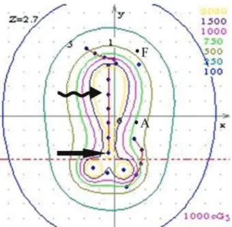
Treatment plan. Stage I patients underwent 252Cf neutron ICBT using the TC applicator alone. Stage II or III patients underwent 252Cf neutron ICBT using the TC applicator combined with EBRT. All patients were hospita-lized throughout the treatment period. External radiation was applied four times per week with opposing fields and a dose per fraction of 2 Gy. Once the total dose reached 20–36 Gy in 10–18 fractions, the central portion of the field was shielded using a 4 cm thick lead block, and the treatment was continued with antero-posterior field irradiation until the total dose reached 46–50 Gy.252Cf neutron ICBT using the TC applicator was delivered after 4–5 fractions of EBRT. ICBT then continued once per week, with the EBRT applied on 4 other days of the week. The total treatment duration was 6-7 weeks. A non-steroidal painkiller or morphine was administered before applicator placement. Barium enemas and bladder catheterizations were prepared prior to injection of the contrast agent into the balloon. Then, between 7 and 12 cervical dilatation rods were used sequentially to grad-ually expand the cervix until it was sufficiently enlarged. The two channels of the applicator were turned to a vertical position such that the two front ends were closed together, followed by insertion of the fundus was inserted into the uterus. The TC applicator was then turned to the horizontal position such that the top of the two channels reached the two horns of the uterus, and an anterolateral image was captured (Figure 1). Points A and F (point F was defined on line A, 2 cm lateral to the top of the TC applicator) were chosen as the dose reference points. The brachytherapy dose was 40–45 Gy to point A and 48–55 Gy to point F in 4–5 fractions. For stage I patients, the dose at point F was higher than that at point A. For stage II or III patients, the dose at point F was approximately the same as that at point A (Figure 2). The dose at the posterior wall of the bladder and the rectum was adjusted based on the isodose contour so that the dose at the bladder and the rectum was below 80% of that at point A. Stage I or II patients were administered a single channel and a vault tube of internal irradiation (Figure 3) to supplement potentially insufficient doses at the uterine fundus and the vaginal fornix. The ratio of the Table 1-Patient characteristics.
Characteristics Number of patients (%)* Total number of patients 31
Age (y)
30–50 5 (16.0%)
51–60 18 (58.0%)
61–72 8 (26.0%)
Mean 55.9
BMI (kg/m2)
Mean 27
Range 20–41
Stage (FIGO)
I 5 (16.0%)
II 22 (71.0%)
III 4 (13.0%)
Morphology
Adenocarcinoma 28 (90.0%)
Clear cell carcinoma 3 (10.0%)
Histopathologic grade
1 11 (35.5%)
2 13 (41.9%)
3 5 (16.1%)
Unknown 2 (6.5%)
Comorbid diseases
Hypertension 11 (35.5%)
Diabetes 11 (35.5%)
Coronary artery disease 3 (9.7%)
Systemic lupus erythematosus 2 (6.4%)
Others** 4 (12.9%)
* Thirty-one patients were enrolled in the study.
dose applied to the cervix to the dose applied to the vaginal fornix was 1.0:(0.8–1.0) for stage I patients (Figure 4) and 1.0: (1.2–1.4) for stage II patients (Figure 5). During252Cf neutron ICBT using the TC applicator, the patients were monitored for symptom alleviation, and gynecological examinations were performed weekly to determine the size of the uterus and the extent of tumor regression. At six months post-treatment, uterine curettage and histopathological examina-tion were performed to examine tumor cell regression.
Follow-ups. Beginning one month post-treatment, a pelvic B-ultrasound was performed monthly. At three months
post-treatment, uterine curettage and histopathological exam-ination of the endometrial tissues were performed. Outpatient follow-up visits were required once every three months beginning three months post-treatment and once every six months beginning one year post-treatment. None of the patients were lost to follow-up.
Figure 1 -A) Antero-posterior view; B) anterolateral view. The anterolateral image was captured when the TC applicator was turned to the horizontal position; this image shows that the top of the two channels reached the two horns of the uterus. A and F denote dose reference points A and F, respectively. The cervix is indicated by arrows, and the uterus is indicated by curled arrows.
Figure 2 -Isodose contour showing that the dose at point F was nearly identical to that at point A in stage II and III patients. A and F denote dose reference points A and F, respectively. The cervix is indicated by arrows and the uterus is indicated by curled arrows.
Statistical analysis. SPSS13.0 software was used for statistical analyses. Discrete data are presented as frequencies (percentages); continuous data are presented as means±SE.
For statistical analyses, Chi-square contingency table analysis
and Kaplan-Meier tests were performed;p-valueso0.05 were
considered statistically significant (Tables 2 and 3).
Ethics. This study was approved by the ethics committee of our hospital, and all patients signed informed consent forms.
’ RESULTS
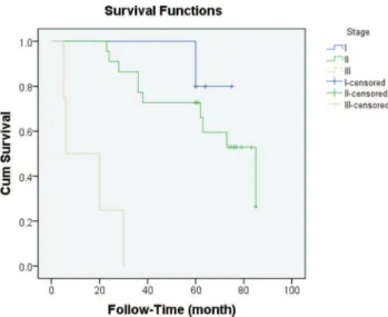
The mean follow-up duration was 54.8 months. Of the 31 patients enrolled in this study, 15 patients died during the follow-up period. One of the stage I patients died as a result of fatal coronary artery disease. Two stage III patients died from the persistent tumor. Three deceased patients had pelvic lymph node recurrence, four deceased patients had local recurrence at the time of death, and five deceased patients had distant metastases. The mean overall survival duration was 61.8±5.2 months, with a 95% confidence
interval of 51.6–72.0 months (Table 3). The 5-year overall survival rate for all patients was 51.6% (Figure 6). The disease-specific survival (DSS) rates for stages I, II, and III were 100%, 54.5%, and 0%, respectively (Table 2).
Following treatment via252Cf neutron ICBT using the TC applicator combined with EBRT, both pelvic B-ultrasound and gynecological examination showed that the uterus had shrunk substantially in all 31 patients and that the uterine cavity depth was reduced to a varying extent (0.5–5.0 cm). Of the two locally uncontrolled patients treated with an additional fraction of 252Cf neutron ICBT using the TC applicator, one experienced locally controlled disease, and the other, who showed clear cell histology, experienced local failure. The most common locations of disease recurrence were distant (16.1%), local recurrence alone (12.9%), and pelvic lymph nodes (9.6%). Of the five patients with distant tumor recurrence, three exhibited a single distant tumor involving the lungs and bone, and two exhibited multi-organ involvement. Additionally, patients with comorbid endome-trial cancer and diabetes were more susceptible to metastasis development and disease recurrence (8/31) (Table 4).
All patients completed the treatment without interruption. All of the late complications were limited to grade 2, and there were no recto-vaginal or vesico-vaginal fistulas. Three patients had frequent urination, urgency, and hematuria at 26 months post-radiotherapy. Cystoscopic examination ruled out other diseases, and these patients were diagnosed with radiation cystitis. Another patient was diagnosed with radiation proctitis. The incidence of late complications was 12.9% (4/31) (Table 5).
’ DISCUSSION
Surgery remains the primary standard treatment for patients with endometrial cancer. Definitive radiotherapy is typically offered to patients who suffer from medical comorbidities and those who have locally advanced tumors, as surgery may be not suitable for these patients. Therefore, patients treated via radiotherapy have a higher mortality rate than those treated via surgery. Nevertheless, radiotherapy remains the best option for patients with inoperable endometrial cancer. Thus, comparisons of survival between surgery and radiotherapy alone are of limited value. Patients with inoperable endometrial cancer are generally treated via ICBT alone or in combination with EBRT.
Figure 5 - Isodose contour showing that the ratio of the dose applied to the cervix to the dose applied to the vaginal fornix was 1.0:(1.2–1.4) in stage II patients. A and F denote dose reference points A and F, respectively. The cervix is indicated by arrows, and the uterus is indicated by curled arrows.
The most common histological type of endometrial cancer is adenocarcinoma, which is not as sensitive to conventional radiation as squamous cell carcinoma. Moreover, locally advanced stage endometrial tumors are often bulky and
contain many hypoxic cells, which are resistant to conven-tional radiation. In contrast to convenconven-tional radioisotopes, the 252Cf neutron represents a high linear energy transfer (LET) ray with distinct radiation and biological character-istics. For example, the252Cf neutron can cause greater direct damage to tumor cell DNA, leading to a higher rate of DNA double-strand breaks, than conventional photon radiation. The252Cf neutron also has a higher RBE and a lower OER than photons (7). Thus, bulky endometrial adenocarcinoma cells may be particularly sensitive to252Cf neutrons, and this form of radiotherapy could be used to treat endometrial adenocarcinoma patients.
However, 252Cf neutron ICBT has not been the main method of choice for endometrial cancer because the neutron source used for ICBT is large and expensive and because it is difficult to achieve precise biological equivalent dose calibration using conventionalg-rays. Thus, 192Ir and137Cs
ICBT are the most commonly used forms of ICBT worldwide, especially in Western Europe and North America. Moreover, the 252Cf neutron source used for ICBT was a neutron TC applicator that was larger than the g-ray TC applicator.
Dilatation of the cervix and the uterus is required to insert the TC applicator. In general, all of the endometrial cancer patients in our study tolerated TC applicator implantation well. Patients with late toxicity to radiotherapy in our study had grade 2 complications and were diagnosed with radiation proctitis or radiation cystitis.
Patients with locally advanced stage endometrial adeno-carcinoma or with inoperable endometrial adenoadeno-carcinoma who are treated via a combination ofg-ray ICBT and EBRT
often suffer from local-regional relapse. In the past two decades, the local control rate for patients with locally advanced stage endometrial adenocarcinoma has remained unsatisfactory, even when a salvage radical hysterectomy was performed. The local control rate was 76–84% for stage I–III endometrial adenocarcinoma patients treated via a combination of g-ray ICBT and EBRT. Alternatively, 252Cf
neutron ICBT has been found to be effective as a radio-therapy and to have radiobiological advantages for the treatment of radio-resistant advanced or bulky endometrial Table 2-Outcomes of endometrial cancer treated via252Cf neutron intracavitary brachytherapy.
Stage No. LCR OS DSS Late toxicity
I 5 100% (5/5) 80.0% (4/5) 100% (5/5) 0% (0/5)
II 22 81.8% (18/22) 54.5% (12/22) 54.5% (12/22) 18.2% (4/22)
III 4 50.0% (2/4) 0% (0/4) 0% (0/4) 0% (0/4)
Total 31 80.6% (25/31) 51.6% (16/31) 54.8% (17/31) 12.9% (4/31)
X2 3.026 5.510 8.496 0.961
P 0.066 0.037 0.004 0.378
No.: number of patients; LCR: local control rate; OS: overall survival; DSS: disease-specific survival.
Table 3-Overall survival duration of endometrial cancer patients receiving252Cf neutron intracavitary brachytherapy.
FIGO stage Mean±SE 95% CI Median±SE 95% CI I 72.0±2.7 (66.7,77.3)
II 66.5±5.4 (56.0,77.0) 85.0±13.1 (59.4, 110.6)
III 15.3±6.0 (3.50, 27.0) 6.00±7.50 (0.00, 20.8)
Total 61.8±5.2 (51.6, 72.0 73.0±6.79 (59.9, 86.1) SE: standard error; CI: confidential interval.
Mean overall survival duration=61.8±5.2 months (95% confidence interval, 5–85 months), X2=31.0, andp=0.000.
Figure 6 - Survival curves of patients with different stages of endometrial cancer.
Table 4-The relationships of morphology and comorbid diseases with metastasis development and disease recurrence.
Site Number Morphology Number Comorbid diseases Number
Local recurrence alone 4 (12.9%) Adenocarcinoma 4 (12.9%) Hypertension Diabetes 2 (6.4%) 2 (6.4%) Pelvic lymph node 3 (9.6%) Adenocarcinoma 3 (9.6%) Diabetes COPD 2 (6.4%) 1 (3.2%) Persistent 2 (6.4%) Adenocarcinoma Clear cell carcinoma 1 (3.2%) 1 (3.2%) Diabetes 2 (6.4%)
Distant 5 (16.1%)
Lung 2 (6.4%) Adenocarcinoma 2 (6.4%) Hypertension Diabetes 1 (3.2%) 1 (3.2%)
Multi-organ 2 (6.4%) Adenocarcinoma 2 (6.4%) Hypertension SLE 1 (3.2%) 1 (3.2%)
Bone 1 (3.2%) Adenocarcinoma 1 (3.2%) Diabetes 1 (3.2%)
tumors (7). Our data showed that the local control rate for stage I–III endometrial adenocarcinoma patients was 80.6%. However, the local relapse rate of patients with stage I–II disease was 14.8% (4/27), and that of patients with stage III disease was 50%.
The selection of an applicator is also important for ICBT of endometrial adenocarcinoma. The TC‘‘Y’’applicator consists of two rigid applicators, which can reach the two uterine horns, permitting coverage of a greater uterine width than a single-channel applicator. The dose distribution of the TC applicator was more optimal than that of the single-channel applicator, such that a satisfactory local control rate might be more effectively achieved using the TC for ICBT of endometrial adenocarcinoma. However, few studies have examined the application of the ‘‘Y’’applicator for treating endometrial cancer. Ohkubo et al. reported a satisfactory local control rate in ten patients with stage I–II endometrial cancer, none of whom suffered from local recurrence at five years (12). The same TC applicator was used by Coon et al. to treat 49 endometrial cancer patients (most of whom had stage I disease) via HDR ICBT; in that study, three patients (6.1%) experienced local failure and the median time to disease recurrence was 17 months. These two studies showing low rates of local recurrence further show that the radiation doses were sufficient for destroying the tumor in the uterus.
Differences in tumor stages and differential tumor grades are associated with distinct outcomes of endome-trial cancer patients. Without question, higher tumor stage, lower differential tumor grade, and unfavorable tumor histological type are associated with poorer prog-nosis. Previous studies had shown that the five-year overall survival rates of stage I–III endometrial cancer patients treated via either 137Cs or 192Ir ICBT, with or without EBRT 1-5(9,13-16). In contrast, in the study by Maruyama et al., 252Cf neutron ICBT combined with EBRT and hysterectomy resulted in five-year overall survival rates of 83% for stage I, 37% for stage II, and 50% for stage III endometrial cancer patients (7). Our current data showed a five-year overall survival rate of 51.6%; this result is consistent with other results of LDR and HDR studies, although it is lower than that from the study by Maruyama et al.
We speculate that the primary reason for the higher survival rate in the study by Maruyama et al. was that nearly half of the patients (14/31) in that study underwent a hysterectomy after receiving radiation therapy; the survival rate of 50% for stage III patients in that study was much higher than that in a previous report, which showed a survival rate of only 17.5% among those undergoing radiotherapy alone (17).
Treatment via252Cf neutron ICBT combined with EBRT as performed in our study may be sufficient for patients with stage I–II endometrial cancer, as these patients showed a five-year overall survival rate of 59.3%. This survival rate is comparable to previous reports showing overall survival rates of 42–61% for stage I–II endometrial cancer patients (18-21). Our treatment outcomes for the four patients with stage III endometrial cancer showed an unsatisfactory mean survival duration of 15.3±6.0
months. Two stage III patients died with persistent tumors. Regarding the other two stage III patients, one died 20 months post-treatment due to lung metastasis, and the other exhibited pelvic lymph node recurrence at 30 months of follow-up. These findings suggest that alternative treatment strategies should be used for patients with stage III endometrial cancer.
In the course of using the ‘‘Y’’ applicator to treat endometrial cancer, two ovoids or a ring applicator and a single-channel applicator should be added to supplement the radiation dose to the vaginal vault; such a treatment generally increases the radiation dose to the bladder and the rectum. In fact, we observed late toxicity of grade 2 in 12.9% (4/31) of the patients; this frequency is similar to the results from most studies of stage I–III patients (Table 6). However, a study of 36 stage I patients by Nguyen et al. using one tandem and one ovoid or cylindrical applicator to perform HDR ICBT alone showed a frequency of late toxicity of up to 21%6(22).
Although much effort has been invested to identify more effective methods for ICBT of endometrial cancer, no completely satisfactory method has been established. Our retrospective study contains several limitations, including the accuracy of cancer diagnosis and staging, the socioeconomic status of the patients, advances in treatment, and nurse-led supportive care. In particular, we used 2-D treatment planning, which is not as effec-tive as 3-D planning for reducing the dose to at-risk organs (23).
252Cf neutron ICBT using a TC applicator combined with
EBRT is effective for the treatment of endometrial cancer, and the incidence of serious late complications related to this therapy is acceptable.
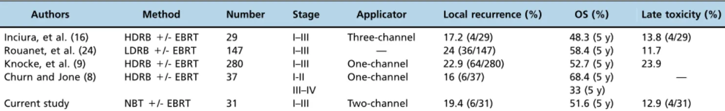
Table 6-Literature review of the outcomes of inoperable stage I-IV endometrial cancer patients receiving radiotherapy.
Authors Method Number Stage Applicator Local recurrence (%) OS (%) Late toxicity (%)
Inciura, et al. (16) HDRB+/- EBRT 29 I–III Three-channel 17.2 (4/29) 48.3 (5 y) 13.8 (4/29) Rouanet, et al. (24) LDRB+/- EBRT 147 I–III — 24 (36/147) 58.4 (5 y) 11.7 Knocke, et al. (9) HDRB+/- EBRT 280 I–III One-channel 22.9 (64/280) 52.7 (5 y) 23.9
Churn and Jone (8) HDRB+/- EBRT 37 I-II One-channel 16 (6/37) 68.4 (5 y) —
III–IV 33 (5 y)
Current study NBT+/- EBRT 31 I–III Two-channel 19.4 (6/31) 51.6 (5 y) 12.9 (4/31)
OS: overall survival; LDRB: low-dose-rate brachytherapy; HDRB: high-dose-rate brachytherapy; EBRT: external beam radiation therapy; NBT: neutron brachytherapy.
Table 5-Late toxicity.
Site Number of grade II complications (%)
Cystitis 3 (9.6%)
’ ACKNOWLEDGMENTS
We first thank the patients and their families. We also thank the
Departments of Obstetrics and Gynecology and of Pathology and the Imaging Center of Daping Hospital.
’ AUTHOR CONTRIBUTIONS
Zhou Q participated in patients care, wrote thefirst draft of the manuscript
and performed the literature review. Tang C, Lei X were responsible for the patient care, study design, draft of the manuscript and intellectual inputs. Zhao KW, Jia L, Shan JL, Xiong YL, Chen S, Xu Wen-Jing were responsible for data collection, data analysis and manuscript writing. All
authors read and approved thefinal version of the manuscript.
’ REFERENCES
1. Siegel R, Ma J, Zou Z, Jemal A. Cancer statistics, 2014. CA Cancer J Clin. 2014;64(1):9-29, http://dx.doi.org/10.3322/caac.21208.
2. Chen W, Zheng R, Zhang S, Zhao P, Zeng H, Zou X. Report of cancer incidence and mortality in China, 2010. Ann Transl Med. 2014;2(7):61. 3. Orton CG. High Dose Rate Versus Low Dose Rate Brachytherapy
for Gynecological Cancer. Semin Radiat Oncol. 1993;3(4):232-239, http:// dx.doi.org/10.1016/S1053-4296(05)80120-1.
4. Bianchi C, Botta F, Conte L, Vanoli P, Cerizza L. Biological effective dose evaluation in gynaecological brachytherapy: LDR and HDR treatments, dependence on radiobiological parameters, and treatment optimisation. Radiol Med. 2008;113(7):1068-78, http://dx.doi.org/10.1007/s11547-008-0291-4. 5. Lei X, Qian CY, Qing Y, Zhao KW, Yang ZZ, Dai N, et al. Californium-252
brachytherapy combined with external-beam radiotherapy for cervical cancer: long-term treatment results. Int J Radiat Oncol Biol Phys. 2011; 81(5):1264-70, http://dx.doi.org/10.1016/j.ijrobp.2010.08.039.
6. Tacev T, Ptackova B, Strnad V. Californium-252 (252Cf) versus conven-tional gamma radiation in the brachytherapy of advanced cervical carci-noma long-term treatment results of a randomized study. Strahlenther Onkol. 2003;179(6):377-84.
7. Maruyama Y, Van Nagell JR, Yoneda J, DePriest P, Kryscio RJ. Clinical evaluation of 252Cf neutron intracavitary therapy for primary endo-metrial adenocarcinoma. Cancer. 1993;71(12):3932-37, http://dx.doi.org/ 10.1002/1097-0142(19930615)71:12o3932::AID-CNCR282071122243.0. CO;2-A.
8. Churn M, Jones B. Primary radiotherapy for carcinoma of the endome-trium using external beam radiotherapy and single line source bra-chytherapy. Clin Oncol (R Coll Radiol). 1999;11(4):255-62, http://dx.doi. org/10.1053/clon.1999.9059.
9. Knocke TH, Kucera H, Weidinger B, Holler W, Potter R. Primary treatment of endometrial carcinoma with high-dose-rate brachytherapy: results of 12 years of experience with 280 patients. Int J Radiat Oncol Biol Phys. 1997;37 (2):359-65, http://dx.doi.org/10.1016/S0360-3016(96)00486-5.
10. Lei X, Shan JL, Tang C, Zhao KW. Follow-up study of clinical effects of californium-252 neutron intracavitary radiotherapy and external beam radiotherapy in endometrial cancer. Zhonghua Fu Chan Ke Za Zhi. 2007;42(11):733-6.
11. Dankulchai P, Petsuksiri J, Chansilpa Y, Hoskin PJ. Image-guided high-dose-rate brachytherapy in inoperable endometrial cancer. Br J Radiol. 2014;87(1039):20140018, http://dx.doi.org/10.1259/bjr.20140018. 12. Varia M, Rosenman J, Halle J, Walton L, Currie J, Fowler W. Primary
radiation therapy for medically inoperable patients with endometrial carcinoma--stages I-II. Int J Radiat Oncol Biol Phys. 1987 Jan;13(1):11-5, http://dx.doi.org/10.1016/0360-3016(87)90253-7.
13. Grigsby PW, Kuske RR, Perez CA, Walz BJ, Camel MH, Kao MS, et al. Medically inoperable stage I adenocarcinoma of the endometrium treated with radiotherapy alone. Int J Radiat Oncol Biol Phys. 1987;13(4):483-8, http://dx.doi.org/10.1016/0360-3016(87)90061-7.
14. Kucera H, Knocke TH, Kucera E, Potter R. Treatment of endometrial carcinoma with high-dose-rate brachytherapy alone in medically inoperable stage I patients. Acta Obstet Gynecol Scand. 1998, 77 (10):1008-12, http://dx.doi.org/10.1080/j.1600-0412.1998.771011.x. 15. Fishman DA, Roberts KB, Chambers JT, Kohorn EI, Schwartz PE, Chambers
SK. Radiation therapy as exclusive treatment for medically inoperable patients with stage I and II endometrioid carcinoma with endometrium. Gynecol Oncol. 1996;61(2):189-96, http://dx.doi.org/10.1006/gyno.1996.0123. 16. Inciura A, Atkocius V, Juozaityte E, Vaitkiene D. Long-term results of
high-dose-rate brachytherapy and external-beam radiotherapy in the primary treatment of endometrial cancer. J Radiat Res. 2010;51(6):675-81, http://dx.doi.org/10.1269/jrr.10080.
17. Ahmad K, Kim YH, Deppe G, Malone J, Herskovic A, Ratanatharathorn V, Sakr WA, Medina A, Malviya V. Results of treatment in locally advanced carcinoma of the endometrium. Acta Oncol. 1990;29(2):203-9, http://dx.doi.org/10.3109/02841869009126546.
18. Wang ML, Hussey DH, Vigliotti AP, Benda J, Wen BC, Doornbos JF, Anderson B: Inoperable adenocarcinoma of endometrium: radiation therapy. Radiology. 1987;165(2):561-5, http://dx.doi.org/10.1148/radiology. 165.2.3659385.
19. Rose PG, Baker S, Kern M, Fitzgerald TJ, Tak WK, Reale Fr, et al. Primary radiation therapy for endometrial carcinoma: a case controlled study. Int J Radiat Oncol Biol Phys. 1993;27(3):585-90, http://dx.doi.org/10.1016/ 0360-3016(93)90383-7.
20. Bond MG, Workman G, Martland J, Clinkard JE, Carey BM, Rothwell RI, et al. Dosimetric considerations in the treatment of inoperable endo-metrial carcinoma by a high dose rate afterloading packing technique. Clin Oncol (R Coll Radiol). 1997;9(1):41-7, http://dx.doi.org/10.1016/ S0936-6555(97)80060-X.
21. Coon D, Beriwal S, Heron DE, Kelley JL, Edwards RP, Sukumvanich P, et al. High-dose-rate Rotte "Y" applicator brachytherapy for definitive treatment of medically inoperable endometrial cancer: 10-year results. Int J Radiat Oncol Biol Phys. 2008;71(3):779-83, http://dx.doi.org/10.1016/ j.ijrobp.2007.10.026.
22. Nguyen TV, Petereit DG. High-dose-rate brachytherapy for medically inoperable stage I endometrial cancer. Gynecol Oncol. 1998;71(2):196-203, http://dx.doi.org/10.1006/gyno.1998.5148.
23. Beriwal S, Kim H, Heron DE, Selvaraj R. Comparison of 2D vs. 3D dosimetry for Rotte ’Y’ applicator high dose rate brachytherapy for medically inoperable endometrial cancer. Technol Cancer Res Treat. 2006;5(5):521-7, http://dx.doi.org/10.1177/153303460600500509. 24. Rouanet P, Dubois JB, Gely S, Pourquier H. Exclusive radiation therapy in