Increased neutrophil-to-lymphocyte ratio predicts
persistent coronary no-flow after wire insertion in
patients with ST-elevation myocardial infarction
undergoing primary percutaneous coronary
inter-vention
Alparslan Kurtul, Sani Namik Murat, Mikail Yarlioglues, Mustafa Duran, Ibrahim Etem Celik, Alparslan Kilic, Adil Hakan Ocek
Ankara Education and Research Hospital, Department of Cardiology, Ankara, Turkey.
OBJECTIVES: Acute ST-segment elevation myocardial infarction patients presenting persistent no-flow after wire insertion have a lower survival rate despite successful mechanical intervention. The neutrophil-to-lymphocyte ratio has been associated with increased mortality and worse clinical outcomes in ST-segment elevation myocardial infarction. We hypothesized that an elevated neutrophil-to-lymphocyte ratio would also be associated with a persistent Thrombolysis In Myocardial Infarction flow grade of 0 after wire insertion in patients undergoing primary percutaneous coronary intervention.
METHODS:A total of 644 patients with ST-segment elevation myocardial infarction who underwent primary percutaneous coronary intervention within 12 hours of symptom onset were included in our study. Blood samples were drawn immediately upon hospital admission. The patients were divided into 3 groups according to their Thrombolysis In Myocardial Infarction flow grade: Thrombolysis In Myocardial Infarction flow grade 0 after wire insertion, Thrombolysis In Myocardial Infarction flow grade 1-3 after wire insertion and Thrombolysis In Myocardial Infarction flow grade 1-3 at baseline.
RESULTS: The neutrophil-to-lymphocyte ratio was significantly higher in the group with Thrombolysis In Myocardial Infarction flow grade 0 after wire insertion compared with the group with Thrombolysis In Myocardial Infarction flow grade 1-3 after wire insertion and the group with Thrombolysis In Myocardial Infarction flow grade 1-3 at baseline. The group with Thrombolysis In Myocardial Infarction flow grade 0 after wire insertion also had a significantly higher in-hospital mortality rate. Persistent coronary no-flow after wire insertion was independently associated with the neutrophil-to-lymphocyte ratio.
CONCLUSIONS: An increased neutrophil-to-lymphocyte ratio on admission is significantly associated with persistent coronary no-flow after wire insertion in patients with ST-segment elevation myocardial infarction undergoing primary percutaneous coronary intervention.
KEYWORDS: Neutrophil-to-Lymphocyte Ratio; TIMI Flow Grade After Wire Insertion; Primary Percutaneous Coronary Intervention; ST-segment Elevation Myocardial Infarction.
Kurtul A, Murat SN, Yarlioglues M, Duran M, Celik IE, Kilic A, et al. Increased neutrophil-to-lymphocyte ratio predicts persistent coronary no-flow after wire insertion in patients with ST-elevation myocardial infarction undergoing primary percutaneous coronary intervention. Clinics. 2015;70(1):34-40.
Received for publication onJuly 23, 2014;First review completed onSeptember 29, 2014;Accepted for publication onNovember 12, 2014 E-mail: [email protected]
& INTRODUCTION
Primary percutaneous coronary intervention (PPCI) is an established therapeutic strategy for ST-elevation myocardial infarction (STEMI) (1), and the Thrombolysis In Myocardial Infarction (TIMI) flow grade is a scoring method for assessing coronary blood flow both before and after reperfusion (2). The baseline TIMI flow before intervention in an infarct-related artery (IRA) has been previously shown to influence mortality in patients with STEMI undergoing PPCI (3). However, the patency of the IRA before PPCI in
Copyrightß2015CLINICS– This is an Open Access article distributed under the terms of the Creative Commons Attribution Non-Commercial License (http:// creativecommons.org/licenses/by-nc/3.0/) which permits unrestricted non-commercial use, distribution, and reproduction in any medium, provided the original work is properly cited.
No potential conflict of interest was reported.
patients with STEMI is a major determinant of TIMI flow 3 after PPCI, which is associated with an improved clinical outcome (4). In contrast, STEMI patients displaying persis-tent no-flow after wire insertion (AWI) have a lower survival rate despite apparently successful PPCI (5).
Atherosclerosis is an inflammatory disease (6). Inflammation promotes coronary atherosclerotic plaque rupture and atherothrombosis (7), which are the main mechanisms in the pathophysiology of acute STEMI. Leukocytosis is a marker of inflammation and is associated with the inflammatory response at plaque sites in patients with STEMI (6-8). As part of this inflammatory reaction, cytokines such as interleukin (IL)-6, IL-8 and CD40 ligand trigger the upregulation of monocyte tissue factor expres-sion, which may facilitate the extrinsic pathway of the coagulation cascade (9). The neutrophil-to-lymphocyte ratio (NLR) has also emerged as an important inflammatory marker for cardiovascular risk stratification (10). Previously, several studies established that an elevated NLR was associated with early IRA patency before PPCI, the development of no-reflow after PPCI, increased mortality and worse cardiovascular outcomes in acute STEMI (11-13). However, the relationship between the admission NLR and coronary flow AWI in STEMI patients has not been assessed. Thus, we investigated whether an elevated NLR, as measured on admission, is associated with a persistent TIMI flow grade of 0 AWI in patients with acute STEMI undergoing mechanical reperfusion.
& METHODS
Between July 2012 and April 2014, consecutive patients who were hospitalized at our institution because of acute STEMI and who underwent PPCI within 12 hours after diagnosis were enrolled in our study. Initially, 669 patients were eligible for this study. In total, 25 patients were excluded from the study for the following reasons: 5 patients had unavailable laboratory data, 2 patients had multivessel intervention, 10 patients had been treated with an urgent coronary artery bypass graft due to failed PPCI or had coronary anatomy that was not amenable to PPCI, 3 patients had severe renal failure and 5 patients had acute or chronic infection/inflammation. Therefore, a total of 644 patients were ultimately included in our study. The mean age was 60¡13 years and 480 (74.5%) of the patients were men.
The definition of STEMI was based on criteria for the classic symptoms of coronary ischemia and the detection of a 1-mm ST-segment elevation in the inferior leads, a 2-mm ST-segment elevation in the anterior chest leads occurring in two contiguous leads (or reciprocal ST depression$1 mm in V1 or V2), or the presence of a new (or presumably new) left bundle branch block. Patients with active infection, previously proven chronic inflammatory disease, known malignancy, advanced-stage liver or renal disorders, or fibrinolytic administration in the previous 30 days were excluded from this study. To avoid the confounding effect of multiple lesions, only patients who underwent PPCI in a single IRA, without the treatment of additional lesions were considered. The patients were divided into 3 groups according to their TIMI flow grade: AWI TIMI flow grade 0 group, AWI TIMI flow grade 1-3 group and baseline TIMI flow grade 1-3 group. The study protocol was approved by
the institutional review board at our hospital and all patients gave written informed consent before study entry. All procedural parameters were evaluated by two independent experienced interventional cardiologists using quantitative cardiovascular angiographic software (Axiom Sensis XP; Siemens, Munich, Germany). The Syntax scores of all patients (except for coronary artery bypass grafting (CABG) patients) were calculated by two independent experienced interventional cardiologists who were blinded to the identities of the patients and to the patients’ clinical information from baseline diagnostic angiography (14). Each lesion with $50% diameter stenosis in vessels
$1.5 mm in diameter was scored using an online calculator, version 2.1, at www.syntaxscore.com. Additionally, the TIMI flow grade was visually assessed in all patients. The TIMI flow grade was measured as follows: 0 = complete vessel occlusion with no angiographic visualization of the vessel beyond the site of stenosis (no perfusion); 1 = penetration without perfusion; 2 = partial reperfusion and 3 = complete filling of the distal vessel by the third cardiac cycle (complete reperfusion) (15). As part of the protocol in this study, operators were requested to film coronary flow before (pre-PPCI flow) and immediately after (TIMI AWI) wire insertion, which was defined as satisfactory position-ing of the wire completely down the length of the IRA, as well as upon removal of the wire after the intervention (post-PPCI flow). Procedural success for coronary stent placement was defined as achieving a minimum stenosis diameter reduction to less than 20% in the IRA, along with TIMI grade 3 coronary flow. The median door-to-balloon interval was 40 (32-55) minutes in our study.
Blood samples were drawn immediately upon hospital admission, before PPCI. Routine complete blood cell counts (XE-2100; Sysmex Inc., Japan) and blood chemistry mea-surements (glucose, creatinine) were then performed at our hospital. Cardiac enzyme (creatinine kinase-myocardial band (CK-MB), troponin T) levels, lipid profiles and high-sensitivity C-reactive protein (hs-CRP) levels were also measured in all patients. The NLR was calculated as the ratio of the neutrophil count to the lymphocyte count. Moreover, to evaluate kidney function, the estimated glomerular filtration rate (eGFR) was obtained by applying the Modification of Diet in Renal Disease Study formula. To assess cardiac function, transthoracic echocardiography was routinely performed on each patient within 48 hours following PPCI (Vivid 3; GE Medical Systems, Horten, Norway). The left ventricular ejection fraction (LVEF) was also measured using the Simpson method according to the recommendations of the American Society of Echocardiography (16).
or an intra-aortic balloon pump were left to the discretion of the operator. Additionally, the use of pre- or post-dilation and thrombus aspiration was left to the discretion of the treating physician. The IRA was the only target of the procedure, except in the case of cardiogenic shock. A 75 mg clopidogrel dose was administered for at least 12 months after the PPCI and 100 mg aspirin was prescribed indefinitely.
Statistical analysis was performed using the SPSS 18.0 statistical package for Windows (SPSS Inc., Chicago, IL, USA). Quantitative variables are expressed as the mean¡
standard deviation or as the median and interquartile range. Continuous variables were analyzed for normal distribution using the Kolmogorov-Smirnov test and analyzed for homogeneity using the Levene test. Comparisons of para-metric values among groups were performed using one-way ANOVA. Comparisons of non-parametric values among groups were performed using the Kruskal-Wallis test. Tukey’s HSD (for parametric variables) and the Bonferroni-adjusted Mann-Whitney U-test (for non-parametric vari-ables) were used as post hoc tests for multiple comparisons between groups. A two-tailedp-value,0.05 was considered statistically significant. A receiver operating characteristic (ROC) analysis was also performed to identify the best cutoff value for the NLR and the sensitivity and specificity at that point were obtained for predicting persistent coronary no-flow AWI. Univariate logistic regression was additionally used to identify independent predictors of TIMI flow grade 0 AWI. After performing the univariate analysis, significant variables (age, smoking, systolic blood pressure (SBP), LVEF, hemoglo-bin, eGFR, total cholesterol, NLR, peak CK-MB, hs-CRP, white blood cell (WBC) count, Syntax score and Killip class$2 on
admission) were used in a multivariate logistic regression analysis. We also performed a multivariate logistic regression analysis to evaluate data the NLRversusin-hospital mortality, coronary no-flow, peak CK-MB and the Syntax score and in-hospital mortality versus the NLR cutoff value, the Syntax score, peak CK-MB and no-flow in our study population.
& RESULTS
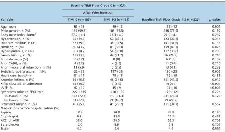
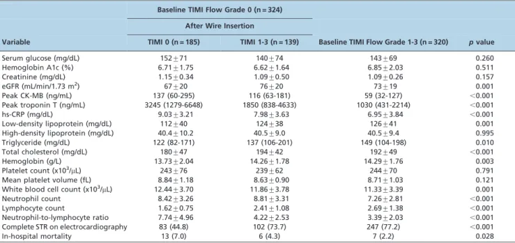
The baseline clinical characteristics of the patients in the 3 groups, divided according to the coronary TIMI flow grade are shown in Table 1. The group with persistent coronary no-flow AWI was older, contained fewer smokers, had a lower initial SBP and lower LVEF values, and more commonly had Killip class $2 on admission. Compared with patients with TIMI flow grade 1-3 at baseline and TIMI flow grade 1-3 AWI, patients with persistent coronary no-flow (TIMI 0) AWI had higher cardiac enzyme (CK-MB, troponin T) and hs-CRP levels, higher WBC and neutrophil counts and a lower lymphocyte count. These patients also had significantly lower hemoglobin, total cholesterol and triglyceride levels (Table 2). The NLR was significantly higher in the AWI TIMI flow 0 group compared with the AWI TIMI flow 1-3 and baseline TIMI flow 1-3 groups (7.74¡4.96, 4.22¡2.53 and 3.39¡2.03, respectively,
p,0.001). Figure 1 and Table 2 show the frequency distribution of the NLR according to the groups.
A total of 26 (4.0%) cases of mortality were documented during hospitalization. The AWI TIMI flow 0 group had a higher rate of in-hospital mortality compared with the AWI TIMI flow 1-3 and baseline TIMI flow 1-3 groups (13 (7.0), 6 (4.3) and 7 (2.2), respectively,p= 0.028) (Table 2). We found
Table 1 -Baseline characteristics and prior medications of the study population.
Baseline TIMI Flow Grade 0 (n = 324) After Wire Insertion
Variable TIMI 0 (n = 185) TIMI 1-3 (n = 139) Baseline TIMI Flow Grade 1-3 (n = 320) pvalue
Age, years 63¡13 59¡13 59¡12 0.001
Male gender, n (%) 129 (69.7) 105 (75.5) 246 (76.9) 0.197
Body mass index, kg/m2 27.2¡4.4 27.3¡4.0 27.9¡4.1 0.237
Hypertension, n (%) 83 (44.9) 53 (38.1) 123 (38.4) 0.311
Diabetes mellitus, n (%) 65 (35.1) 34 (24.5) 101 (31.6) 0.116
Smoking, n (%) 80 (43.2) 81 (58.3) 159 (49.7) 0.028
Hyperlipidemia, n (%) 56 (30.2) 50 (36.0) 117 (36.6) 0.293
Family history, n (%) 43 (23.2) 44 (31.7) 86 (26.9) 0.240
Prior stroke, n (%) 6 (3.2) 0 (0) 6 (1.9) 0.102
Prior CABG, n (%) 4 (2.2) 4 (2.9) 11 (3.4) 0.716
Prior myocardial infarction, n (%) 11 (5.9) 3 (2.2) 13 (4.1) 0.239
Systolic blood pressure, mmHg 122¡25 127¡26 130¡23 0.006
Heart rate, beats/min 81¡17 78¡15 79¡15 0.185
Anterior infarct, n (%) 86 (46.5) 48 (34.5) 151 (47.2) 0.019
Killip class$2 on admission 29 (15.7) 7 (5.0) 14 (4.4) ,0.001
LVEF, % 42¡10 45¡9 47¡10 ,0.001
Symptoms prior to PPCI, min 222¡115 210¡136 175¡121 0.225
,6 hours, n (%) 134 (72.4) 113 (81.3) 241 (75.3) 0.176
$6 hours, n (%) 51 (27.6) 26 (18.7) 79 (24.7)
Preinfarct angina, n (%) 46 (25.0) 41 (29.7) 111 (34.7) 0.557
Medications before hospitalization (%)
Aspirin 18.5 20.8 23.8 0.190
Clopidogrel 9.3 12.5 14.2 0.458
ACEI or ARB 32.0 28.3 33.3 0.708
Beta-blocker 12.0 8.9 7.8 0.707
Statin 4.0 4.4 4.4 0.991
that in-hospital mortality was independently associated with an increased NLR and increased no-flow (OR 1.52, p= 0.004 and OR 3.1,p,0.001, respectively). In addition, the percentage of complete ($70%) ST-segment resolution on electrocardiography after PPCI was significantly lower in the AWI TIMI flow 0 group compared with the AWI TIMI flow 1-3 and baseline TIMI flow 1-3 groups (p,0.001).
The baseline angiographic and procedural characteristics of the study population are presented in Table 3. The number of diseased vessels and the prevalence of multi-vessel disease were similar among the groups (p= 0.252 and p= 0.277, respectively). The IFAs of patients with TIMI flow grade 1-3 AWI were less frequently located in the left anterior descending coronary artery, whereas in patients Table 2 -Baseline biochemical and hematologic measurements for the study population.
Baseline TIMI Flow Grade 0 (n = 324) After Wire Insertion
Variable TIMI 0 (n = 185) TIMI 1-3 (n = 139) Baseline TIMI Flow Grade 1-3 (n = 320) pvalue
Serum glucose (mg/dL) 152¡71 140¡74 143¡69 0.260
Hemoglobin A1c (%) 6.71¡1.75 6.62¡1.64 6.85¡2.03 0.511
Creatinine (mg/dL) 1.15¡0.34 1.09¡0.50 1.09¡0.26 0.157
eGFR (mL/min/1.73 m2) 67¡20 76¡20 73¡19 0.001
Peak CK-MB (ng/mL) 137 (60-295) 116 (63-181) 59 (32-127) ,0.001
Peak troponin T (ng/mL) 3245 (1279-6648) 1850 (838-4633) 1030 (431-2214) ,0.001
hs-CRP (mg/dL) 9.03¡3.21 7.98¡3.63 6.95¡3.84 ,0.001
Low-density lipoprotein (mg/dL) 112¡40 124¡38 126¡41 0.001
High-density lipoprotein (mg/dL) 40.4¡10.2 40.5¡9.0 40.5¡9.4 0.995
Triglyceride (mg/dL) 122 (82-171) 137 (106-201) 149 (104-198) 0.010
Total cholesterol (mg/dL) 180¡47 194¡42 192¡49 ,0.001
Hemoglobin (g/L) 13.73¡2.04 14.26¡1.78 14.29¡1.76 0.003
Platelet count (x103/mL) 243¡76 239¡62 244¡70 0.791
Mean platelet volume (fL) 8.84¡1.18 8.63¡0.90 8.71¡1.03 0.121
White blood cell count (x103/mL) 12.44¡3.70 11.86¡3.78 11.33¡3.39 0.001
Neutrophil count 8.42¡3.26 8.81¡3.31 7.26¡2.81 ,0.001
Lymphocyte count 1.62¡0.75 2.41¡1.08 2.69¡1.38 ,0.001
Neutrophil-to-lymphocyte ratio 7.74¡4.96 4.22¡2.53 3.39¡2.03 ,0.001
Complete STR on electrocardiography 83 (44.8) 102 (73.7) 247 (77.2) ,0.001
In-hospital mortality 13 (7.0) 6 (4.3) 7 (2.2) 0.028
TIMI: Thrombolysis In Myocardial Infarction; eGFR: estimated glomerular filtration rate; CK-MB: creatine kinase-myocardial band; hs-CRP: high-sensitivity C-reactive protein; STR: ST-segment resolution.
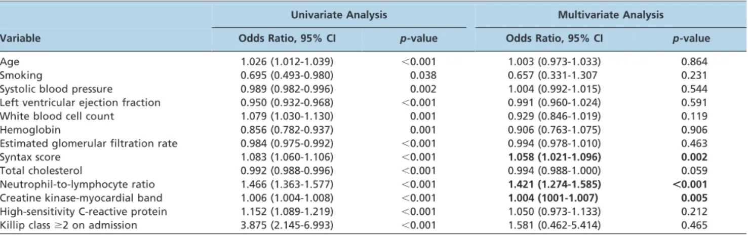
with baseline TIMI flow grade 0, the culprit lesions were more commonly located in the right coronary artery (p= 0.024). TIMI flow grade 0 was present before PPCI in 51.9% (n = 334) of infarct vessels and suboptimal flow (TIMI flow grade 1-2) was found in 25.0% (n = 161), whereas the remainder of infarct arteries presented with TIMI flow grade 3. As shown in Table 3, a final TIMI flow grade of 3 was achieved in 56.2% (n = 104) of patients with TIMI flow grade 0 AWI and in 87.8% (n = 122) of patients with TIMI flow grade 1-3 AWI; the remainder of patients had TIMI flow grade 1-3 at baseline. However, patients with TIMI flow grade 0 with persistent coronary no-flow after passage of the wire had a significantly lower prevalence of stent implantation, a lower rate of direct stenting, a higher total stent length and higher rate of balloon predilatation compared with those with TIMI flow grade 1-3 AWI and TIMI flow grade 1-3 at baseline (p,0.001 in post hoc analysis) (Table 3). Additionally, patients presenting with TIMI flow grade 0 had a significantly higher prevalence of tirofiban use compared with those with TIMI flow grade 1-3 at baseline (p= 0.006 in post hoc analysis). The multivariate logistic regression analysis showed that persistent coronary no-flow (TIMI 0) AWI was independently associated with the NLR (OR 1.421, 95% CI 1.274-1.585, p,0.001), peak CK-MB (OR 1.004, 95% CI 1001-1.007,p= 0.005) and the Syntax score (OR 1.058, 95% CI 1.021-1.096,p= 0.002) (Table 4). In the ROC curve analysis, an NLR.4.34 independently predicted TIMI flow 0 AWI, with 72% sensitivity and 70% specificity (AUC 0.801, 95% CI 0.765-0.837) (Figure 2). We also found that an NLR.4.34 was independently associated with the Syntax score (OR 1.035,p= 0.004), no-flow (OR 3.044,p,0.001) and peak CK-MB (OR 1.003,p= 0.001).
& DISCUSSION
To the best of our knowledge, this is the first study to identify an association between the NLR and coronary flow AWI in patients with STEMI undergoing PPCI. The main
finding in this study was that an increased NLR value on admission was a strong and independent predictor of persistent coronary no-flow AWI in patients with STEMI undergoing mechanical reperfusion. In addition, the patients with TIMI flow grade 0 AWI had a higher rate of in-hospital mortality.
The early restoration of coronary flow in an IRA recovers ventricular performance and decreases mortality in patients with STEMI (3). Brodie et al. (3) were the first to report a different survival pattern in patients presenting with TIMI flow grade 2 or 3 before intervention compared with patients with baseline TIMI flow grade 0 or 1. Moreover, STEMI patients presenting persistent no-flow AWI have a lower survival rate despite apparently successful mechan-ical intervention (5). Atherosclerosis is an inflammatory process (6) and inflammatory markers have been identified as useful predictors of clinical outcomes. In particular, the NLR has emerged as an important inflammatory marker for cardiovascular risk stratification (10). Akpek et al. (13) showed that the NLR was independently associated with the development of no-reflow and in-hospital major adverse cardiac events in patients with STEMI undergoing PPCI. In a recently published study, we also demonstrated that the NLR is associated with early patency of the IRA before PPCI in patients with STEMI (11).
There are several possible explanations for why an increased NLR value on admission was an independent predictor of persistent coronary no-flow AWI in patients with STEMI undergoing PPCI in our study.
First, the NLR is a sign of balance between neutrophil and lymphocyte counts in the body and is an indicator of systemic inflammation (17). Increased inflammation in patients with coronary artery disease may be linked to more extensive atheroma (18,19) and the association between thrombotic and inflammatory pathways in acute
coronary syndromes has been previously shown.
Atherosclerotic plaque rupture is an inflammatory process mediated by the complex interplay between innate neutro-phil-mediated reactive immune responses and subsequent Table 3 -Baseline angiographic and procedural characteristics of the study population.
Baseline TIMI Flow Grade 0 (n = 324) After Wire Insertion
Variable TIMI 0 (n = 185) TIMI 1-3 (n = 139) Baseline TIMI Flow Grade 1-3 (n = 320) pvalue
Multivessel disease, n (%) 105 (56.8) 73 (52.5) 158 (49.4) 0.277
Single-vessel disease, n (%) 81 (43.8) 66 (47.5) 165 (51.6) 0.550
Double-vessel disease, n (%) 62 (33.5) 43 (30.9) 95 (29.7)
Triple-vessel disease, n (%) 42 (22.7) 30 (21.6) 60 (18.8)
Number of diseased vessels, n (%) 1.79¡0.79 1.74¡0.79 1.72¡0.78 0.252
Infarct-related artery, n (%)
Left anterior descending 86 (46.5) 49 (35.3) 160 (50.0) 0.024
Left circumflex 24 (13.0) 20 (14.4) 57 (17.8)
Right 75 (40.5) 69 (49.6) 101 (31.6)
Left main 0 (0) 1 (0.7) 2 (0.6)
Chronic total occlusion, n (%) 32 (16.3) 16 (11.5) 45 (14.1) 0.329
Thrombus aspiration, n (%) 30 (16.2) 21 (15.0) 44 (13.8) 0.560
Stent deployed, n (%) 155 (83.8) 136 (97.8) 302 (94.4) ,0.001
Number of stents used 1.32¡0.60 1.26¡0.54 1.23¡0.45 0.232
Total stent length, mm 27.42¡11.84 23.86¡11.87 20.91¡8.52 ,0.001
Direct stenting, n (%) 5 (2.7) 78 (56.1) 229 (71.6) ,0.001
Stent with balloon predilatation, n (%) 147 (79.5) 56 (40.3) 68 (21.3) ,0.001
Tirofiban therapy, n (%) 91 (49.2) 68 (48.9) 117 (36.6) 0.006
Procedural success, n (%) 104 (56.2) 122 (87.8) 275 (89.6) ,0.001
lymphocyte-mediated adaptive immune responses. Neutrophils are the first leukocytes found in the damaged myocardial area. Procoagulants are secreted locally by the leukocytes, increasing the oxidative and proteolytic damage. Lymphocytes also have an essential role in modulating the inflammatory response at different stages of the athero-sclerotic process (20). In an acute setting, lymphopenia is a common finding during a stress response, secondary to increased levels of corticosteroids (21). Under pathologic conditions, defective clearance of apoptotic cells due to poor phagocytosis of apoptotic cells results in secondary, necrosis-inducing secretion of proinflammatory cytokines (tumor necrosis factor-a and IL-6). In addition, lymphopenia has
been reported in critical inflammatory states due to increased lymphocyte apoptosis (22).
Second, neutrophil infiltration can contribute to no-reflow by increasing blood viscosity and hypercoagulability. As part of this inflammatory reaction, cytokines such as IL-6, IL-8 and CD40 ligand trigger the upregulation of monocyte tissue factor expression, which may facilitate the extrinsic pathway of the coagulation cascade (23). Additionally, the distal embolization of leukocytes and platelet-leukocyte aggregates might contribute to reduced downstream micro-vascular perfusion as well as to thrombosis and widespread myocardial inflammation (8).
Third, it has been shown that a higher WBC count is correlated with the infarct size (24). After AMI, the release of chemoattractants draws neutrophils into the infarct zone during the first 6 hours of myocardial reperfusion; during the next 24 hours, the cells migrate into the myocardial Table 4 -Univariate and multivariate predictors of Thrombolysis In Myocardial Infarction 0 flow grade in ST-segment elevation myocardial infarction after wire insertion.
Univariate Analysis Multivariate Analysis
Variable Odds Ratio, 95% CI p-value Odds Ratio, 95% CI p-value
Age 1.026 (1.012-1.039) ,0.001 1.003 (0.973-1.033) 0.864
Smoking 0.695 (0.493-0.980) 0.038 0.657 (0.331-1.307 0.231
Systolic blood pressure 0.989 (0.982-0.996) 0.002 1.004 (0.992-1.015) 0.544
Left ventricular ejection fraction 0.950 (0.932-0.968) ,0.001 0.991 (0.960-1.024) 0.591
White blood cell count 1.079 (1.030-1.130) 0.001 0.929 (0.846-1.019) 0.119
Hemoglobin 0.856 (0.782-0.937) 0.001 0.906 (0.763-1.075) 0.906
Estimated glomerular filtration rate 0.984 (0.975-0.992) ,0.001 0.994 (0.978-1.010) 0.463
Syntax score 1.083 (1.060-1.106) ,0.001 1.058 (1.021-1.096) 0.002
Total cholesterol 0.992 (0.988-0.996) ,0.001 0.994 (0.988-1.000) 0.059
Neutrophil-to-lymphocyte ratio 1.466 (1.363-1.577) ,0.001 1.421 (1.274-1.585) ,0.001
Creatine kinase-myocardial band 1.006 (1.004-1.008) ,0.001 1.004 (1001-1.007) 0.005
High-sensitivity C-reactive protein 1.152 (1.089-1.219) ,0.001 1.050 (0.973-1.133) 0.212
Killip class$2 on admission 3.875 (2.145-6.993) ,0.001 1.581 (0.462-5.414) 0.465
AWI: after wire insertion; TIMI: Thrombolysis In Myocardial Infarction; STEMI: ST-segment elevation myocardial infarction.
tissue (24). Neutrophil infiltration is regulated through a complex sequence of molecular steps involving selectins and integrins, which mediate leukocyte rolling and adhe-sion to the endothelium (25). These neutrophils cause proteolytic and oxidative damage to the endothelial cells, plug the microvasculature, and induce hypercoagulability and may promote infarct expansion (24,25). As we have found that peak CK-MB and troponin T levels were elevated and the LVEF was decreased in patients with TIMI flow grade 0 AWI, the NLR may act as a combined surrogate marker for both the reactive and the adaptive components of the inflammatory response that results in plaque rupture, ischemic myocardial damage, adverse ventricular remodel-ing and consequent LV dysfunction (26).
Our study has certain limitations. First, inflammatory markers other than hs-CRP were not analyzed and compared with the NLR. Second, the myocardial blush score, which is a well-known prognostic indicator in STEMI patients (27), was not assessed in our study. Finally, the NLR was measured only at the time of admission to evaluate its prognostic impact. In conclusion, the NLR is a strong independent predictor of persistent coronary no-flow AWI in patients with STEMI undergoing mechanical revascularization. Patients with STEMI in whom persistent no-flow AWI is detected during PPCI are at greater risk for in-hospital mortality. As an early and readily available prognostic marker, the NLR may thus be useful in the early risk stratification of STEMI patients treated via PPCI.
& AUTHOR CONTRIBUTIONS
Kurtul A, Murat SN and Yarlioglues M wrote the manuscript, performed the statistical analyses and were responsible for the final editing. Duran M, Celik IE, Kilic A and Ocek AH collected the data.
& REFERENCES
1. Kushner FG, Hand M, Smith SC Jr, King SB 3rd, Anderson JL, Antman EM, et al. 2009 Focused Updates: ACC/AHA Guidelines for the Management of Patients With ST-Elevation Myocardial Infarction (updating the 2004 Guideline and 2007 Focused Update) and ACC/ AHA/SCAI Guidelines on Percutaneous Coronary Intervention (updat-ing the 2005 Guideline and 2007 Focused Update): a report of the American College of Cardiology Foundation/American Heart Association Task Force on Practice Guidelines. Circulation. 2009;120(22):2271-306, http://dx.doi.org/10.1161/CIRCULATIONAHA.109.192663.
2. TIMI Study Group. The Thrombolysis in Myocardial Infarction (TIMI) trial. Phase I findings. N Engl J Med. 1985;312(14):932-6.
3. Brodie BR, Stuckey TD, Hansen C, Muncy D. Benefit of coronary reperfusion before intervention on outcomes after primary angioplasty for acute myocardial infarction. Am J Cardiol. 2000;85(1):13-8, http://dx. doi.org/10.1016/S0002-9149(99)00598-6.
4. Mehta RH, Harjai KJ, Cox D, Stone GW, Brodie B, Boura J, et al. Primary Angioplasty in Myocardial Infarction (PAMI) Investigators. Clinical and angiographic correlates and outcomes of suboptimal coronary flow in patients with acute myocardial infarction undergoing primary percuta-neous coronary intervention. J Am Coll Cardiol. 2003;42(10):1739-46, http://dx.doi.org/10.1016/j.jacc.2003.07.012.
5. Valgimigli M, Campo G, Malagutti P, Anselmi M, Bolognese L, Ribichini F, et al. Persistent coronary no flow after wire insertion is an early and readily available mortality risk factor despite successful mechanical intervention in acute myocardial infarction: a pooled analysis from the STRATEGY (Single High-Dose Bolus Tirofiban and Sirolimus-Eluting Stent Versus Abciximab and Bare-Metal Stent in Acute Myocardial Infarction) and MULTISTRATEGY (Multicenter Evaluation of Single High-Dose Bolus Tirofiban Versus Abciximab With Sirolimus-Eluting Stent or Bare-Metal Stent in Acute Myocardial Infarction Study) trials. JACC Cardiovasc Interv. 2011;4(1):51-62, http://dx.doi.org/10.1016/j.jcin.2010.09.016.
6. Korkmaz L, Kul S, Korkmaz AA, Akyu¨z AR, Ag˘ac¸ MT, Erkan H, et al. Increased leucocyte count could predict coronary artery calcification in patients free of clinically apparent cardiovascular disease. Turk Kardiyol Dern Ars. 2012;40(3):223-8, http://dx.doi.org/10.5543/tkda.2012.37801.
7. Hoffman M, Blum A, Baruch R, Kaplan E, Benjamin M. Leukocytes and coronary heart disease. Atherosclerosis. 2004;172(1):1-6, http://dx.doi. org/10.1016/S0021-9150(03)00164-3.
8. van der Wal AC, Becker AE, van der Loos CM, Das PK. Site of intimal rupture or erosion of thrombosed coronary atherosclerotic plaques is characterized by an inflammatory process irrespective of the dominant plaque morphology. Circulation. 1994;89(1):36-44, http://dx.doi.org/10. 1161/01.CIR.89.1.36.
9. Marx N, Neumann FJ, Ott I, Gawaz M, Koch W, Pinkau T, Scho¨mig A. Induction of cytokine expression in leukocytes in acute myocardial infarction. J Am Coll Cardiol. 1997;30(1):165-70, http://dx.doi.org/10. 1016/S0735-1097(97)00116-2.
10. Tamhane UU, Aneja S, Montgomery D, Rogers EK, Eagle KA, Gurm HS. Association between admission neutrophil to lymphocyte ratio and outcomes in patients with acute coronary syndrome. Am J Cardiol 2008;102(6):653-7, http://dx.doi.org/10.1016/j.amjcard.2008.05.006. 11. Kurtul A, Murat SN, Yarlioglues M, Duran M, Karadeniz M, Ergun G,
et al. The relationship between neutrophil/lymphocyte ratio and infarct-related artery patency before mechanical reperfusion in patients with ST-elevation myocardial infarction. Coron Artery Dis. 2014;25(2):159-66, http://dx.doi.org/10.1097/MCA.0000000000000067.
12. Park JJ, Jang HJ, Oh IY, Yoon CH, Suh JW, Cho YS, et al. Prognostic value of neutrophil to lymphocyte ratio in patients presenting with ST-elevation myocardial infarction undergoing primary percutaneous coronary intervention. Am J Cardiol. 2013;111(5):636-42, http://dx.doi. org/10.1016/j.amjcard.2012.11.012.
13. Akpek M, Kaya MG, Lam YY, Sahin O, Elcik D, Celik T, et al. Relation of neutrophil/lymphocyte ratio to coronary flow to in-hospital major adverse cardiac events in patients with ST-elevated myocardial infarc-tion undergoing primary coronary interveninfarc-tion. Am J Cardiol. 2012;110(5):621-7, http://dx.doi.org/10.1016/j.amjcard.2012.04.041. 14. Serruys PW, Onuma Y, Garg S, Sarno G, van den Brand M, Kappetein
AP, et al. Assessment of the SYNTAX score in the Syntax study. EuroIntervention. 2009;5(1):50-6, http://dx.doi.org/10.4244/EIJV5I1A9. 15. Gibson CM, Cannon CP, Daley WL, Dodge JT Jr, Alexander B Jr, Marble
SJ, et al. TIMI frame count: a quantitative method of assessing coronary artery flow. Circulation. 1996;93(5):879-88, http://dx.doi.org/10.1161/ 01.CIR.93.5.879.
16. Schiller NB, Shah PM, Crawford M, DeMaria A, Devereux R, Feigenbaum H, et al. Recommendations for quantitation of the left ventricle by two-dimensional echocardiography. American Society of Echocardiography Committee on Standards, Subcommittee on Quantitation of Two-Dimensional Echocardiograms. J Am Soc Echocardiogr. 1989;2(5):358-67.
17. Zahorec R. Ratio of neutrophil to lymphocyte counts-rapid and simple parameter of systemic inflammation and stress in critically ill. Bratisl Lek Listy. 2001;102(1):5-14.
18. Arbel Y FA, Halkin A, Birati EY, Revivo M, Zuzut M, Shevach A, et al. Neutrophil/lymphocyte ratio is related to the severity of coronary artery disease and clinical outcome in patients undergoing angio-graphy. Atherosclerosis. 2012;225(2):456-60, http://dx.doi.org/10.1016/ j.atherosclerosis.2012.09.009.
19. Arroyo-Espliguero R, Avanzas P, Cosı´n-Sales J, Aldama G, Pizzi C, Kaski JC. C-reactive protein elevation and disease activity in patients with coronary artery disease. Eur Heart J. 2004;25(5):401-8, http://dx.doi.org/ 10.1016/j.ehj.2003.12.017.
20. Ducloux D, Challier B, Saas P, Tiberghien P, Chalopin JM. CD4 cell lympho-penia and atherosclerosis in renal transplant recipients. J Am Soc Nephrol. 2003;14(3):767-72, http://dx.doi.org/10.1097/01.ASN.0000048718.43419.44. 21. Onsrud M, Thorsby E. Influence of in vivo hydrocortisone on some human blood lymphocyte subpopulations. I. Effect on natural killer cell activity. Scand J Immunol. 1981;13(6):573-9.
22. Hotchkiss RS, Karl IE. The pathophysiology and treatment of sepsis. N Engl J Med. 2003;348(2):138-50.
23. Ott I, Neumann FJ, Kenngott S, Gawaz M, Scho¨mig A. Procoagulant inflammatory responses of monocytes after direct balloon angioplasty in acute myocardial infarction. Am J Cardiol. 1998;82(8):938-42, http://dx. doi.org/10.1016/S0002-9149(98)00509-8.
24. Yellon DM, Hausenloy DJ. Myocardial reperfusion injury. N Engl J Med. 2007;357(11):1121-35.
25. Frangogiannis NG, Smith CW, Entman ML. The inflammatory response in myocardial infarction. Cardiovasc Res. 2002;53(1):31-47, http://dx. doi.org/10.1016/S0008-6363(01)00434-5.
26. Mazzone A, De Servi S, Ricevuti G, Mazzucchelli I, Fossati G, Pasotti D, et al. Increased expression of neutrophil and monocyte adhesion molecules in unstable coronary artery disease. Circulation. 1993;88(2):358-63, http://dx.doi.org/10.1161/01.CIR.88.2.358.