1 Yüzüncü Yıl Üniversitesi Dursun Odabaşı Tıp Merkezi Göğüs Hastalıkları, Van, Türkiye 2 Van Bölge Eğitim ve Araştırma Hastanesi Göğüs Hastalıkları, Van, Türkiye
Yazışma Adresi /Correspondence: Buket Mermit Çilingir,
Van Bölge Eğiim ve Araşırma Hastanesi Göğüs Hastalıkları Bölümü, Van, Türkiye Email: buketmermitcilingir@gmail.com Geliş Tarihi / Received: 15.05.2015, Kabul Tarihi / Accepted: 30.07.2015
ORIGINAL ARTICLE / ÖZGÜN ARAŞTIRMA
The relationship between neutrophil-to-lymphocyte ratio and platelet-to-lymphocyte
ratio in patients with obstructive sleep apnea syndrome
Obstruktif uyku apne sendromunda platelet lenfosit oranı ve nötrofil lenfosit oranı ilişkisi
Hülya Günbatar1, Selami Ekin1, Aysel Sünnetçioğlu1, Ahmet Arısoy1, Buket Mermit Çilingir2, Selvi Aşker1, Bünyamin Sertoğullarından1
ÖZET
Amaç: Obstruktif Uyku Apne Sendromu (OSAS) ile Kardi-yovasküler Hastalıklar (KVH) arasında güçlü bir ilişki var-dır. Kronik intermittan hipoksi, inlamasyon, oksidatif stres ve endotelyal disfonksiyon OSAS ve KVH arasındaki et-yolojik mekanizmaları ortaya çıkarmış olabilir. İnlamas-yon KVH gelişiminde önemli rol oynar. Platelet Lenfosit Oranı (PLO) ve Nötrofil Lenfosit Oranı (NLO) inlamasyo-nu gösteren yeni belirteçlerdir. Bu çalışma PLO ve NLO arasındaki ilişkiyi ve OSAS şiddeti, polisomnografik para-metreler ve PLR arasındaki ilişkiyi araştırmaktadır. Yöntemler: OSAS ön tanısı ile tüm gece polisomnografisi yapılan hastaların çalışmaya dahil edildiği bir kohort çalış-ması planlandı. Hastalar apne hipopne indeksi skorlarına göre sınılandırıldı. Basit horlama (Grup 1; AHİ <5 adet/ saat), hafif OSAS (Grup 2; AHİ: 5-15), orta derece OSAS (Grup 3; AHİ: 15-30) ve ağrı OSAS (Grup 4; AHİ >30). Bulgular: 111 hasta çalışmaya dahil edildi. Grup 1, 2 ve 3 ‘te sırasıyla 26, 22 ve 63 hasta vardı. PLO gruplar ara-sında anlamlı düzeyde değişiklik göstermekteydi. Grup 1’de 87,12, Grup2’de 103,6, Grup 3’te 112,5 idi (p < 0.05). PLO ile NLO, AHİ, oksijen desaturasyon indeksi, ortalama ve minimum oksijen saturasyon değeri arasında anlamlı derecede ilişki vardı (p<0.05). Çoklu regresyon analizi ile PLO’ nun KVH için bağımsız bir belirleyici olduğu saptan-dı. KVH varlığında, PLO cut off değerinin 86,03 olduğu gösterildi.
Sonuç: Tüm bu bulgular ışığında PLO ile OSAS şiddeti arasında güçlü ilişki vardır. PLO OSAS hastalarında KVH varlığını belirlemede bir belirteç olarak kullanılabilir. Anahtar kelimeler: obstruktif uyku apnesi, inlamasyon, platelet lenfosit oranı
ABSTRACT
Objective: There is a strong relationship between ob-structive sleep apnea syndrome (OSAS) and cardiovas-cular disease (CVD). Chronic intermittent hypoxia, inlam-mation, oxidative stress, and endothelial dysfunction may create etiologic mechanisms, connection between OSAS to CVD. Inlammation play an important role in the devel-opment of CVD. Platelet- Lymphocyte Ratio (PLR) and Neutrophil-lymphocyte Ratio (NLR) are new biomarkers showing inlammation. This study was designed to inves-tigate the association between PLR, NLR and relationship between severity of OSAS, polysomnographic param-eters and PLR.
Methods: This was a cohort study in which patients who had undergone a full night polysomnography for diagnosis of OSA were recruited. Patients were divided according to their apnea hypopnea index (AHI) scores into OSAS neg-ative simple snoring (Group 1; AHI <5 times/hours), mild (Group 2; AHI: 5-15), moderate (Group 3; AHI:15-30), and severe OSAS (Group 4; AHI >30) groups.
Results: A total of 111 patients were included in this study. There were 26, 22 and 63 patients in Groups 1, 2 and 3, respectively. PLR were signiicantly different be-tween groups (Group 1: 87.12, Group 2: 103.6, Group 3: 112.5, p < 0.05). PLR were signiicantly correlated with NLR, AHI, oxygen desaturation index, average and mini-mum O2 saturation values (p < 0.05). Multiple regression analysis demonstrated that PLR is an independent pre-dictor of CVD. PLR cut-off value for demonstrating the presence of CVD is higher than 86.03.
Conclusion: In the light of these indings, PLR is strongly associated with the severity of OSAS. PLR might be used as a biomarker to predict CVD in OSAS patients.
INTRODUCTION
Obstructive sleep apnea syndrome (OSAS) is char-acterized by collapse of the upper airway during sleep, recurring apnea, intermittent hypoxemia, and daytime sleepiness, associated with a decreased daytime performance and impaired quality of life [1]. It is a common disorder of middle-aged adults, affecting 4% of men and 2% of women [2].
Recent studies have indicated that OSAS is associated with multiple causal factors of endothe-lial damage and atherosclerosis due to oxidative
stress, systemic inlammation and increased levels
of soluble adhesion molecules and coagulation fac-tors [3,4]. One routinely available marker of the
systemic inlammatory response is the
neutrophil-lymphocyte ratio (NLR). The leukocyte count and
its subtypes are well-known inlammatory markers
[5,6]. Since the response to physiological stress of leukocytes in circulation leads to an increase in the number of neutrophils and a decrease in the num-ber of lymphocytes, the ratio of these subgroups
to each other is used as a marker of inlammation. During the inlammatory response, changes occur in
the ratios of leukocyte subgroups in the circulation. Neutrophilia is accompanied by relative lympho-penia [7]. In recent years, neutrophilia and relative lymphocytopenia were shown to be an independent predictor of mortality in patients with acute heart failure [4,8]. As NLR, platelet-to-lymphocyte ratio (PLR) was found to associated with all cause mor-tality in patients with acute myocardial infarction. Thrombocytosis is caused by the stimulation of
megakaryocytes by proinlammatory cytokines [9],
and its association with prognosis shown in other re-lated studies may be explained based on an elevated platelet count being an indicator of the severity of
inlammation. However, to date, the data about PLR and its association with inlammation are lacking in
OSAS patients. Therefore, we aimed to determine
the relationship between PLR, NLR, and inlamma -tion in OSAS patients.
METHODS
One hundred eleven patients between ages 27 and
69 who had undergone polysomnography exami -nation at the sleep laboratory of the pulmonology department at the Faculty of Medicine at Yüzüncü
Yıl University Hospital between February 2014 and January 2014 were retrospectively evaluated. Poly -somnography examinations at the sleep laboratory were conducted prior to inclusion in this study. Pa-tients were excluded from the study if they had acute
inlammation, infection, diabetes mellitus, systemic
hypertension, hyperlipidemia, congestive heart failure, chronic renal failure, chronic liver disease, chronic obstructive pulmonary disease, connective
tissue disease, or inlammatory bowel disease. Ac
-cording to the apnea hypopnea index (AHI), sub -jects were divided into three groups. Group 1
con-tained the control subjects without OSA (AHI < 5
times/hours, n = 26) Group 2 contained the patients
with moderate OSA (AHI = 15–30 times/hours, n =
22), and Group 3 contained the patients with severe
OSA (AHI > 30 times/hours, n = 63).
Polysomnography
Overnight polysomnography was performed with 16 channel Embla (Medcare Inc, Iceland) continu-ous sleep technician monitoring. The system con-sists of 4 channels of electroencephalogram (EEG) (with electrode placements at C4-A1, C3-A2, O2-A1, and O1-A2), 2 channels of electrooculography (EOG), submental electromyography(EMG),
oro-nasal air low, thoracic and abdominal movements,
pulse oximeter oxygen saturation, tibial EMG, body position detector, electrocardiogram and tracheal
sound. Apnea was deined as the complete cessation of airlow lasting more than 10 seconds. Hypopnea was deined as a reduction >30% in airlow lasting more than 10 seconds accompanied by >4% desatu -ration and/or arousal. The average number of epi-sodes of apnea and hypopnea per hour of sleep were
measured as AHI. The OSAS diagnosis was made on the basis of an apnea/ hypopnea index (AHI) >
5 times/hours. Sleep stages were scored following
standard criteria with 30-second epochs and were reviewed and veriied by a certiied sleep physician.
Biochemical measurement
per-formed with a Coulter LH 750 hemogram device
(Beckman Coulter, Fullerton, California).
Statistical Analysis
The results were explained as the mean ± standard deviation. The parametric variables were compared using Student’s t-test. The non-parametric con-tinuous variables were compared with the
Mann-Whitney U-test. Pairwise Pearson correlation was
executed to estimate linear relationships between characteristics. All statistical calculations were
per-formed using SAS version 9.3 (SAS, 2014). The re
-sults were considered statistically signiicant when the p value was <0.05.
RESULTS
A total of 111 patients examined by polysomnog-raphy were included in the analysis. The patients
with OSA included 66 men (78.4%) and 19 women
(21.6%). The OSA and control groups were similar
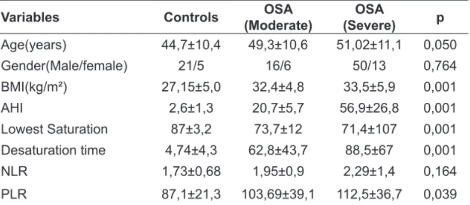
in terms of sex and age (p>0.05). Clinical charac -teristics of the patients and controls are shown in
Table 1. There was no statistically signiicant dif -ference between the two groups regarding the age, BMI, Epworth sleep score, there was no statistically
signiicant difference on the gender distribution. The mean AHI was 20.7 ± 5.7 in the moderate OSA group and 56.9 ± 26.8 in the severe OSA group. The difference in AHI and minimum oxygen desat
-uration between groups was statistically signiicant (Table 1). There was no signiicant statistical differ -ence between NLR in the controls and patients with moderate and severe OSA but the NLR values were higher in patients with severe and moderate OSA
than in the control group (respectively, 2.29 ± 1.43 and 1.95 ± 0.93, 1.73 ± 0.68, p>0.05). The PLR val
-ues were statistically signiicant higher in patients
with severe and moderate OSA than in the control
group (respectively, 112.5 ± 36.7 and 103.69 ± 39.1, 87.1 ± 21.3 p=0.039). Additionally, correlation of
NLR and PLR with parameters of sleep was noted. NLR and PLR were positively correlated with BMI
(NLR; r=0.540, p<0.01; PLR; r=0.514, p<0.05) and
negatively correlated with oxygen desaturation time
under 90 % (NLR: r=0.602, p<0.01; PLR: r = 0.680 p<0.01). PLR cut-off value for demonstrating the severity of OSAS is higher than 86.03. (sensitivity, 67.1% and speciity, 61.5%) (Figure 1).
Figure 1. Roc curve for Platelet-to-Lymphocyte Ratio
Variables Controls OSA
(Moderate)
OSA
(Severe) p
Age(years) 44,7±10,4 49,3±10,6 51,02±11,1 0,050
Gender(Male/female) 21/5 16/6 50/13 0,764
BMI(kg/m²) 27,15±5,0 32,4±4,8 33,5±5,9 0,001
AHI 2,6±1,3 20,7±5,7 56,9±26,8 0,001
Lowest Saturation 87±3,2 73,7±12 71,4±107 0,001
Desaturation time 4,74±4,3 62,8±43,7 88,5±67 0,001
NLR 1,73±0,68 1,95±0,9 2,29±1,4 0,164
PLR 87,1±21,3 103,69±39,1 112,5±36,7 0,039
BMI: Body mass index, AHI: Apnea-hypopnea index, NLR: Neutrophil-lympho-cyte ratio, PLR: Platelet-to-lymphoNeutrophil-lympho-cyte ratio
DISCUSSION
There were two main indings of the present study. First, inlammation markers including PLR, NLR,
were increased in OSA patients when compared with simple snoring patients. Second, PLR was positively correlated with NLR, and PLR were
sig-niicantly correlated with AHI, oxygen desaturation
index, average and minimum O2 saturation values
(p < 0.05).
The NLR was introduced as a cost-effective
potential inlammatory marker with prognostic and predictive values in systemic inlammatory diseases
such as cardiovascular disease, kidney disease,
in-lammatory bowel disease, and familial Mediter
-ranean fever [10-14]. In many studies, white blood
cell counts and their subtypes have been found to
be classic inlammatory markers, especially in car -diovascular disease [15,16]. OSA is not a simple respiratory abnormality that occurs during sleep;
the systemic inlammatory response generated by
OSA can be associated with cardiovascular disease and considered a new, independent cardiovascular risk factor [17,18]. Various studies have
demon-strated elevated inlammatory marker levels in OSA
patients compared with matched controls, with a
signiicant fall after effective treatment with con
-tinuous positive airway pressure [19]. Intermittent
hypoxia and sleep deprivation may explain the
re-lationship between inlammatory markers and AHI
in OSA and is a trigger for the cardiovascular and metabolic alterations. Many studies have reported
that patients with OSA develop systemic inlam -mation, with increased levels of mediators of the
systemic inlammatory response, including inter -cellular adhesion molecules (ICAM), coagulation factors (Factor VIII, tissue factor), and C-reactive
protein (CRP) [20-22]. In this study, increased NLR could be a new inlammatory marker for inlamma -tion in OSA patients. Furthermore, NLR might also
be inluenced by many different factors, including
atherosclerosis, hypertension, and diabetes, and can even be affected by atherosclerotic risk factors [23]. In this study, our patient population was free of con-ditions that increase NLR, such as cardiovascular disease, hypertension, hypercholesterolemia, and diabetes mellitus.
As new inlammation markers, PLR and NLR
were introduced in cardiac and noncardiac disorders [24]. Recent studies demonstrated that activated platelets could be an important part of increased
atherogenesis especially in the period of inlam -mation [25,26]. Platelets can interact with a kind of different cell types including endothelial cells, dendritic cells, T-lymphocytes, neutrophils, and mono-nuclear phagocytes, the relationship of plate-lets with these cells mentioned above might initiate
and exacerbate the inlammation in the arterial wall.
There has been an increasing evidence demonstrat-ed that activatdemonstrat-ed platelets could incite leukocyte
re-cruitment to the vessel wall and initiate the inlam -mation that can mainly seen in the pathogenesis of atherosclerosis [27].
This study demonstrates a relationship between the severity of OSA and PLR that is dependent on
inlammation and intermittent hypoxia. PLR was signiicantly increased in patients with moderate
to severe OSA compared to patients with no OSA. Only one study have suggested the optimal cut-off
value for PLR at >108.6 in OSAS patients for car -diovascular risk factors (28). Multiple regression analysis demonstrated that PLR is an independent predictor of CVD. In this study PLR cut-off value for demonstrating the presence of CVD is higher
than 86.03. PLR could be considered a new inlam
-matory marker for inlammation in OSA patients.
This study also found a positive association be-tween NLR, PLR and the lowest oxygen
desatura-tion time and desaturadesatura-tion time under 90% in OSA,
which could be explained by the effect of hypoxia on NLR and PLR.
Calculation of PLR and NLR are quite simple and inexpensive methods when compared other
in-lammatory cytokines including IL-6, IL-1b, and TNF-a. Our results conirm that PLR and NLR can predict inlammation in OSAS patients. Therefore,
these simple, relatively cheap and universally
avail-able methods can be used by clinicians for the irst assessment of inlammation in OSAS patients be -fore applying other expensive and invasive proce-dures.
This study had some limitations. First, the study
-tory markers such as CRP and ibrinogen were not
analyzed and compared with NLR and PLR in the study population.
In conclusion, novel biomarkers are needed
to indicate the level inlammation of disease in pa -tients with OSA. Therefore, PLR could be
consid-ered a new inlammatory marker for inlammation
in OSA patients, as it is a quick, cheap, and easily measurable property on routine CBC analysis.
REFERENCES
1. Young T, Palta M, Dempsey J, et al. The occurrence of sleep-disordered breathing among middle- aged adults. N Engl J
Med 1993;328:1230-1235.
2. Lattimore JD, Celermajer DS, Wilcox I. Obstructive sleep apnea and cardiovascular disease. J Am Coll Cardiol
2003;41:1429-1437.
3. Ryan S, Taylor CT, McNicholas WT. Systemic inlammation:
a key factor in the pathogenesis of cardiovascular com-plications in obstructive sleep apnoea syndrome? Thorax
2009;64:631-636.
4. Yokoe T, Minoguchi K, Matsuo H, et al. Elevated levels of
C-reactive protein and interleukin-6 in patients with obstruc-tive sleep apnea syndrome are decreased by nasal
continu-ous positive airway pressure. Circulation
2003;107:1129-1134.
5. Thomsen M, Ingebrigtsen TS, Marott JL, et al. Inlammatory
biomarkers and exacerbations in chronic obstructive
pul-monary disease. JAMA 2013;309:2353–2361.
6. Hotchkiss RS, Karl IE. The pathophysiology and treatment of sepsis. NEJM 2003;348:138–150.
7. Mehta J, Dinerman J, Mehta P, et al. Neutrophil function in
ischemic heart disease. Circulation 1989;79:549–546. 8. Rudiger A, Burckhardt OA, Harpes P, et al. The relative
lymphocyte count on hospital admission is a risk factor for long-term mortality in patients with acute heart failure. Am
J Emerg Med. 2006;24:451–454.
9. Alexandrakis MG, Passam FH, Moschandrea IA, et al.
Lev-els of serum cytokines and acute phase proteins in patients with essential and cancer-related thrombocytosis. Am J
Clin Oncol 2003; 26:135–140.
10. Ahsen A, Ulu MS, Yuksel S, et al. As a new inlammatory marker for familial Mediterranean fever: Neutrophil-to-lymphocyte ratio. Inlammation. 2013;36:1357-1362.
11. Núñez J, Núñez E, Bodí V, et al. Usefulness of the neutro
-phil to lymphocyte ratio in predicting long-term mortality in ST segment elevation myocardial infarction. Am J
Car-diol 2008;101:747–752.
12. Ertaş G, Sönmez O, Turfan M, et al. Neutrophil/lympho
-cyte ratio is associated with thromboembolic stroke in
patients with non-valvular atrial ibrillation. J Neurol Sci 2013;324:49–52.
13. Celikbilek M, Dogan S, Ozbakir O, et al. Neutrophil–lym
-phocyte ratio as a predictor of disease severity in ulcerative
colitis. J Clin Lab Analysis 2013;27:72–76.
14. Okyay GU, Inal S, Oneç K, et al. Neutrophil to lymphocyte ratio in evaluation of inlammation in patients with chronic
kidney disease. Renal Failure 2013;35:29–36.
15. Akpek M, Kaya MG, Lam YY, et al. Relation of neutrophil/
lymphocyte ratio to coronary low to in-hospital major ad
-verse cardiac events in patients with ST-elevated
myocar-dial ınfarction undergoing primary coronary ıntervention. Am J Cardiol 2012;110:621-627.
16. Gibson PH, Cuthbertson BH, Croal BL, et al. Usefulness of
neutrophil/lymphocyte ratio as predictor of new-onset atrial
ibrillation after coronary artery bypass grafting. Am J Car
-diol 2010;105:186-191.
17. Pack AI, Gislason T. Obstructive sleep apnea and
cardio-vascular disease: a perspective and future directions. Prog Cardiovasc Dis 2009;51:434-451.
18. Horne BD, Anderson JL, John JM, et al. Which white blood
cell subtypes predict increased cardiovascular risk? J Am
Coll Cardiol 2005;45:1638–1643.
19. Kent BD, Ryan S, McNicholas WT. Obstructive sleep apnea
and inlammation: Relationship to cardiovascular comor
-bidity. Respir Physiol Neurobiol 2011;178:475–481.
20. Yokoe T, Minoguchi K, Matsuo H, et al. Elevated levels
of C-reactive protein and interleukin-6 in patients with obstructive sleep apnea syndrome are decreased by na-sal continuous positive airway pressure. Circulation.
2003;107:1129–1134.
21. Shamsuzzaman AS, Winnicki M, Lanfranchi P, et al. El-evated C-reactive protein in patients with obstructive sleep
apnea. Circulation 2002;105:2462–2464.
22. Kokturk O, Ciftci TU, Mollarecep E, et al. Elevated C-re
-active protein levels and increased cardiovascular risk in
patients with obstructive sleep apnea syndrome. Int Heart J 2005;46:801–809.
23. Szkandera J, Pichler M, Gerger A, et al (2013b) Reply:
comment on ‘Elevated preoperative neutrophil/lymphocyte ratio is associated with poor prognosis in soft-tissue sar-coma patients’. Br J Cancer 108:2627.
24. Dotsenko O, Chaturvedi N, Thom SA, et al. Platelet and
leukocyte activation, atherosclerosis and inlammation in European and South Asian men.J Thromb Haemost 2007;5:2036–2042.
25. Koyama H, Maeno T, Fukumoto S, et al. Platelet P-selectin
expression is associated with atherosclerotic wall thickness
in carotid artery in humans. Circulation 2003;108:524–529. 26. Shoji T, Koyama H, Fukumoto S, et al. Platelet activation
is associated with hypoadiponectinemia and carotid
ath-erosclerosis. Atherosclerosis 2006;188:190–195.
27. Langer HF, Gawaz M. Platelet-vessel wall interactions in atherosclerotic disease. Thromb Haemost 2008;99:480–
486.
28. Koseoglu HI, Altunkas F, Kanbay A, et al. Platelet-lym
-phocyte ratio is an independent predictor for cardiovascu-lar disease in obstructive sleep apnea syndrome. J Thromb