DOI: 10.1590/0004-282X20160157 ARTICLE
Refractory epilepsy in children with brain
tumors. The urgency of neurosurgery
Epilepsia refratária em crianças com tumores cerebrais. A urgência de neurocirurgia
Marília Rosa Abtibol Bernardino1, Carolina Funayama1, Ana Paula Andrade Hamad1, Hélio Machado1,
Américo Sakamoto1, Ursula Thome1, Vera CristinaTerra1, Antonio Carlos dos Santos1,2,3, Luciano Nader
Serafani4, Nathalia Cunha Calixto2, Huria Shalom Monturil de Carvalho Silva4
Refractory epilepsy, mainly for those patients resistant to
drug treatment, has a great impact, causing signiicant dam
-age to the brain, afecting the development, cognition, learn
-ing, behavior and quality of life. Tumors are among the con -ditions associated with refractory epilepsy, with satisfactory surgical results in children1,2,3,4.
A group of a highly epileptogenic brain tumors, called “long-term epilepsy-associated tumors”, usually manifest with early onset of spontaneous seizures, and occur pre-dominately in the temporal lobe5. Long-term epilepsy-asso
-ciated tumors are mostly classiied as glio-neuronal tumors
by the World Health Organization (WHO), are likely to occur
during brain development and can be associated with focal cortical dysplasia (International League Against Epilepsy
(ILAE) classiication: Type IIIb)6,7,8. A tumor manifesting in
the temporal lobe in children or adolescents should be care-fully examined, as these neoplasms usually belong to the spectrum of long-term epilepsy-associated tumors and not
to malignant gliomas. Seizure control should be the primary treatment target to reduce the risk of progressive cognitive impairment or adverse efects of medication and, in order to
obtain the best seizure control, surgical treatment is man-datory in these cases5,9.
Knowledge about the proile and outcome of patients
with epilepsy and brain tumors can assist in making
deci-sions, emphasizing the beneits of a broad discussion
between oncology, pediatric neurology, epilepsy and
neuro-surgery teams.
1Universidade de São Paulo, Faculdade de Medicina de Ribeirão Preto, Departamento de Neurociências e Ciências do Comportamento, Ribeirão Preto SP, Brasil; 2Universidade de São Paulo, Faculdade de Medicina de Ribeirão Preto, Departamento de Neuroradiologia, Seção de Ressonância Magnética, Ribeirão Preto SP, Brasil; 3Universidade de São Paulo, Faculdade de Medicina de Ribeirão Preto, Hospital das Clínicas, Centro de Ciências das Imagens e Física Médica, Ribeirão Preto SP, Brasil; 4Universidade de São Paulo, Faculdade de Medicina de Ribeirão Preto, Departamento de Patologia e Medicina Legal, Ribeirão Preto SP, Brasil.
Correspondence: Marília Rosa Abtibol Bernardino; Faculdade de Medicina de Ribeirão Preto da USP, Departamento de Neurologia; Avenida Bandeirantes, 3900; 14040-900 Ribeirão Preto SP, Brasil; E-mail: [email protected]
Conflict of interest: There is no conlict of interest to declare.
Received 16 May 2016; Received in inal form 26 July 2016; Accepted 16 August 2016.
ABSTRACT
In order to verify indications for surgery, 27 patients with refractory epileptic seizures and brain tumor, aged up to 19 years at the time of surgery, were studied between 1996 and 2013 and followed up for at least one year. The mean interval between the onset of seizures and the diagnosis of the tumor was 3.6 years, and from diagnosis to the surgery, 18 months. The location of the tumor was in the temporal lobe in 16, with ganglioglioma and dysembryoplastic neuroepithelial tumors being the most frequent. Among the patients, 92.5% and 90.4% were seizure-free in the irst and ifth year after surgery, respectively. Twelve of 16 children were successful in becoming drug-free, with complete withdrawal by 3.2 years. Surgery proved to be potentially curative and safe in these cases, suggesting that the tumor diagnosis and surgery cannot be postponed.
Keywords: brain neoplasms; epilepsy; child; adolescent; neurosurgery.
RESUMO
A im de veriicar os aspectos da indicação cirúrgica, vinte e sete pacientes com epilepsia refratária secundária a tumor cerebral, com idade de até 19 anos na cirurgia, operados entre 1996 e 2013 e seguidos por pelo menos um ano, foram estudados. O intervalo médio entre o início das crises e o diagnóstico do tumor foi de 3,6 anos, e deste para a cirurgia, 18 meses. A localização do tumor foi lobo temporal em 16, sendo ganglioglioma e DNET os tipos mais frequentes. Entre os pacientes, 92,5% e 90,4% estavam livres de crises no primeiro e no quinto ano após a cirurgia, respectivamente. Doze de 16 crianças obtiveram sucesso na retirada de drogas, com a média de tempo de 3,2 anos após o procedimento. A cirurgia provou ser potencialmente curativa e segura nestes casos, o que sugere que perante o diagnóstico de tumor esta não pode ser adiada.
METHODS
his is a retrospective cross-sectional observational study. Subjects inclusion criteria were: age less than 19 years at the time of surgery; histopathological exam conirming the
nature of the tumor; epilepsy refractory to drug treatment;
preoperative evaluation and surgery by the Epilepsy Surgery Center staf of the Hospital of the Ribeirão Preto Medical School, University of São Paulo (CIREP - HCFMRP- USP),
surgery between the years 1996 and 2013; follow-up period of a minimum of one year after surgery; medical records con-taining personal data, clinical and surgical histories recorded
clearly and completely.
he use of anti-epileptic drugs (AEDs) was considered, according to the ILAE, as ‘appropriate’, when AEDs dem
-onstrated eicacy for the epileptic patient’s syndrome; ‘adequate’ when in suicient doses and for long enough, used alone or in combination, to leave the patient persistently free
of seizures; and pharmacoresistant as the failure of two anti-epileptic drugs appropriately chosen and tolerated10.
he option and choice of a surgical procedure was decided by clinical data analysis and tests (video-EEG, structural and
functional neuroimaging) with the participation of a multi-disciplinary team of neurologists, neuroradiologists, neuro-surgeons, clinical neurophysiologists, neuropsychologists
and social workers. Families, legal guardians and patients were informed about the risks and beneits of surgery. he
patients were operated on by the same team of
neurosur-geons qualiied for surgical treatment of tumor and epilepsy. When indicated, intraoperative electrocorticography or inva
-sive preoperative investigation were performed. he resected tissue was sent to the Pathology Service of the HCFMRP-USP. For tumor classiication, we used the histological scoring sys
-tem of the WHO, based on the malignancy level that predicts the biological behavior of cancer.
To categorize the preoperative response to AEDs, the lev
-els set by the ILAE were used: level 1 – patients free of sei
-zures; level 2 – not seizure free; and undetermined – when the information is not suicient to determine the category10.
For analysis of outcomes in the incidence of seizures, we relied on the Engel classiication, adapted for use after the irst year of surgery11,12.his includes four classes: class I - free
of seizures, class II - rare disabling seizures, class III - marked
improvement, but with seizures, and class IV- without a clear improvement. he irst, ifth and tenth postoperative years were deined as landmarks for analysis, with the purpose of
tracing the clinical outcome of crisis control from the surgery
in the short and long term. Descriptive analysis of the data is presented in the Tables.
his project was approved by the Research Ethics Committee of the HCFMRP-USP/Plataforma Brazil, with certiicate of introduction to Ethics Assessment: 37388114.6.0000.5440. he
researchers had undertaken to comply with the principles
expressed in the resolution 12/466 of the Medical Ethics Code.
RESULTS
Forty-six patients underwent brain surgery in the period between 1996 and 2013. Of these, 19 did not meet the inclu -sion criteria for this study, as they were older than 19 years at the time of surgery (two patients), the result of the
histopath-ological examination did not conirm the neoplastic nature of the lesion (eight patients), clinical postoperative follow-up
was less than one year (two patients), there was no refractory epilepsy ( four patients) and incomplete data records (three
patients). herefore, 27 patients were included in the study. he mean number of AEDs used in the period prior to surgery was 3.6, and at the time of surgery, 2.1 drugs. Clinical features of the patients are presented in Table 1. he char -acteristics of seizures and drug treatments and the
pres-ence of associated lesions are found in Table 2. Table 3 shows the tumor characteristics. Video-EEG inds are listed in Table 4. Previous invasive monitoring to detect the epilepto
-genic zone was performed in two patients (7.4%).
Electrocorticography was performed in 10 children (37%) and monitoring of the motor area in two (7.4%). he resection was complete in 24 patients (88.8%). Two patients had par
-tial resection to preserve motor areas, and in a third patient, because of the aggressive behavior of the tumor, an anaplas
-tic oligodendroglioma, that had already iniltrated extensive brain areas. Eleven patients (40,7%) were treated with lesio
-nectomy and seven (26%) with extended lesio-nectomy. Nine (33,3%) were submitted to temporal lobectomy and all of them had their mesial temporal lobes structures removed.
Transient neurological changes after surgery were pres
-ent in ive pati-ents (18.5%): headache and hemiparesis in one child with a high-grade tumor, hemiparesis in two, nerve cra
-nial VI and VII paresis in one, and neuralgic headache at the surgical site in another.
hree patients (11.1%) had recurrence of the tumor and needed further surgery. Osteomyelitis occurred in one child (3.7%) and was the only surgical complication. here were no intraoperative deaths. Death occurred in one child (3.7%),
due to the high degree of tumor (anaplastic
oligodendrogli-oma), two years after surgery.
Follow-up to evaluate recurrence of seizures in the period after the surgery was for an average of 6.8 years (1.6 to 14.1 years). As there was a signiicant drop in the return of the patients after the ifth year, only the irst ive years of follow-up were consid
-ered for this analysis. During the irst year, 25 of the 27 patients had been classiied on the Engel scale as class I and, at the end of ifth year, 19 of 21 patients were class I. he two patients, who showed no improvement in the irst and ifth year postopera
-tively, had the highest frequency of seizures in the preoperative evaluation. Five patients (two with ganglioglioma and three with dysembryoplastic neuroepithelial tumors (DNET)) had associ
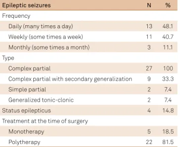
Table 2. Epileptic seizures and treatment before surgery.
Epileptic seizures N %
Frequency
Daily (many times a day) 13 48.1
Weekly (some times a week) 11 40.7
Monthly (some times a month) 3 11.1
Type
Complex partial 27 100
Complex partial with secondary generalization 9 33.3
Simple partial 2 7.4
Generalized tonic-clonic 2 7.4
Status epilepticus 4 14.8
Treatment at the time of surgery
Monotherapy 5 18.5
Polytherapy 22 81.5
Table 3. Tumor characteristics.
Tumor characteristics N %
Location
Supratentorial Right Left 26 96.3
Temporal 7 9 16 59.3
Parietal 2 5 7 25.9
Frontal 2 0 2 7.4
Insula 1 0 1 3.7
Infratentorial (4th ventricle) 1 3.7
Histological type [Graduation according to WHO]
Ganglioglioma [I] 9 33.3
Dysembryoplastic neuroepithelial (DNET) [I] 9 33.3
Oligodendroglioma low grade [II] 3 11.2
Oligodendroglioma anaplastic [III] 1 3.7
Pilocytic astroytoma [I] 1 3.7
Fibrillary astrocytoma [II] 1 3.7
Pleomorphic xanthoastrocytoma [II] 1 3.7
Ependymoma tanycytic [II] 1 3.7
Angiocentric glioma [I] 1 3.7
Gradation WHO
Low grade [I] and [II] 26 96.3
High grade [III] 1 3.7
Eloquent area involvement
Yes 5 18.5
No 22 81.5
Cortical dysplasia associated
Yes 7 26.0
No 20 74.0
Other lesions associated
Yes 6 22.3
Astrocytic reaction 2 7.5
Hippocampal neuronal loss 1 3.7
Angiomatosis 1 3.7
Hippocampal gliosis 1 3.7
Gliosis and giant cell reaction to foreign
body (previous surgery) 1 3.7
No 21 77.7
WHO: World Health Organization
Table 4. Electroencephalographic indings before surgery.
Findings N %
Video-electroencephalography
Abnormal 26 96.3
Normal 1 3.7
EEG background activity
Abnormal 9 33.3
Normal 18 66.7
Focal slowing
Present 17 63.0
Absent 10 37.0
Epileptiform activity in relation to the tumor location
Focal - corresponding to the tumoral area 23 85.2
Multifocal 1 3.7
Generalized 1 3.7
Absent epileptiform activity 2 7.4
Table 1. Clinical features of the sample (27 patients).
Clinical features
Gender - N (%)
Male 16 (59.2)
Female 11 (40.8)
First signs of the brain tumor - N (%)
Seizures 26 (96.2)
Other (nausea and vomiting) 1 (3.7)
Comorbidities – N (%)
Migraine 3 (11.1)
Panic disorder and separation anxiety with
somatic symptoms 1 (3.7)
Attention deicit hyperactivity disorder 1 (3.7)
Obesity due to food intake 1 (3.7)
Previous medical history - N (%) Perinatal hypoxic-ischemic
encephalopathy 2 (7.4)
Developmental delay 1 (3.7)
Family history of epilepsy 12 (44.4)
Family history of cancer 1 (3.7)
Mental retardation 1 (3.7)
Physical examination - N (%)
Normal 24 (88.8)
Abnormal 3 (11.1)
Dysphasia 1 (3.7)
Impairment in short and long-term
memories 1 (3.7)
Ages and intervals (means) - Years (range)
Onset of symptoms 6 y (3 m -15.8 y)
Age at the brain tumor diagnosis 9.7 y (10 m-16.8 y) Interval between the onset of seizures
and the diagnosis of the tumor. 3.6 y (1 m - 14.5 y) Interval between the brain tumor
diagnosis and surgery 1.5 y (days - 7 y)
After surgery, 16 patients (59.2%) started the process of withdrawal of AEDs. he mean time and mode for with
-drawal of AEDs in these patients was 3.2 years and two years after surgery, respectively, ranging from 1.7 years to seven years. In these 16 patients, 12 (44.4%) succeeded in staying drug-free, two (7.4%) were still in the process of withdrawal, with monotherapy, and the other two (7.4%) had failed the attempt, with return of the seizures.
DISCUSSION
he patients in this study were children from the Refractory Epilepsy Outpatient Clinic, and did not relect routine patients found in the oncology clinic. his explains
seizure as the initial manifestation of brain tumor in the
majority (96.2%); only one patient had nausea and vomiting as initial symptoms.
All patients had complex partial seizures. he literature
refers to the possibility of brain tumors in a scenario of refrac-tory complex partial seizures, associated with normal
neu-rological examination and preserved intelligence13. Epilepsy
associated with tumor has great potential for
refractori-ness to AED treatment14. In the present study, the selected
patients had epilepsy resistant to drug treatment. However,
we should not exclude the possibility of a tumoral etiology in cases of good response to drug treatment, as occurred in four
children found during the search for patients in this study. he interval between the onset of seizures and tumor diagnosis of 3.6 years is above the average delay in diagnosis
in relation to other brain tumors15. his can be explained by
the fact that the vast majority of tumors in the study (96.3%) were low grade (grade I and II according to WHO). Due to the slow growth of these tumors, seizures can occur even before the tumor is detected by imaging. High-grade tumors are diagnosed in a shorter time due to the more signiicant symptoms and increased growth rate.
he occurrence of seizures is more common in a supraten
-torial lesion. Studies difer on the lobe that is more prone to
epileptogenesis, but most agree that occipital lesions are less associated with seizure16,17. he indings in the present study
are consistent with the literature, since the temporal location
was the most frequent, and none occurred in the occipital lobe. here was involvement of eloquent cortex in 18.5% of children. In the case of low-grade tumors, sometimes in functionally viable brain regions, surgical intervention for
tumor treatment is often prohibited or delayed by
oncol-ogy specialists. On the other hand, for these cases, the work
of neuropediatric and epileptology teams is based on the
refractoriness of the epilepsy and its impact on the devel
-oping child, motivating the preoperative evaluation for epi -lepsy treatment so that the epileptogenic zone is bounded
and removed. Electroencephalographic monitoring tech
-niques are of great importance in further investigation of
patients with brain tumor in childhood, taking into account not only the tumor area as a cause of epilepsy, but aiming also to locate peritumoral regions that are also enabling the
generation of ires. hese techniques also enable a better delineation of the area to be removed, contributing to more favorable outcomes after surgical treatment. All patients in this study were submitted to extended video-EEG, as part of the preoperative assessment protocol, and 3.7% presented with no changes.
Typical indings of focal epileptic activity correspond -ing to the area of tumor location and focal slow-ing were
the most prevalent. However, multifocal and generalized epileptic activity were also detected, and in 7.4% of the patients the epileptic activity was absent. In these cases of nonlocalized changes or those with missing epileptic activ -ity, surgical treatment was indicated because of the
neo-plastic nature of the lesion. hus, it can be stated that mul -tifocal or generalized abnormalities, as well as the absence
of abnormalities in the EEG, do not exclude the possibility
of tumor occurrence2,18.
Tumors of glioneural origin, gangliogliomas and DNET were the most frequent in our series and they are included
in the long-term epilepsy-associated tumors6,7,8. he lat
-ter, largely occurring in children and young adults, have the highest rate of association with epilepsy, being 85% to 92% in DNETs and 63% to 91% in gangliogliomas14,16. As its ori
-gin suggests, the glioneural tumors consist of dysplastic
neu-rons and neoplastic glial cells. Within these, tumors develop
in hyperexcitable regions of dysplastic neurons, which are
believed to favor their potential epileptogenesis19.
Among the low-grade glial tumors, low-grade
oligoden-droglioma (11.2%) was another with signiicant occurrence in the study. In the group of gliomas, these low-grade tumors are the most likely to generate ires. Recent studies have shown seizures in 50-81% of astrocytomas and 46-78% in oli -godendrogliomas16,20,21. hese slowly growing tumors invade
the surrounding tissue causing gliosis and chronic inlamma
-tion, with changes in peritumoral regions. Evidence of these inlammatory changes is detectable using immunohisto
-chemical techniques, where there is a signiicant increase in reactive astrocytes in peritumoral cortical tissue of patients
with chronic epilepsy compared to peritumoral tissues of patients without seizures17.
The only high-grade tumor detected was the anaplas
-tic oligodendroglioma (WHO grade III). The high-grade gliomas (WHO grades III and IV) are associated with a lower rate of seizures. This can be explained more by the short survival time of patients due to the tumor’s aggres
-sive nature, than by its epileptogenesis potential16. Despite
the seizures being less frequent, these are the most dif -ficult to treat and they demonstrate increased refractori-ness to clinical treatment and are likely to persist after the
ischemia and necrosis. Because of their growth rate, they
can also cause changes in peritumoral regions through their mass effect or disruption of neural networks17.
Complete tumor resection in 88.8% of patients, tran
-sient focal neurological deicits after surgery, absence of intra-operative deaths, only one occurrence of surgical com
-plication and the favorable clinical outcomes related to seizure control are indings that encourage the indication for surgery in a tertiary center. Lesionectomy was the surgical technique most used (40.7%). he seizures caused by long-term epilepsy-associated tumors usually have excellent outcome following
appropriate surgical resection, using not only a tumoral, but also an epilepsy surgery-oriented strategy5.
he postoperative seizure control was consistent with the lit
-erature, where the range of Engel classiication class I was from 55% to 79%, class II from 4% to 25% and class III from 4% to 15%1,2,3.
Two of these publications cited 5%3 and 12.2%2 with class IV.
he present work is the second review of cases handled in CIREP. he irst22 included patients seen up to 2010, with
all tumor types, including non-neoplastic indings, in addi
-tion to the refractory cases, and a postoperative follow-up period of six months. he most common tumors found in this review were also gangliogliomas and DNETs, together covering 45% of the total number of tumors diagnosed, and the outcome was signiicantly positive, both types corre
-sponding to Engel class I in 89.3%. his suggests that the favorable postoperative outcomes in relation to seizures control have remained.
he correlation between the preoperative control of sei
-zures and favorable outcomes after surgery has been well
established in the literature20. We found that children who
showed no postoperative improvement had an increased fre
-quency of seizures in the preoperative evaluation.
A negative outcome in relation to seizure control and death was observed in a child with anaplastic oligodendro
-glioma, due to the aggressive nature of the tumor (grade III). he patient had a high frequency of seizures in the preop
-erative period, refractory to anticonvulsant treatment, with a brief interval between the onset of seizures, tumor diag
-nosis, and surgery. here was no improvement in seizure control after surgery and radiotherapy; this child had Engel class IV one year after surgery, and died in the second year. Another tumor that showed unfavorable outcome regarding seizure control was a ibrillary astrocytoma. his was a type of difuse astrocytoma that despite being well-diferentiated (grade II), the tendency was to become more iniltrative, with evolution to Engel class IV.
Contrary to being a low-grade tumor (grade I), with a
good prognosis after surgical treatment, two cases of DNET did not progress favorably in the long-term outcome regard
-ing seizure control. he irst case had good response ini -tially, becoming free of seizures in the two years after surgery
(Engel class I at one year postoperatively), but destabilized clinically after attempted withdrawal of AEDs in this period,
maintaining improvement in relation to their preoperative
status, but with recurrence of crippling crises (class III in the
ifth postoperative year). he second patient remained free
of seizures in the six years following the surgery (class I in
the irst and ifth postoperative years). He stopped taking two AEDs after the second year, remaining on monotherapy until the sixth postoperative year. At this time, there was clinical
decompensation due to tumor growth in residual areas and associated cortical dysplasia, making it necessary to undergo
electrocorticography and a new surgery. However, he main
-tained poor control, with class IV in the 10th year. hese two
cases illustrate the general trend in epilepsy surgery, when the short-term results tend to be higher than those of the
long term. Particularly in the case of children whose seizures are very common and can impact their development, incur -ring encephalopathies, this temporary seizure control can be
considered a positive prognosis.
In the present study, 59.2% of patients attempted AED withdrawal and among these, 7.4% relapsed. Successful AED withdrawal occurred in 44.4% of all patients, without recurrence of seizures in an average of 3.2 years after sur
-gery. here is a need for controlled studies with the goal of examining the best time for withdrawal of AEDs and which factors are involved in the success of the attempts for these cases. he withdrawal of AEDs is a question in many other conditions, each one with particularities to be considered.
Recent meta-analysis23 enrolled ive studies unrelated to
epilepsy surgery, showing that for children, the variables associated with higher risk of seizure relapse were: abnor
-mal EEG indings, especially epileptiform activity; epilepsy
onset before two years or after 10 years of age; history of sta-tus epilepticus; intellectual disability (IQ < 70); and a high
seizure frequency before and during treatment. Gender and family history did not show any signiicant inluence over seizure relapse. However, these authors state that the trials included were classiied as low or unclear risk of bias where
methodological information was not reported and could
not be provided by the original study authors.
In conclusion, surgical treatment for refractory
epi-lepsy secondary to tumor etiology had favorable out
-comes related both to seizure control, and by improving
the response to drug treatment in those who persisted with
epilepsy after surgery. he surgery proved to be a poten
-tially curative treatment modality, with temporary postsur
-gical neurolo-gical traits, with no record of intraoperative deaths. Tumor diagnosis cannot be postponed, considering
the possibility of curing epilepsy, or in non-curable cases,
providing a better response to AEDs, promoting positive impact on the quality of life of children sufering from this debilitating condition. Full preoperative investigation at a
referral center for proper location of the epileptogenic zone
and electrocorticographic intraoperative monitoring help to assess the origin of the seizures and to prevent impair
References
1. Alexiou GA, Varela M, Sfakianos G, Prodromou N. Benign lesions accompanied by intractable epilepsy in children. J Child Neurol. 2009;24(6):697-700. doi:10.1177/0883073808331079
2. Fattal-Valevski A, Nissan N, Kramer U, Constantini S. Seizures as the clinical presenting symptom in children with brain tumors. J Child Neurol. 2013;28(3):292-6. doi:10.1177/0883073812445786 3. Gaggero R, Consales A, Fazzini F, Mancardi MM, Baglietto MG,
Nozza P et al. Epilepsy associated with supratentorial brain tumors under 3 years of life. Epilepsy Res. 2009;87(2-3):184-9. doi:10.1016/j.eplepsyres.2009.08.012
4. Cross JH, Jayakar P, Nordli D, Delalande O, Duchowny M, Wieser HG et al. Proposed criteria for referral and evaluation of children for epilepsy surgery: recommendations of the Subcommission for Pediatric Epilepsy Surgery. Epilepsia. 2006;47(6):952-9. doi:10.1111/j.1528-1167.2006.00569.x
5. Blumcke I, Aronica E, Urbach H, Alexopoulos A, Gonzalez-Martinez JA. A neuropathology-based approach to epilepsy surgery in brain tumors and proposal for a new terminology use for long-term epilepsy-associated brain tumors. Acta Neuropathol. 2014;128(1):39–54. doi:10.1007/s00401-014-1288-9
6. Louis DN, Ohgaki H, Wiestler OD, Cavenee WK, Burger PC, Jouvet A et al. The 2007 WHO classiication of tumours of the central nervous system. Acta Neuropathol. 2007;114(2):97-109. doi:10.1007/s00401-007-0243-4
7. Blümcke I, Löbach M, Wolf HK, Wiestler OD. Evidence for
developmental precursor lesions in epilepsy-associated glioneuronal tumors. Microsc Res Tech. 1999;46(1):53–8. doi:10.1002/(SICI)1097-0029(19990701)46:1<53::AID-JEMT5>3.0.CO;2-0
8. Palmini A, Paglioli E, Silva VD. Developmental tumors and adjacent cortical dysplasia: single or dual pathology? Epilepsia. 2013;54(9):18-24. doi:10.1111/epi.12438
9. Santos MV, Oliveira RS, Machado HR. Approach to cortical dysplasia associated with glial and glioneuronal tumors (FCD type IIIb). Childs Nerv Syst. 2014;30(11):1869- 74. doi:10.1007/s00381-014-2519-z 10. Kwan P, Arzimanoglou A, Berg AT, Brodie MJ, Allen Hauser W, Mathern
G et al. Deinition of drug resistant epilepsy: consensus proposal by the ad hoc Task Force of the ILAE Commission on Therapeutic Strategies. Epilepsia. 2010;51(6):1069-77. doi:10.1111/j.1528-1167.2009.02397.x 11. Engel J Jr et al. Surgical treatment of the epilepsies. 2nd ed. New
York: Raven; 1993. p. 609-21.
12. Wieser HG, Blume WT, Fish D, Goldensohn E, Hufnagel A, King D et al. Proposal for a new classiication of outcome with respect to epileptic seizures following epilepsy surgery. Epilepsia. 2001;42(2):282-6. doi:10.1046/j.1528-1157.2001.35100.x
13. Blume WT, Girvin JP, Kaufmann JC. Childhood brain tumors presenting as chronic uncontrolled focal seizure disorders. Ann Neurol. 1982;12(6):538-41. doi:10.1002/ana.410120606 14. Aronica E, Leenstra S, Veelen CW, Rijen PC, Hulsebos TJ,
Tersmette AC et al. Glioneuronal tumors and medically intractable epilepsy: a clinical study with long-term follow-up of seizure outcome after surgery. Epilepsy Res. 2001;43(3):179-91. doi:10.1016/S0920-1211(00)00208-4
15. Reulecke BC, Erker CG, Fiedler BJ, Niederstadt TU, Kurlemann G et al. Brain tumors in children: initial symptoms and their influence on the time span between symptom onset and diagnosis. J. Child Neurol.2008;23(2):178-83. doi:10.1177/0883073807308692
16. Hamasaki T, Yamada K, Kuratsu J. Seizures as a presenting symptom in neurosurgical patients: a retrospective single-institution analysis. Clin Neurol Neurosurg. 2013;115(11):2336-40. doi:10.1016/j.clineuro.2013.08.016
17. Berntsson SG, Malmer B, Bondy ML, Qu M, Smits A. Tumor-associated epilepsy and glioma: are there common genetic pathways? Acta Oncol. 2009;48(7):955-63. doi:10.1080/02841860903104145
18. Williams BA, Abbott KJ, Manson JI. Cerebral tumors in children presenting with epilepsy. J Child Neurol. 1992;7(3):291-4. doi:10.1177/088307389200700309
19. Ferrier CH, Aronica E, Leijiten FS, Spliet WG, Huffelen AC, Rijen PC et al. Electrocorticographic discharge patterns in glioneuronal tumors and focal cortical dysplasia. Epilepsia. 2006;47(9):1477-86. doi:10.1111/j.1528-1167.2006.00619.x
20. Kahlenberg CA, Fadul CE, Roberts DW, Thadani VM, Bujarski KA, Scott RC et al. Seizure prognosis of patients with low-grade tumors. Seizure Eur J Epilepsy. 2012;21(7):540-5. doi:10.1016/j.seizure.2012.05.014
21. Englot DJ, Berger MS, Barbaro NM, Chang EF. Predictors of seizures freedom after resection of supratentorial low-grade gliomas. J Neurosurg. 2011;115(2):240-4. doi:10.3171/2011.3.JNS1153
22. Pinto KGFD. Epilepsia na criança com tumor cerebral: Peril clínico e evolução dos pacientes tratados cirurgicamente [dissertação]. Ribeirão Preto: Faculdade de Medicina de Ribeirão Preto, Universidade de São Paulo; 2011.
ERRATUM https://doi.org/10.1590/0004-282x20170004err
Erratum
Refractory epilepsy in children with brain tumors. he urgency of neurosurgery. Arq Neuropsiquiatr 2016;74(12):1008-1013. DOI: 10.1590/0004-282X20160157
he authors:
Marília Rosa Abtibol Bernardino1, Carolina Funayama1, Ana Paula Andrade Hamad1, Hélio Machado1, Américo Sakamoto1,
Ursula home1, Vera Cristina Terra1, Antonio Carlos dos Santos1,2,3
Should be:
Marília Rosa Abtibol Bernardino1, Carolina Funayama1, Ana Paula Andrade Hamad1, Hélio Machado1, Américo Sakamoto1,
Ursula home1, Vera CristinaTerra1, Antonio Carlos dos Santos1,2,3, Luciano Nader Serafani4, Nathalia Cunha Calixto2,
Huria Shalom Monturil de Carvalho Silva4
he ailiations:
1Universidade de São Paulo, Faculdade de Medicina de Ribeirão Preto, Departamento de Neurociências e Ciências do
Comportamento, Ribeirão Preto SP, Brasil;
2Universidade de São Paulo, Faculdade de Medicina de Ribeirão Preto, Departamento de Neuroradiologia, Seção de
Ressonância Magnética, Ribeirão Preto SP, Brasil;
3Universidade de São Paulo, Faculdade de Medicina de Ribeirão Preto, Hospital das Clínicas, Centro de Ciências das Imagens
e Física Médica, Ribeirão Preto SP, Brasil.
Should be:
1Universidade de São Paulo, Faculdade de Medicina de Ribeirão Preto, Departamento de Neurociências e Ciências do
Comportamento, Ribeirão Preto SP, Brasil;
2Universidade de São Paulo, Faculdade de Medicina de Ribeirão Preto, Departamento de Neuroradiologia, Seção de
Ressonância Magnética, Ribeirão Preto SP, Brasil;
3Universidade de São Paulo, Faculdade de Medicina de Ribeirão Preto, Hospital das Clínicas, Centro de Ciências das Imagens
e Física Médica, Ribeirão Preto SP, Brasil;
4Universidade de São Paulo, Faculdade de Medicina de Ribeirão Preto, Departamento de Patologia e Medicina Legal, Ribeirão
Preto SP, Brasil.
Aliocha Dostoevski´s death during an epileptic seizure.
Arq Neuropsiquiatr 2016; 74 (11): 944-946. DOI: 10.1590/0004-282x20160147
he paragraph:
However, Anna panicked and called their pediatrician, Dr. A. Tchochin, who lived nearby, picked up the phone immediately. (Page 945)
Should be:
However, Anna panicked and called their pediatrician, Dr. A. Tchochin, who lived nearby.
Fat embolism showing restriction on difusion sequence in brain magnetic resonance imaging. Arq Neuropsiquiatr 2016;74(7):597-598. DOI: http://dx.doi.org/10.1590/0004-282X20160052
he authors:
Henry Koiti Sato1, Pedro André Kowacs1, Josep Dalmau2, Paulo Sergio Faro Santos2
Should be:
Henry Koiti Sato1, Pedro André Kowacs1, Paulo Sergio Faro Santos1
he ailiation “2Universitat de Barcelona, Institut D’Investigacions Biomédiques August Pi I Sunyer, Hospital Clínic,