741 Rev Soc Bras Med Trop 50(5):741-742, September-October, 2017 doi: 10.1590/0037-8682-0275-2016
Images in Infectious Diseases
Dengue fever with thrombocytopenia
and gingival bleeding
Fred Bernardes Filho
[1],[2], Caio Cavalcante Machado
[2],[3]and Andreia de Oliveira Alves
[4][1]. Divisão de Dermatologia, Departamento de Clínica Médica, Faculdade de Medicina de Ribeirão Preto, Universidade de São Paulo, Ribeirão Preto, SP, Brasil.
[2]. Departamento de Emergência, Hospital Imaculada Conceição da Sociedade Portuguesa de Beneicência, Ribeirão Preto, SP, Brasil.
[3]. Divisão de Reumatologia, Departamento de Clínica Médica, Faculdade de Medicina de Ribeirão Preto, Universidade de São Paulo, Ribeirão Preto, SP, Brasil. [4]. Faculdade de Medicina, Centro Universitário Barão de Mauá, Ribeirão Preto, SP, Brasil.
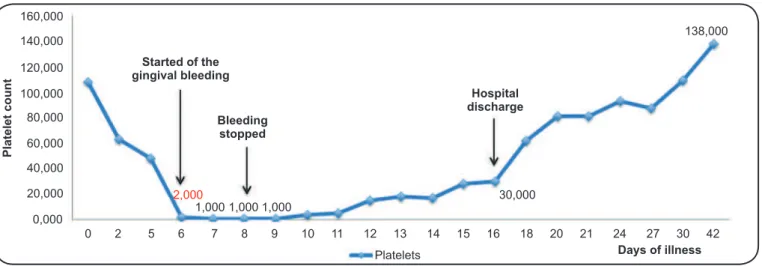
A 45-year-old man was admitted on the sixth day owing to classic dengue symptoms that included spontaneous bleeding from the gums and a purpuric rash. His white blood cell count on admission was 3.1 × 10³/µL with a low platelet count of
2,000. On physical examination, gingival bleeding, scattered petechiae, and ecchymoses were observed (Figures 1A, 1B, 1C and 1D). Results of cardiorespiratory, gastrointestinal, and neurological examinations were normal. Chest radiography showed no pleural effusion and an abdominal ultrasound showed
no free luid in the cavity. The lowest platelet count (1.0 × 10³/
µL) was reported from the seventh to ninth day of the illness. Laboratory tests revealed positivity for immunoglobulin M (IgM) antibodies to dengue virus. Subsequently, treatment for thrombocytopenic purpura due to dengue was initiated. Hydrocortisone 500mg/day was administered intravenously for 6 days, and, prednisone 20mg/day, for an additional 10 days.
Two days after initiating treatment, the bleeding stopped, and by
the ninth day post-treatment, platelet count increased to 30,000 cells/mm3. The patient’s hospital course was uncomplicated, he
remained clinically well, and he was discharged with instructions to return for follow-up in the outpatient clinic. No spontaneous hemorrhagic events have occurred since, and platelet counts have remained within normal levels (Figure 2 and Figure 3).
Acute immune thrombocytopenia can be linked to underlying conditions like connective tissue disease, lymphoproliferative
disease, immune-deicient states, and viral infections, or to
medications administered1-3. Thrombocytopenia associated with
viral infection results from both lowered platelet production from megakaryocytes, and decrease in platelet half-life.
Conlict of interest
The authors declare that there is no conlict of interest.
FIGURE 1 - Dengue fever. A: Acute gingival bleeding (white arrows).
B: Scattered ecchymoses and petechiae. C: Hematoma on the left arm after
blood pressure cuff inlation. D: Multiple ecchymoses on the right arm.
Corresponding author:Dr. Fred Bernardes Filho.
e-mail: [email protected] Received 30 June 2016
742
Bernardes Filho F et al - Dengue fever
FIGURE 3 - Disappearance of the hematoma as observed at the inal
follow-up.
REFERENCES
1. Cines DB, Blanchette VS. Immune thrombocytopenic purpura. N Engl J Med 2002;346(13):995-1008.
2. Kumar S, Khadwal A, Verma S, Singhi SC. Immune thrombocytopenic purpura due to mixed viral infections. Indian J Pediatr. 2013;80(5):421-2.
3. Fonseca T, Segarra-Torres A, Jaume-Anselmi F, Ramírez-Rivera J. Dengue fever: a rare cause of immune thrombocytopenia. Bol Asoc Med P R 2015;107(2):51-3.
160,000
Started of the gingival bleeding
Bleeding stopped
Hospital discharge
140,000
120,000
100,000
80,000
60,000
40,000
20,000
0,000
2,000
1,000 1,000 1,000
30,000
Platelets Days of illness
138,000
0 2 5 6 7 8 9 10 11 12 13 14 15 16 18 20 21 24 27 30 42
Platelet count