ARTICLE
Stroke caused auditory attention deficits
in children
Acidente vascular cerebral causa défices da atenção seletiva auditiva em crianças
Karla Maria Ibraim da Freiria Elias,Maria Valeriana Leme de Moura-Ribeiro
he functional consequences of the stroke during infancy and childhood are considerably broad, involving motor, cog-nitive, sensory, behavioral, speech and language abilities1-5.
With regard to auditory function, there are practically no data about stroke in children, although it is known that perception or processing may be compromised6.
he mechanisms and processes performed by the auditory system allow the recognition and interpretation of all kind of sounds. It encompasses a variety of abilities mediated by audi-tory centers located in the brainstem, the subcortex and the cortex, that could be afected by the stroke, causing deicits in auditory processing to some degree, even in patients whose evolution seems satisfactory. he aim of the present study was
to assess the Central Auditory Processing (CAP) in children that had a favorable outcome after the vascular event. By ana-lysing these cases, we wanted to verify if the stroke could cause a decrease in performance in any ear, which might collaborate for some diiculties in communication, learning and socializa-tion, sometimes described in these children3,7-9.
Of all the auditory abilities, selective attention seems to be the one with most relevance to the processing of stimuli in environments with unfavorable hearing conditions, such as parallel information or background noise10. his study applies
dichotic verbal and non-verbal stimuli to evaluate the audito-ry selective attention in children afected by stroke when per-forming tasks related to binaural separation and integration.
Department of Neurology, Faculty of Medical Sciences, Universidade Estadual de Campinas (UNICAMP), Campinas, SP, Brazil.
Correspondence: Maria Valeriana Leme de Moura-Ribeiro; Departamento de Neurologia, Faculdade de Ciências Médicas, UNICAMP; Rua Tessália Vieira de Camargo 126 / Caixa Postal 6.111; 13083-970 Campinas SP - Brasil; E-mail: [email protected]
Support: This work was supported by grants from the Conselho Nacional de Desenvolvimento Científico e Tecnológico (CNPq). Conflict of interest: There is no conflict of interest to declare.
Received 13 April 2012; Received in final form 20 July 2012; Accepted 27 July 2012
ABSTRACT
Objective: To verify the auditory selective attention in children with stroke. Methods: Dichotic tests of binaural separation (non-verbal and consonant-vowel) and binaural integration — digits and Staggered Spondaic Words Test (SSW) — were applied in 13 children (7 boys), from 7 to 16 years, with unilateral stroke confirmed by neurological examination and neuroimaging.Results:The attention performance showed significant differences in comparison to the control group in both kinds of tests. In the non-verbal test, identifications the ear opposite the lesion in the free recall stage was diminished and, in the following stages, a difficulty in directing attention was detected. In the conso-nant-vowel test, a modification in perceptual asymmetry and difficulty in focusing in the attended stages was found. In the digits and SSW tests, ipsilateral, contralateral and bilateral deficits were detected, depending on the characteristics of the lesions and demand of the task.
Conclusion:Stroke caused auditory attention deficits when dealing with simultaneous sources of auditory information.
Key words: stroke, auditory perception, clinical outcome, infancy, childhood. RESUMO
Objetivo: Verificar a habilidade de atenção seletiva em crianças com acidente vascular cerebral (AVC). Métodos: Foram aplicados testes dicóticos de separação (não verbal e consoante-vogal) e integração — dígitos e Staggered Spondaic Words Test (SSW) — binaural em 13 cri-anças (7 meninos), entre 7 e 16 anos, com AVC unilateral confirmado por neuroimagem. Resultados: O desempenho atencional diferiu entre os grupos na realização de ambos os tipos de tarefa. Ao teste não verbal, houve menor quantidade de identificações com a orelha contralat-eral à lesão em atenção livre e dificuldade de focalizar a atenção nas etapas direcionadas. No teste consoante-vogal, houve modificação da assimetria perceptual e dificuldade de focalizar a atenção nas etapas direcionadas. Nos testes de dígitos e SSW, foram constatados défices ipsilaterais, contralaterais e bilaterais dependendo das características da lesão e da demanda da tarefa.Conclusão:As crianças com AVC apresentaram défices na habilidade de atenção seletiva em presença de fontes simultâneas de informação auditiva verbal e não verbal.
METHODS
he study was approved by the Ethic Research Committee of the Medical Sciences Faculty at the Universidade Estadual de Campinas, in accordance with the Regional Health Counsel’s Resolution nº 196/96 (Protocol nº 372/2001). Parents signed the relevant consent form.
Thirteen children (seven boys) with unilateral stroke, between ages of 7 and 16 years, were evaluated. They were all right-handed before the vascular episode and/or presented a negative family history of left-handedness. They were all followed by a multidisciplinary team at the Childhood and Adolescence Stroke Research Group at Universidade Estadual de Campinas from the acute phase of the disease onwards. This group will be referred to as the study group (SG).
he stroke diagnosis was conirmed by clinical and neu-roimaging investigations and, for the purposes of the study, the deinition of the areas afected by the vascular lesion used the magnetic resonance as reference. his image was done up to six months before the auditory evaluation, and the scans were reviewed by a neuroradiologist.
he SG was matched with a control group (CG) consist-ing of 13 healthy, right-handed children, with matchconsist-ing age, sex and socio-economic level. In the match by age, a maxi-mum diference of six months was permitted. he children in both groups went to regular public schools.
All participants had normal peripheral hearing, language and cognition abilities compatible with the tasks required by the CAP tests. hese abilities were conirmed through mul-tidisciplinary evaluations. he speech-language assessment was based on standardized tests using screening procedure and thematic images in spontaneous and semi-spontaneous conversation11. he hearing assessment used standard
base-line audiometric tests (pure-tone audiometry, speech audi-ometry, tympanaudi-ometry, ipsilateral and contralateral acous-tic relex threshold). All children performed an inventory on auditory behavior and this questionnaire also was admin-istered before CAP testing. he neuropsychological assess-ment included Luria-Nebraska, Wechsler Intelligence Scale for Children-WISC, Bender Visual Motor Gestalt test and the Test of School Performance.
he exclusion criteria were: bilateral stroke, recurrent vascular episodes, Moyamoyadisease, sickle cell disease, epi-lepsy and psychiatric disorders. Children with impairments related to receptive or expressive language, auditory sensitiv-ity, ossicular mobility in the middle ear and relex responses to acoustic stimulation were also excluded, as well as those with an IQ below 70.
Evaluation of selective attention auditory ability
he auditory evaluation occurred after the minimum period of six months following the stroke and consisted of the application of four CAP tests: non-verbal dichotic,
consonant-vowel, dichotic digits and Staggered Spondaic Word test (SSW).
he irst two tests aimed at the investigation of selec-tive attention in binaural separation task. In this test, difer-ent stimuli are presdifer-ented simultaneously to both ears, only one of which should be repeated (in the free recall, the child had to respond to the irst item perceived and, in the subse-quent stages, respond only to stimuli perceived by the spec-iied ear — right or left)12.
he last other two tests evaluate selective attention in binaural integration task, so the children are asked to re-peat, respectively, all stimuli presented simultaneously to both ears12.
hese tests have normative values available for Brazilian Portuguese speakers12. All tests were applied in a
sound-treated room, using a two-channel audiometer connected to a CD player — calibration standard ANSI-69. All test material was presented via TDH-39 earphones at a 50 dB sensation level, referenced to the pure-tone audiogram average (at 0.5, 1, 2 and 4 kHz).
Statistical analysis included analysis of variance (ANOVA) for repeated measures with conversion into ranks, proile test for contrasts and the Wilcoxon test for paired measurements with signiicance level of 5% (p<0.05) and signiicant values marked with an asterisk.
RESULTS
he data for identiication and unilateral structural brain impairment are presented in Table 1. he magnetic reso-nance scans of the brain revealed the involvement of the middle cerebral artery and its branches, anterior cerebral ar-tery and vertebro-basilar system. Eight children were found to have impairments in the right hemisphere (Table 1).
At interview with parents, they were raised complaints referred to children’s diiculty in attending promptly to calls, especially if engaged in other activities; diiculty in follow-ing oral instructions, requirfollow-ing repetition of the messages; need to help to understand the topics taught in class and/or solve homework assignments; grades below average in one or more subjects, among others. In the CAP assessment, the average age in the SG was 11 years and 7 months, and in the CG, it was 11 years and 4 months. he auditory assess-ment for children in the SG was done approximately 5 years and 3 months after stroke.
Neuroimaging studies, conducted at follow up, con-irmed the involvement of areas associated with central au-ditory processes, with lesions situated cortically in the left and right temporal and parietal lobes and/or subcortically in thalamus and/or basal ganglia and, in one patient, a mes-encephalic lesion.
Table 1. Stroke in children – identification data and cerebral impairment.
Subject Sex Age SG/Stroke Age SG/CAP Age CG/CAP Artery Side Type Site
S1 M 7y 8y 8y 5m MCA R I Cortical-Subcortical
S2 F 1y 1m 10y 5m 10y 6m MCA L I Subcortical
S3 M 3y 12y 9m 13y 1m MCA R I Subcortical
S4 M 5y 4m 14y 7m 14y 7m MCA L I Cortical-Subcortical
S5 F 6y 6m 16y 16y 1m MCA R I-H Cortical-Subcortical
S6 M 13y 1m 16y 7m 16y 2m MCA/ACA R I Cortical-Subcortical
S7 M 3y 8m 7y 8m 7y 3m MCA R I-H Cortical-Subcortical
S8 M 4y 11m 12y 3m 11y 10m MCA R I Cortical
S9 F 6y 1m 10y 6m 10y 11m VB R H Midbrain
S10 F 10y 0m 14y 6m 14y 2m MCA L I Subcortical
S11 F 7y 5m 13y 2m 12y 8m ACA L I-H Cortical-Subcortical
S12 F 10y 6m 15y 15y 2m MCA R I Subcortical
S13 M 4y10m 8y 1m 8y 7m MCA/ACA L I-H Cortical-Subcortical
SG: study group; CG: control group; S: subject; M: male; F: female; y: years; m: months; MCA: middle cerebral artery; ACA: anterior cerebral artery; VB: vertebral-basilar system; R: right; L: left; I: ischemic; I-H: secondary hemorrhagic conversion of ischemia; H: hemorrhagic; Age SG/Stroke: age at time of stroke; Age SG/CAP: age at auditory assessment; Age CG/CAP: control group’s age at auditory assessment.
Table 2. Stroke in children – neuroimage and clinical manifestations of acute and chronic phases.
Subject Magnetic resonance
Evolution
Acute phase Chronic phase
S1 PR IFG I STG PrCG SPG CR
Hemiplegia Hemiparesis
LN CN IC
S2 Pt Hemiplegia No residual
impairment* Behavior
disorder
S3 T LN CN IC Hemiplegia No residual impairment* S4 FTP LN CN CI Hemiplegia Hemiparesis Aphasia Speech disorder
S5 TP Hemiplegia No residual
impairment*
S6 TP Hemiplegia No residual
impairment*
S7 PrCG SFG
MFG
Speech disorder
No residual impairment* CSO I IC CN
LN
S8 SP Hemiplegia Hemiparesis
Dysarthria
S9 InC SC Ptosis Visual deficit
Visual deficit
S10 LN CN EC IC Hemiplegia Hemiparesis
Aphasia
S11 SoMG CG Hemiplegia Behavior disorder
CN IC T Behavior disorder
Learning difficulty
S12 IC Hemiplegia No residual
impairment* S13 LOG IFG PO P
MTG STG
Hemiplegia Hemiparesis
PhG I LN CN EC IC
Aphasia
S: subject; CG: cingulate gyrus; CN: caudate nucleus; CR: corona radiata; CSO: centrum semiovale; EC: external capsule; FTP: fronto-temporoparietal; I: insula; IC: internal capsule; InC: inferior colliculus; IFG: inferior frontal gyrus; LN: lenticular nucleus; LOG: lateral orbital gyrus; MFG: middle frontal gyrus; MTG: middle temporal gyrus; P: parietal; Pt: putamen; PCG: postcentral gyrus; PrCG: precentral gyrus; PhG: parahippocampal gyrus; PO: pars opercularis; PR: perisylvian region; SC: superior colliculus; SFG: superior frontal gyrus; SoMG: superior orbitary medial gyrus; SP: superior parietal; STG: superior temporal gyrus; T: thalamus; TP: temporoparietal. *No residual impairments in physical and cognitive aspects of outcome.
language (5 children) and alterations in vision and/or behav-ior. Motor abnormalities persisted in most children. In regard to speech and language abilities, only one patient, S4, lacked luency; the rest evolved favorably (Table 2).
he results, in absolute values, of SG and CG for the dich-otic tests applied were included in Table 3.
In the non-verbal dichotic test, diferences between the groups were not identiied in relation to free recall. However, at this stage of the evaluation, the SG had fewer correct re-sponses when the ear with direct access to the injured hemi-sphere was tested; in contrast, the CG had a similar amount of correct responses for each ear (Table 3).
In the stage to measure attention in the right ear, the SG scored signiicantly lower than the GC in the identiication of stimuli presented in the target ear (p=0.0254*), Wilcoxon (Fig 1). In the stage to measure attention in the left ear, there was no diference between the groups. he children in the CG performed on a level compatible with the established range for their age.
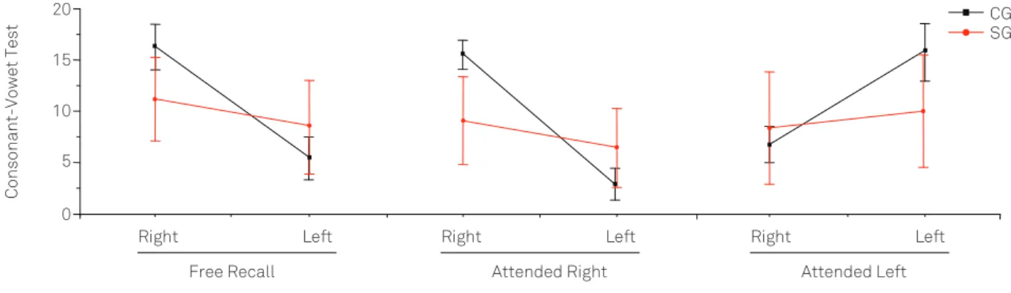
When the groups were compared in the consonant-vowel test of the free recall stage, children in the SG had inferior performance (p=0.0001*), ANOVA.
At this stage of the test, it was also checked wheth-er thwheth-ere was a difwheth-erence in the numbwheth-er of identiications made with one of the ears, either right or left. It was found that in the SG there was no diference between ears in the ability to identify items (Table 3). However, in the CG, there was a signiicant amount of identiication of the items pre-sented in the right ear, a condition called right perceptual asymmetry or right ear advantage (p<0.0001*).
In the focused attention stage, the SG showed inferior performance, with p=0.0001* and p=0.0059* (Wilcoxon) re-spectively for the right and left conditions of the test (Fig 2).
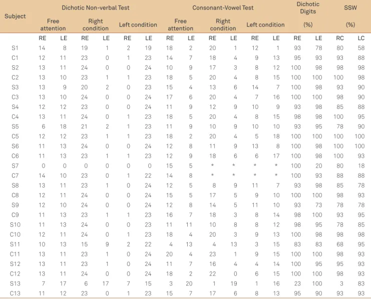
Table 3. Dichotic tests – individual results in absolutes values.
Subject
Dichotic Non-verbal Test Consonant-Vowel Test Dichotic
Digits SSW Free
attention
Right
condition Left condition
Free attention
Right
condition Left condition (%) (%) RE LE RE LE RE LE RE LE RE LE RE LE RE LE RC LC
S1 14 8 19 1 2 19 18 2 20 1 12 1 93 78 80 58
C1 12 11 23 0 1 23 14 7 18 4 9 13 95 93 93 88
S2 13 11 24 0 0 24 10 9 17 3 8 12 100 98 98 98
C2 13 10 23 1 1 23 18 5 20 4 8 15 100 100 100 98
S3 13 9 20 2 0 23 15 4 13 6 14 7 100 98 93 90
C3 13 10 24 0 0 24 17 6 20 4 7 16 100 100 98 90
S4 12 12 23 0 0 24 11 9 12 9 10 9 93 98 85 88
C4 13 11 24 0 1 23 18 5 20 4 8 15 98 98 100 95
S5 6 18 21 2 1 23 11 9 10 9 10 10 93 95 78 90
C5 12 12 23 1 1 23 18 2 20 4 5 18 100 100 100 100
S6 11 13 24 0 0 24 12 8 11 9 13 8 100 98 100 100
C6 11 13 23 1 1 23 12 9 18 6 6 17 100 98 100 93
S7 0 0 0 0 0 0 15 5 * * * * 100 20 80 18
C7 14 10 23 0 1 22 14 8 * * * * 100 93 88 88
S8 13 11 23 1 0 24 12 5 8 9 11 7 93 98 85 78
C8 12 11 24 0 0 24 15 5 17 5 9 10 100 100 98 93
S9 12 10 24 0 0 24 12 8 14 5 11 10 93 73 78 78
C9 11 13 23 1 1 23 16 7 18 3 8 14 98 100 93 95
S10 11 13 24 0 0 23 11 11 10 8 8 12 98 95 78 85
C10 12 11 24 0 1 23 18 4 20 3 9 13 100 98 98 98
S11 10 13 15 9 2 22 4 13 4 13 3 15 83 83 68 95
C11 13 11 23 1 0 24 20 4 23 1 9 15 100 100 98 93
S12 13 11 23 1 0 24 11 7 16 4 4 14 100 95 95 93
C12 13 11 24 0 0 24 18 2 22 0 6 15 100 100 98 93
S13 7 17 6 17 7 15 3 20 1 19 1 16 23 100 3 83
C13 11 12 23 0 1 23 15 7 17 6 8 13 95 90 93 93
SSW: staggered spondaic words test; S: subject; C: control; RE: right ear; LE: left ear; *not assess.
CG SG
Group Stimuli
Attended Right
25
20
15
10
5
0
-5
Fig 1. Dichotic non-verbal – study group and control group performances at attended right condition.
SG: study group; CG: control group.
Children in both groups showed diferences in compe-tence for binaural integration tasks (p=0.0038*) (ANOVA). In the proile test for contrasts, considering the perfor-mance of both ears in each group, no diferences were found; both in the SG and in the CG the number of identii-cations per ear did not vary. In the same test, when compar-ing the performance of both groups, the SG’s performance was below the CG regardless of the ear. We found a difer-ence for the right ear with p=0.0048* and for the left ear, p=0.0311* was obtained (Fig 3).
In the SSW test, in relation to laterality of lesion, they were found contralateral (S1, S7, S11 and S13), ipsilateral (S4) and bilateral (S4, S8, S9 and S10) changes (Table 3).
comparing the groups, the SG performance was signiicantly lower than the CG in both competitive conditions, the right having p=0.0001* and the left, p=0.0271* (Fig 4).
DISCUSSION
In the present study, patients with stroke presented pre-dominantly with ischemic lesions of the middle cerebral ar-tery and cortico-subcortical involvement. In pediatric stroke, the involvement of the middle cerebral artery and its lenticu-lar branches are the most commonly reported13-19 and,
con-sidering the course and distribution, there could cause al-terations in blood low of primary and secondary areas and others related to the processing and integration of auditory information. In one of the patients, S9, the lesion was mes-encephalic. However, the afected structure, the inferior col-liculus is required for the processing of complex stimuli due to the way in which it processes information10. Also because
of the complexity of its interaction, the inferior colliculus has profound implications for binaural abilities, notably for selec-tive attention10.
he type of lesion (ischemic, ischemic with secondary hemorrhagic transformations and hemorrhage) is frequent-ly associated at functional outcome, sometimes inluencing physical and cognitive abilities in a wide spectrum, however this aspect did not make any diference within the SG. hese children were evaluated in the chronic phase of the disease, presenting stability from a neurological point of view, good cognitive performance and displaying maintenance or re-covery of linguistic ability. he requirement that these abili-ties be intact was a limiting factor for the number of patients in the study. Sequelae are generally common and intense in stroke patients, as shown in the literature1-5.
In the free recall stage of the dichotic non-verbal test, the SG identiied more items with one ear than the other. his is a lateralization efect and it is a reduction in the amount of identiications made with the contralateral ear
SSW: staggered spondaic words test; SG: study group; CG: control group.
Fig 4. Staggered Spondaic Words test – study group and control group performances in right and left competitive conditions.
Right Left
0.5 0.6 0.7 0.8 0.9 1.0 1.1
CG SG
SSW
Condition %
Right Left
0.6 07 08 09 10 11
iti %
DD: Dichotic digits; SG: study group; CG: control group.
Fig 3. Dichotic digits – study group and control group performances in right and left competitive conditions.
SG: study group; CG: control group.
Fig 2. Consonant-vowel – study group and control group performances in free recall, attended right, attended left conditions.
0 5 10 15
20 CG
SG
Right Left
Free Recall
Right Left
Attended Right
Right Left
Attended Left
to the afected hemisphere, since most of the stimuli be-comes lateralized to the ear with direct access to the intact hemisphere, in accordance with the paradigm of dichotic listening10,20. In situations in which there was competition,
the information carried by the ipsilateral auditory pathway was suppressed by the contralateral pathway, which is more strongly represented. Historically, the combination of hemi-spheric specialization and contralateral dominance would justify the advantages of one ear over another, depending on the type of stimulus used, verbal or non-verbal10,20-21. In
this stage, in normal people, symmetry between the ears is expected as a result of the contribution of the two cere-bral hemispheres in the identiication of this speciic type of stimuli10,13,22, as was the case found in the CG. In the
di-rected attention stages of the dichotic non-verbal test, the SG made more correct identiications with the ear request-ed, however, without reaching the amount of identiications expected for their age. he errors detected indicate that, al-though the subjects of the SG used the requested ear, the diiculty to recognize auditory information was the same or worse. his fact can be interpreted as a modiication in the ability for directed attention in children with stroke.
In the consonant-vowel test, the inding of a right percep-tual asymmetry10,14-15 is expected, since the stimuli
present-ed to this ear have direct and quick access to the dominant hemisphere for language. In this test, children with lesions in the right hemisphere had an advantage with the right ear, but on a lesser magnitude compared to the children in the CG. he decreased right advantage in the SG can conceivably be explained by the processing of the stimuli, which essentially depends on the left hemisphere, but also requires the assis-tance of the right hemisphere. Considering that the stimuli are syllables, correct identiication requires the extraction of acoustic parameters ( frequency, duration, etc.), a capacity attributed to the right hemisphere10,20. In children with a
le-sion in the left hemisphere, the perceptual asymmetry took a diferent direction: right ear advantage, left ear advantage or symmetry between the ears. his is generally related to a reor-ganization of language after the vascular event23-26. hese
dif-ferences in degree and functional pattern are due to the inter-action of multiple factors, including the localization, extent and type of injury, age at onset, epilepsy and integrity of sur-rounding and contralateral brain areas2,24-26. Usually, lesions
involving the anterior and posterior temporal lobe result in inter-hemispheric reorganization and consequent reversal of dichotic verbal asymmetry, as seen in S13. Small perisylvian lesions can be followed by intra-hemispheric reorganization, and thus the right ear advantage is maintained24,25. In some
patients in the SG, these areas were spared and, in these, at-tenuation or even reversal of the right ear advantage could be a result of the reorganization of subcortical auditory struc-tures, or changes in subsequent stages of cortical process-ing27,28. In focused attention, the SG had diiculty responding
to stimuli in the two evaluation conditions, difering consis-tently from the CG. his is because the individuals in the SG, in the majority and independent of the side in which the le-sion was located, failed to eiciently alter the focus of atten-tion. he amount of errors and inversions made by the SG indicate that, in evolution, children with stroke were not able to use the required auditory canal, nor were they efective in suppressing the information received with the other ear.
In the two tests of binaural integration, dichotic dig-its and SSW, the SG had a decrease in the number of cor-rect identiications with both ears, attributed to ipsilateral, contralateral and bilateral alterations. In general, deicits are observed in the ear contralateral to the afected hemi-sphere, as identiied in three of our patients in the dichotic digits, and in four in the SSW. However, variable results are observed depending on the location and extent of the le-sion10,25. Ipsilateral deicits may be related to deep parietal
lesions or parieto-occipital lesions of either hemisphere when they afect the inter-hemispheric auditory ibers20, as
noted in one of patients of this study who had ipsilateral deicits in both tests of binaural integration – dichotic digits and SSW. Bilateral changes are usually observed in lesions of the dominant hemisphere for the applied stimulus. In our study, bilateral deicits were conirmed as much in children with lesion in the left as in the right hemisphere. he poor bilateral performance may be attributed to the binaural in-tegration task itself. his is because the subject is required to divide his attention between two sources of information simultaneously, imposing a substantial burden on the lis-tener. he lower performance could be a relection of the efort required for the task in detriment of the next task1,29.
he proof of this efect is the inding of worse performance in SSW than in dichotic digits, veriied by the increased amount of errors and in the change of default conigura-tion, going from unilateral deicit in the dichotic digits test to bilateral deicit in the SSW, regardless of the hemisphere afected by the injury.
Evidence of change in the ability for selective attention could justify complaints, such as failure to understand in-structions and explanations at unfavorable acoustic envi-ronments, below average academic performance, poor peer relations, among others aspects of communication, learning and behavior, in spite of the normal intellectual and cognitive abilities of patients in this study. Using the CAP, we were able to understand aspects of auditory processing in the evolution of children with stroke, conirming our expectations and the propositions of the Paediatric Stroke Working Group6.
topic to date and provide strong evidence that children af-ter stroke of central auditory system could present auditory functional limitations that afect them in everyday life. hese indings have implications for the clinical management of childhood stroke by recognizing that auditory processing im-pairments may limit the ability of the patient to recover from stroke and must be routinely evaluated. Further research is required to replicate and extend these indings.
In conclusion, this study of 13 children affected by uni-lateral stroke, with predominant involvement of the mid-dle cerebral artery, mostly ischemic and cortico-subcor-tical, enabled the characterization of selective attention auditory ability.
In the dichotic non-verbal test, there was a compromised ability to direct attention, as demonstrated by the lateraliza-tion of stimuli and diiculty in directing attenlateraliza-tion in the di-rected steps, especially in the right condition. In the conso-nant-vowel test, patients made errors and inversions, being unable to efectively alter the focus of attention in relation to controls. In the binaural integration tests, dichotic digits and SSW, ipsilateral, contralateral and bilateral alterations were found depending on the characteristics of the lesion and the level of demand in the task.
Children with stroke showed impairments in the selective attention ability in activities that required separation and in-tegration of verbal and non-verbal auditory information.
1. Kolk A, Ennok M, Laugesaar R, Kadoja ML, Talvik T. Long-term cognitive outcomes after pediatric stroke. Pediatr Neurol 2011;44:101-109.
2. Westmacott R, Askalan R, Macgregor D, Anderson P, DeVeber G. Cognitive outcome following unilateral arterial ischaemic stroke in childhood: effects of age at stroke and lesion location. Dev Med Child Neurol 2010;52:387-393.
3. Pavlovic J, Kaufmann K, Boltshauser E, et al. Neuropsychological problems after paediatrics stroke: two year follow-up of Swiss children. Neuropediatrics 2006;37:13-19.
4. Cnossen MH, Aarsen FK, Akker SLJ, et al. Paediatric arterial ischaemic stroke: functional outcome and risk factors. Dev Med Child Neurol 2010;52:394-399.
5. Braun KPJ, Bulder MMM, Chabrier S, et al. The course and outcome of unilateral intracranial artheriopathy in 79 children with ischaemic stroke. Brain 2008;132:544-557.
6. Paediatric Stroke Working Group. Stroke in childhood: clinical guidelines for diagnosis, management and rehabilitation. The Royal College of Physicians 2004;1:31-41.
7. Blom I, De Schryver ELLM, Kappele LJ, Rinkel GJE, Jennekens-Schinkel A, Peters ACB. Prognosis of haemorrhagic stroke in childhood: a long-term follow-up study. Dev Med Child Neurol 2003;45:233-239.
8. Hogan AM, Kirkham FJ, BChir MB, Isaacs EB. Intelligence after stroke in childhood: review of the literature and suggestions for future research. J Child Neurol 2000;15:325-332.
9. Ganesan V, Hogan A, Shack N, Gordon A, Isaacs E, Kirkham FJ. Outcome after ischemic stroke in children. Dev Med Child Neurol 2000;42:455-461.
10. Bellis TJ. Assessment and management of central auditory processing disorders in the educational setting: from science to practice. 2nd ed. Clifton Park, New York: Delmar Learning; 2003.
11. Yavas M, Hernandorena CM, Lamprecht RR. Avaliação fonológica da criança: reeducação e terapia. Porto Alegre: Artmed, 2001.
12. Pereira LD, Schochat E. Processamento auditivo central: manual de avaliação. São Paulo: Lovise; 1997.
13. Lemos SMA. Análise de sons não-verbais sobrepostos por escolares: influência dos distúrbios da comunicação e audição [dissertação de mestrado]. Universidade Federal de São Paulo, São Paulo, Brasil; 2000.
14. Tedesco MLF. Audiometria verbal: teste dicótico consoante-vogal em escolares de 7 a 12 anos de idade [dissertação de mestrado]. Universidade Federal de São Paulo, São Paulo, Brasil; 1995.
15. Sauer LO. Teste dicótico consoante-vogal e indivíduos de 8 a 12 anos
de idade [monografia de especialização]. Universidade Federal de São Paulo, São Paulo, Brasil; 1997.
16. Moura-Ribeiro MVL, Ferreira LS, Montenegro MA, et al. Doença cerebrovascular na infância: aspectos clínicos em 42 casos. Arq Neuropsiquiatr 1999;57:594-598.
17. Amlie-Lefond C, Sébire G, Fullerton HJ. Recent developments in childhood arterial ischaemic stroke. Lancet Neurol 2008;7:425-435.
18. Raju TNK, Nelson KB, Ferriero D, Lynch JK. Ischemic perinatal stroke: summary of workshop sponsored by the Institute of Child Health and Human Development and the National Institute of Neurological Disorders and Stroke. Pediatrics 2007;120:609-616.
19. Krleza JL, Duranovic V, Lujic L, et al. The burden of paediatric stroke and cerebrovascular disorders in Croatia. Int J Stroke 2009;4:390-394.
20. Bamiou DE, Sisodiya S, Musiek FE, Luxon LM. The role of interhemispheric pathway in hearing. Brain Res Rev 2007;56:170-182.
21. Crinion JT, Lambon-Ralph MA, Warburton EA, Howard D, Wise RJS. Temporal lobe regions engaged during normal speech comprehension. Brain 2003;126:1193-1201.
22. Ortiz KZ, Pereira LD, Borges ACLC, Vilanova LCP. Verbal and non-verbal auditory processing: a comparative study. Iran Audiol 2003;2:52-60.
23. Bamiou DE, Musiek FE, Luxon LM. The insula (Island of Reil) and its role in auditory processing: literature review. Brain Res Rev 2003;42:143-154.
24. Isaacs E, Christie D, Vargha-Khadem F, Mishkin M. Effects of hemisphere side of injury, and presence of seizure disorder on functional ear and hand asymmetries in hemiplegic children. Neuropsychologia 1996;34:127-137.
25. Brizzolara D, Pecini C, Brovedani P, Ferretti G, Cipriani P, Cioni G. Timing and type of congenital brain lesion determine different patterns of language lateralization in hemiplegic children. Neuropsychologia 2002;40:620-632.
26. Chilosi AM, Cipriani P, Pecini C, et al. Acquired focal brain lesions in childhood: effects on development and reorganization of language. Brain Lang 2008;106:211-225.
27. Sidtis JJ. Predicting brain organization from dichotic listening performance: cortical and subcortical functional asymmetries contribute to perceptual asymmetries. Brain Lang 1982;17:287-300.
28. Wexler BE, Halwes T. Dichotic listening tests in studying brain-behavior relationships. Neuropsychologia 1985;23:545-559.
29. Mackersie CL, Boothroyd A, Prida T. Use of simultaneous sentence perception test to enhance sensitivity to ease of listening. J Speech Lang Hear Res 2000;43:675-682.