Quantification of Left Ventricular Dilatation in Myocardial Perfusion
Scintigraphy
Mauren B. Azambuja Gonzalez
2,3, Roberto Alves Azambuja
3, Luiz Carlos Bodanese
1,2Serviço de Cardiologia do Hospital São Lucas da PUC-RS1, Programa de Pós-Graduação em Medicina e Ciências da Saúde/Clínica Médica da
PUC-RS2, Porto Alegre, RS; Hospital São Vicente de Paulo3, Passo Fundo, RS - Brazil
Mailing address: Mauren B. Azambuja Gonzalez •
Departamento de Medicina Nuclear do Hospital São Vicente de Paulo - Rua Teixeira Soares, 808 - 99010-080 - Passo Fundo, RS - Brasil E-mail: [email protected] ou [email protected] Manuscript received June 23, 2010; revised manuscript received October 24, 2010; accepted on January 11, 2011.
Abstract
Background: The rate of transient dilatation can be determined by exercise testing or pharmacological stress test. It is unknown whether the type of stress has an impact on average transient dilatation index values.
Objective: To compare average transient dilation index values in 99mTc-sestamibi scintigraphy in patients undergoing treadmill stress test, versus dipyridamole stress test. The secondary purpose was to evaluate the impact on the average index value by demographic characteristics, risk factors for coronary artery disease and severity of ischemia.
Methods: The cross-sectional study included 200 patients between 40 and 70 years old, with or without risk factors for ischemic heart disease, with or without a previous diagnosis of ischemic heart disease. The separation between groups was sequential. The software 4D-MSPECT calculated the transient dilatation index and provided a scoring system for perfusion analysis.
Results: The average transient dilation index value of the group undergoing exercise stress test was 1.06 (±0.23). For the group undergoing the dipyridamole stress test, it was 1.10 (±0.22); (p = 0.200). There was no association between the type of stress and the average transient dilatation index values. An association was found between the average index values and age only for those patients from the exercise test group (p = 0.009).
Conclusion: The results of our study demonstrate that the transient dilation index does not differ when patients undergo exercise stress test on a treadmill or pharmacological stress by dipyridamole. (Arq Bras Cardiol 2011;96(5):363-369)
Keywords: Dilatation; ventricular function, left; exercise test; radionuclide imaging; dipyridamole.
of specificity in the diagnosis of coronary stenosis from 90% of the vascular lumen5. It was also reported with the protocols 99mTc-sestamibi stress/rest6 and 99mTc-tetrofosmin stress/ rest7. In the exam associated with exercise stress, the images are performed between 30 to 60 minutes after the stress to allow the myocardium some time to recover from the mechanical dysfunction caused by it. But in the exam associated with dipyridamole, when the myocardium is subjected to a real stress, the TDI was also described8.
Although the TDI been described for years, it has not yet been assessed whether the type of stress, exercise or pharmacological, affects its average value in homogeneous groups.
In clinical practice, comparison between the methods may direct patients to the type of stress that most affects the average TDI value, which may minimize the constraints of PMS, such as in cases of patients with three-vessel balanced coronary artery disease and stenosis of left coronary artery trunk. In the visual analysis of perfusion, these two situations are established causes of false negative in scintigraphy2,3.
Methods
The study was submitted to the Ethics Committee of the Pontifícia Universidade Católica de Porto Alegre (record 08/04461) and the participants signed an informed consent.
Introduction
Myocardial perfusion scintigraphy (MPS) is associated with stress testing performed on a treadmill or pharmacological stress with dipyridamole, adenosine, or dobutamine1.
The MPS provides a relative evaluation of perfusion in the degree of radiopharmaceutical uptake by ventricular walls. In balanced triple vessel stenosis, perfusion defects induced by exercise may be distributed evenly through the myocardium, and may cause a false negative2,3.
MPS evaluates the dilation of the left ventricular cavity, induced by stress, measured by the transient dilatation index (TDI). We consider that dilation is present when the left ventricular cavity is increased after stress, compared to the image at rest4.
The operating assumption says that effort mechanisms induce different left ventricular responses upon stress.
The main purpose of the study was to compare the mean values of TDI upon PMS in patients undergoing exercise stress test on a treadmill with the protocol Ellestad, and the average TDI values in patients undergoing dipyridamole stress test.
The secondary purpose was to assess the impact on the average TDI value upon standard scintigraphy or not, according to the summed stress score (SSS). The analysis included the impact of the following factors: sex, age, body mass index, hypertension, diabetes mellitus, dyslipidemia and smoking on the average TDI of each of these parameters in the groups.
The sample was recruited from the patients referred by their assistant physicians to perform scintigraphy in the service routine.
To compare the effect of the pharmacological stress versus exercise stress test, based on a minimum significant difference of 0.5 standard deviation, we estimated a sample of 86 patients per group, keeping α = 0.05 and statistical power of 90% (β = 0.10). To allow statistical adjustment and estimate the impact of confounding factors, the sample size was increased by approximately 15%, totaling 100 patients per group.
Scintigraphy was performed in patients with previous diagnosis of coronary ischemic heart disease, or not. We included patients of both sexes, aged between 40 and 70 years, with or without diabetes mellitus, hypertension and dyslipidemia, smokers or not. All patients who reported the use of oral hypoglycemic agents or insulin were considered to have diabetes. All patients who reported using hypotensive medications were considered hypertensive, and patients using lipid-lowering drugs were considered dyslipidemic. Smokers were those who said they had smoked more than 100 cigarettes in their entire life.
Patients were separated into two groups undergoing scintigraphy associated with exercise stress test on a treadmill versus the second group, undergoing scintigraphy associated with dipyridamole stress test. The separation between groups was sequential. Randomization was not possible, because we analyzed the patients who underwent scintigraphy in the service routine. We performed 100 scintigraphy sessions in a row for each group.
Scintigraphy was performed according to the first guideline of the Brazilian Society of Cardiology on Nuclear Cardiology1.
Patients were instructed to suspend: aminophylline and drugs with xanthine 36 hours before scintigraphy; beta-blockers and calcium channel antagonists 72 hours before; nitrates 24 hours before; and drugs with caffeine 24 hours before. Patients were instructed not to consume caffeine and follow a diet low in xanthine 24 hours before1.
In both groups, scintigraphy was performed using the one-day protocol with 99mTc-sestamibi, administered intravenously in activity from 296 to 370 MBq at rest and in activity from 925 to 1,110 MBq during stress, for patients of both groups1.
Approximately two hours after the resting study, patients in the first group underwent exercise stress test on a treadmill according to the protocol by Ellestad. We administered the
radiopharmaceutical drug when the patient reached at least 85% of maximum heart rate for the age, determined by the formula (220 - age)1.
Approximately two hours after the resting study, patients in the second group received 0.14 mg/kg/min dipyridamole intravenously for 04 minutes. We administered the radiopharmaceutical drug between the 7th and 9th minute after the infusion of dipyridamole1.
The image acquisition occurred within 15 to 30 minutes after exercise stress, 45 to 60 minutes after the pharmacological stress and from 45 to 60 minutes after injection of tracer at rest1.
The scintigraphy was performed on gamma camera E-Cam Siemens™ with LEHR collimator and energy window centered at 140 keV (±15%), matrix of 64 by 64 pixels and rotation by the step-by-step method. Patients were positioned in the supine position, with arms raised. Tomographic imaging was performed with a 180o rotation arch in an elliptical orbit and counterclockwise, with the acquisition of 64 projections of 20 seconds of duration each, at rest, and 64 projections of 25 seconds each, after each stress. The images of the two phases were synchronized to the electrocardiogram (ECG). Each cardiac cycle was divided into 16 portions. ECG determined the RR interval of each patient during the examination. Based on the mean RR interval, it created a window of acceptance of 15%, excluding the heart beats out of this window. The image processing used the Butterworth filter, followed by image reconstruction by filtered back projection technique1.
We used the software 4D-MSPECT™ in scintigraphy processing by a blind operator for the automatic calculation of the TDI, by dividing the volume of the endocardial surface of the left ventricular cavity after the stress by the volume of the endocardial surface of the cavity at rest. The software estimated the left ventricular volume from three-dimensional images provided by the scintigraphy. The algorithm segments the left ventricle and determines the limits of the endocardial and epicardial surfaces and valve plane for the images of stress and rest. Then, the TDI is calculated according to the ratio (ventricular cavity volume after stress/ventricular cavity volume at rest)5,9.
Perfusion analysis used a scoring system that divides the myocardium into 17 segments and considers a numerical scale with the following values: zero = normal; a = lightly low uptake of the radiopharmaceutical drug; two = moderately low uptake; three = severely low uptake; 04 = lack of radiopharmaceutical drug uptake. The scores three and 04 are associated with coronary stenosis greater than 90%. Each of the 17 segments is assigned a value between zero and 04. Then, thesum of the values assigned to each segment are calculated to represent the stress phase called SSS (summed stress score).Numerical values of SSS smaller than or equal to three are considered normal, between 04 and 08, discreetly abnormal, between 09 and 13, moderately abnormal, and from 14, frankly abnormal1.
Scintigraphy was not performed when the cardiologist judged the patient unable to undergo stress. The only absolute contraindication was pregnancy.
Patients were not exposed to radiation for academic purposes. We analyzed patients who underwent scintigraphy at the request of their doctor, without funding.
Results
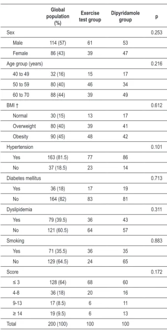
The degree of obesity of the patients was assessed by calculating body mass index (BMI) obtained by the ratio of the patient’s weight in kilograms over height in square meters. Patients with BMI between 18.5 and 24.9 were considered normal weight; between 25.0 and 29.9 were considered overweight; individuals with BMI greater than or equal to 30.0 were considered obese. Out of these patients, 64% had normal perfusion scintigraphy, according to the SSS. Table 1 shows that the groups were homogeneous (p > 0.05).
To describe the quantitative data, we used mean ± standard deviation. Categorical data were expressed as counts and percentages. The TDI between groups was compared using multiple linear regression. Categorical data were compared by the chi-square procedure. The level of significance adopted was α = 0.05. The data were analyzed and processed with the aid of the program SPSS, version 17.
Table 2 shows that the study found no association between type of stress and the average TDI values. An association was found between the average TDI values and age only for those patients from the exercise test group (p = 0.009). There was also a significant association between the mean TDI values for those patients in the youngest tertile, whose TDI of the dipyridamole had a higher average value (p = 0.010).
Discussion
PMS is associated with treadmill stress test, or stress with dipyridamole, adenosine, or dobutamine1. The presence of extensive and severe perfusion defects, reduced left ventricular ejection fraction after stress and left ventricular dilation induced by stress are indicators of poor prognosis on scintigraphy10.
Exercise has positive inotropic and chronotropic effects on the heart and increases arterial blood pressure. In healthy coronary arteries, the increased need for oxygen is compensated by arterial physiological vasodilation, which increases blood flow to blood two to three times above the baseline. Thus, the cardiac stress causes ischemia in stenosed coronary arteries or those arteries unable to dilate (with impairment of coronary flow reserve), resulting in disturbances of kinesis, and perfusion of the ventricular walls and electrocardiogram tracing, as well as symptoms like chest pain11.
Dipyridamole has an indirect vasodilatory for blocking the reuptake of adenosine into the intracellular medium. In coronary arteries with obstruction from 50% of the vascular lumen, to maintain the flow, there is intense arteriolar dilatation distal to the obstruction at rest. In obstructed coronary arteries, dipyridamole causes a little dilation. The arterioles of the normal coronary arteries will have maximum dilation. The coronary reserve starts to decrease due to stenosis of 45% to
Table 1 - Demographic data
Global population
(%)
Exercise test group
Dipyridamole group p
Sex 0.253
Male 114 (57) 61 53
Female 86 (43) 39 47
Age group (years) 0.216
40 to 49 32 (16) 15 17
50 to 59 80 (40) 46 34
60 to 70 88 (44) 39 49
BMI † 0.612
Normal 30 (15) 13 17
Overweight 80 (40) 39 41
Obesity 90 (45) 48 42
Hypertension 0.101
Yes 163 (81.5) 77 86
No 37 (18.5) 23 14
Diabetes mellitus 0.713
Yes 36 (18) 17 19
No 164 (82) 83 81
Dyslipidemia 0.311
Yes 79 (39.5) 36 43
No 121 (60.5) 64 57
Smoking 0.883
Yes 71 (35.5) 36 35
No 129 (64.5) 24 65
Score 0.172
≤ 3 128 (64) 68 60
4-8 36 (18) 20 16
9-13 17 (8.5) 6 11
≥ 14 19 (9.5) 6 13
Total 200 (100) 100 100
† -BMI - body mass index.
also been described in stimulus situations with dipyridamole4. As no study had evaluated the impact of the type of stress on the average values of TDI, this was the main goal of our study. MPS evaluates the dilation of the left ventricular cavity, induced by stress, measured by the transient dilatation index (TDI). We consider that dilation is present when the left ventricular cavity is increased after stress, compared to the image at rest4.
Several pathophysiological mechanisms may be associated with increased TDI. The TDI may be associated with subendocardial hypoperfusion and systolic ventricular dysfunction. Multivessel coronary artery disease is associated with diffuse subendocardial ischemia, which compromises the perfusion of the endocardium, resulting in a lower endocardial concentration of the tracer. Another explanation could be that the ischemia causes a systolic dysfunction would result in increased ventricular cavity due to increased end-systolic volume of left ventricle. Systolic dysfunction also gives the impression of ventricular cavity dilation4.
Other studies suggest that severe hypertensive heart disease is another cause of diffuse subendocardial ischemia in the absence of coronary stenosis, since it is associated to delayed diastolic relaxation, endothelial dysfunction of coronary arteries, reduction of capillary density in myocardial hypertrophy and increased final left ventricular diastolic pressure. Although in cases of severe ventricular hypertrophy there can be a compensatory dilation of epicardial coronary arteries, the myocardium seems to suffer transmural ischemia unrelated to any coronary stenosis12.
Robinson et al13 evaluated 237 consecutive scintigraphies and found a transient ventricular dilation in 23 of them. Nine out of 23 patients had dilatation in the absence of segmental perfusion defects upon image and in the absence of coronary stenoses. In 07 of these 09 patients, dilation is solely attributed to left ventricular hypertrophy. Thus, the transient dilatation is not a specific marker for CAD in populations with high prevalence of hypertensive heart disease, associated with ventricular hypertrophy13. Our study did not assess the prevalence of ventricular hypertrophy in the subgroup of patients with hypertension of this sample. However, as the prevalence of hypertension was similar in both groups, we believe that this factor has not affected the results.
Emmet et al10 have assessed the impact of left ventricular hypertrophy and diabetes mellitus in the prevalence of TDI. The sample (n = 103) underwent scintigraphy, transthoracic echocardiography and catheterization. TDI ≥ 1.22 and ventricular wall thickness ≥ 11 mm were considered abnormal. Factors SSS ≥ 14, hypertrophy and diabetes were independent predictors of abnormal TDI. In patients with stenosis ≥ 90% of the vascular lumen, the presence of hypertrophy or diabetes increased significantly the prevalence of abnormal TDI (p < 0.005).
Thus, increased TDI is associated with severe CAD and the association is magnified in the presence of left ventricular hypertrophy or diabetes. Hypertrophy could be associated with diffuse subendocardial ischemia. Diabetes was associated with impairment of coronary reserve secondary to microvascular disease10. Our study found no such association
Table 2 - Results
Variable Global‡TDI ‡
TDI Exercise test group
TDI Dipyridamole
group p
Sex
Male 1.07 ± 0.20 1.05 ± 0.20 1.08 ± 0.18 0.369
Female 1.10 ± 0.26 1.08 ± 0.28 1.12 ± 0.25 0.419
p 0.318 0.641 0.413
Age group (years)
40 to 49 1.04 ± 0.21 0.95 ± 0.18 1.13 ± 0.20 0.010
50 to 59 1.04 ± 0.20 1.03 ± 0.20 1.05 ± 0.21 0.644
60 to 70 1.13 ± 0.24 1.14 ± 0.25 1.13 ± 0.23 0.775
p 0.016 0.009 0.281
BMI †
Normal 1.14 ± 0.22 1.14 ± 0.30 1.14 ± 0.17 0.979
Overweight 1.08 ± 0.22 1.06 ± 0.22 1.10 ± 0.22 0.372
Obesity 1.06 ± 0.22 1.04 ± 0.22 1.08 ± 0.23 0.378
p 0.223 0.382 0.629
Hypertension
Yes 1.09 ± 0.24 1.07 ± 0.25 1.10 ± 0.22 0.490
No 1.06 ± 0.16 1.02 ± 0.15 1.13 ± 0.15 0.053
p 0.503 0.274 0.534
Diabetes mellitus
Yes 1.14 ± 0.23 1.15 ± 0.28 1.13 ± 0.18 0.773
No 1.07 ±0.22 1.04 ± 0.21 1.09 ± 0.22 0.127
p 0.112 0.157 0.532
Dyslipidemia
Yes 1.10 ± 0.22 1.09 ± 0.25 1.10 ± 0.20 0.801
No 1.07 ± 0.22 1.04 ± 0.22 1.10 ± 0.23 0.174
p 0.376 0.338 0.906
Smoking
Yes 1.07 ± 0.25 1.05 ± 0.26 1.10 ± 0.25 0.480
No 1.08 ± 0.21 1.06 ± 0.22 1.10 ± 0.20 0.284
p 0.742 0.802 0.847
Score
3 1.08 ± 0.24 1.06 ± 0.24 1.11 ± 0.23 0.216
4 to 8 1.10 ± 0.21 1.09 ± 0.24 1.13 ± 0.17 0.624
9 to 13 1.07 ± 0.23 0.95 ± 0.11 1.14 ± 0.25 0.115
14 1.03 ± 0.16 1.10 ± 0.12 1.00 ± 0.16 0.229
p 0.709 0.626 0.349
Total 1.08 ± 0.23 1.06 ± 0.23 1.10 ± 0.22 0.200
† -BMI - body mass index; ‡ - transient dilation index.
in the subgroup of diabetics, perhaps due to the small number of diabetic patients in this sample (n = 36).
In the absence of epicardial coronary stenoses, the transient dilation was described in patients with dilated cardiomyopathy. It is suggested that the limitation of coronary reserve in these patients is the explanation14. Our study excluded patients with prior diagnosis of any cardiomyopathy.
Dilation of the stress-induced ventricular cavity was first described in scintigraphy at rest with thallium-201 and exercise stress test (Bruce protocol) with 99mTc-sestamibi. This study examined the association between the increase in the TDI and the presence of coronary stenoses on catheterization. The value of positive TDI corresponded to 1.22 (±2). In the diagnosis of coronary stenosis from 70% of the vascular lumen, the TDI equal or greater than 1.22 had 40% sensitivity and specificity exceeding 90%. The TDI greater than 1.22 had 71% sensitivity and 95% specificity in the diagnosis of coronary stenosis greater than or equal to 90% of the lumen.
Thirty-three percent of patients with SSS between 04 and 08 showed high TDI, 38% with SSS between 9 and 13 and 48% of patients with SSS higher or equal to 14. Among those patients with stenoses greater than or equal to 90% in two coronary arteries, 34% had SSS above 8 and 76% presented an increased TDI. Therefore, the TDI allowed the identification of patients with extensive and severe coronary stenoses, which would have been underestimated in the isolated analysis of SSS5.
Our study found average TDI values lower than those reported in this study with double isotope, even in patients with abnormal perfusion according to SSS, which can be attributed to the fact that two studies have been conducted with different radiopharmaceutical drugs3. Nevertheless, as our research has made no cardiac catheterization to confirm the presence of coronary stenosis in patients in this sample, the presence and extent of any stenoses could not be evaluated. Our research found no significant difference between the average TDI values in patients with abnormal SSS, in the different stress methods. Perhaps this is due to the small number of patients in these subgroups, since 64% of patients showed normal perfusion (SSS ≤ 3.0).
One study evaluated the average TDI value in 356 patients undergoing MPS with adenosine, the stress protocol with 99mTc-sestamibi and rest with thallium-201. TDI of 1.36 (± had 71% sensitivity and 86% specificity in the diagnosis of coronary stenosis greater than or equal to 70% of the lumen. The visual analysis of perfusion had a sensitivity and specificity of 87% and 49%, respectively. Perfusion analysis was through the association of SSS and percentage extension of hypoperfused myocardium. TDI was abnormal in: 12.2% of patients with normal perfusion; in 28.2% of patients with discreet hypoperfusion; in 29.4% of patients with discreet hypoperfusion; and in 40.3% of patients with severe hypoperfusion15. Thus, the TDI associated with the presence of CAD was higher than the numerical value described in the protocol with the treadmill stress test (1.22)5. In our study, the overall average values of TDI were numerically higher in the dipyridamole group, confirming the findings of previous studies.
An association was found between the average TDI values and age for those patients from the exercise test group (p = 0.009). There was also a significant association between the mean TDI values for those patients in the youngest tertile, whose TDI of the dipyridamole had a higher average value (p = 0.010). These findings had not yet been reported in the literature and we found no theories that could explain them. Perhaps studies with cardiac magnetic resonance imaging may elucidate the prevalence and the relative importance of the mechanisms associated with increased TDI in pharmacological stress16.
Ventricular perfusion can be assessed by positron emission tomography with rubidium-82, combined with dipyridamole. In this protocol, the administration of pharmacological stressor precedes the administration of rubidium-82 and the images can be acquired during peak stress17. A study has measured the TDI in patients undergoing pharmacological stress examination with rubidium-82, subsequently undergoing coronary angiography within 15 days. The TDI upper limit of normality was 1.15 (±2) with high specificity in the diagnosis of single vessel stenosis and multivessel stenosis (100% and 93%, respectively). Therefore, it was reported that the dilation associated with pharmacological stress is evident before the usual time of image acquisition of conventional PMS18.
There should be caution in the analysis of scintigraphy associated with transient left ventricular dilation, since the TDI cannot be associated with coronary diseases such as dilated or hypertrophic cardiomyopathy. The TDI can be potentiated by diabetes mellitus. In addition, the TDI may be falsely increased in small hearts, due to technical limitations inherent in scintigraphy19.
Our study had some limitations such as non-randomization, because some patients could not develop any stress due to physical limitations. The patients did not undergo routine coronary angiography because they were doing tests to evaluate myocardial ischemia and also due to lack of resources. Thus, the findings do not relate to the presence and degree of coronary involvement.
Conclusion
The transient dilatation index does not differ when patients undergo exercise stress test on a treadmill compared to pharmacological stress by dipyridamole, demonstrating the same evaluative capacity.
Potential Conflict of Interest
No potential conflict of interest relevant to this article was reported.
Sources of Funding
There were no external funding sources for this study.
Study Association
References
1. Chalela WA, Camargo EE, Marin-Neto JA, Nicolau JC, Meneghetti JC/ / Sociedade Brasileira de Cardiologia. I Diretriz sobre cardiologia nuclear. Arq Bras Cardiol. 2002;78(supl. 3):5-42.
2. Duarte PS, Smanio PE, Oliveira CA, Martins LR, Mastrocolla LE, Pereira JC. Clinical significance of transient left ventricular dilation assessed during myocardial Tc-99m sestamibi scintigraphy. Arq Bras Cardiol. 2003;81(5):474-82.
3. Berman DS, Shaw LJ, Hachamovitch R, Friedman JD, Polk DM, Hayes SW, et al. Comparative use of radionuclide stress testing, coronary artery calcium scanning, and noninvasive coronary angiography for diagnostic and prognostic cardiac assessment. Semin Nucl Med. 2007;37(1):2-16.
4. McLaughlin MG, Danias PG. Transient ischemic dilation: a powerful diagnostic and prognostic finding of stress myocardial perfusion imaging. J Nucl Cardiol. 2002;9(6):663-7.
5. Mazzanti M, Germano G, Kiat H, Kavanagh PB, Alexanderson E, Friedman JD, et al. Identification of severe and extensive coronary artery disease by automatic measurement of transient ischemic dilation of the left ventricle in dual-isotope myocardial perfusion SPECT. J Am Coll Cardiol. 1996;27(7):1612-20.
6. Marcassa C, Galli M, Baroffio C, Campini R, Giannuzzi P. Transient left ventricular dilation at quantitative stress-rest sestamibi tomography: clinical, electrocardiographic, and angiographic correlates. J Nucl Cardiol. 1999;6(4):397-405.
7. Kinoshita N, Sugihara H, Adachi Y, Nakamura T, Azuma A, Kohno Y, et al. Assessment of transient left ventricular dilatation on rest and exercise on Tc-99m tetrofosmin myocardial SPECT. Clin Nucl Med. 2002;27(1):34-9.
8. Chouraqui P, Rodrigues EA, Berman DS, Maddahi J. Significance of dipyridamole-induced transient dilation of the left ventricle during thallium-201 scintigraphy in suspected coronary artery disease. Am J Cardiol. 1990;66(7):689-94.
9. Ficaro EP, Lee BC, Kritzman JN, Corbett JR. Corridor4DM: the Michigan method for quantitative nuclear cardiology. J Nucl Cardiol. 2007;14(4):455-65.
10. Emmett L, Magee M, Freedman SB, Van der Wall H, Bush V, Trieu J, et al. The role of left ventricular hypertrophy and diabetes in the presence of transient ischemic dilation of the left ventricle on myocardial perfusion SPECT images. J Nucl Med. 2005;46(10):1596-601.
11. Vesely MR, Dilsizian V. Nuclear cardiac stress testing in the era of molecular medicine. J Nucl Med. 2008;49(3):399-413.
12. Robinson VJ, Corley JH, Marks DS, Eberhardt LW, Eubig C, Burke GJ, et al. Causes of transient dilatation of the left ventricle during myocardial perfusion imaging. AJR Am J Roentgenol. 2000;174(5):1349-52.
13. Robinson VJB, Corley JH, Gunter LE. Transient ischemic dilation occurs in patients without severe multivessel epicardial stenoses. J Investig Med. 1997;45:226A.
14. Inoue T, Sakai Y, Morooka S, Hayashi T, Takayanagi K, Yamanaka T, et al. Coronary flow reserve in patients with dilated cardiomyopathy. Am Heart J. 1993;125(1):93-8.
15. Abidov A, Bax JJ, Hayes SW, Cohen I, Nishina H, Yoda S, et al. Integration of automatically measured transient ischemic dilation ratio into interpretation of adenosine stress myocardial perfusion SPECT for detection of severe and extensive CAD. J Nucl Med. 2004;45(12):1999-2007.
16. Panting JR, Gatehouse PD, Yang GZ, Grothues F, Firmin DN, Collins P, et al. Abnormal subendocardial perfusion in cardiac syndrome X detected by cardiovascular magnetic resonance imaging. N Engl J Med. 2002;346(25):1948-53.
17. Strauss HW, Miller DD, Wittry MD, Cerqueira MD, Garcia EV, Iskandrian AS, et al. Procedure guideline for myocardial perfusion imaging 3.3. J Nucl Med Technol. 2008;36(3):155-61.
18. Shi H, Santana CA, Rivero A, Sanyal R, Esteves FP, Verdes L, et al. Normal values and prospective validation of transient ischaemic dilation index in 82Rb PET myocardial perfusion imaging. Nucl Med Commun. 2007;28(11):859-63.