Association between Uric Acid and Cardiovascular Risk Variables in a
Non-Hospitalized Population
Monica Cristina Campos Barbosa, Andréa Araujo Brandão, Roberto Pozzan, Maria Eliane Campos Magalhães,
Érika Maria Gonçalves Campana, Flavia Lopes Fonseca, Oswaldo Luiz Pizzi, Elizabete Viana de Freitas, Ayrton
Pires Brandão
Universidade do Estado do Rio de Janeiro, Rio de Janeiro, RJ - Brazil
Mailing address: Andréa Araujo Brandão •
Rua General Tasso Fragoso, 24/503 - Lagoa - 22470-170 - Rio de Janeiro, RJ - Brazil
E-mail: [email protected], [email protected]
Manuscript received March 09, 2010; revised manuscript received June 24, 2010; accepted July 16, 2010.
Abstract
Background: The association between uric acid (UA) and cardiovascular risk variables remains a controversial issue in epidemiological studies.
Objective: To evaluate the association between UA, blood pressure (BP), anthropometric indices and metabolic variables in a non-hospitalized population stratified by UA quintiles.
Methods: A cross-sectional observational study evaluated 756 individuals (369 males), mean aged 50.3 ± 16.12 years, divided in UA quintiles. BP, body mass index (BMI), abdominal circumference (AC), UA, glucose, insulin, HOMA-IR, total cholesterol (TC), LDL-c, HDL-c, triglycerides (TG) and creatinine (C) levels were obtained. The estimated glomerular
filtration rate (eGFR) was calculated and arterial hypertension (AH) was considered when BP ≥ 140x90 mmHg, overweight/obesity (OW/O) was considered when BMI ≥ 25 kg/m² and metabolic syndrome (MS) was established
according to the I Brazilian Guideline of MS.
Results: 1) there was no difference between the groups regarding the distribution by sex and age range; 2) the highest UA quintiles presented higher mean age (p < 0.01), BMI, AC (p < 0.01), SBP, DBP (p < 0.001), TC, LDL-c, TG (p < 0.01), C and eGFR (p < 0.001) and lower mean HDL-c (p < 0.001); 3) the group with the highest UA quintile showed higher prevalence of AH, OW/O and MS (p < 0.001); 4) higher percentages of the lowest quintiles of insulin (p < 0.02) and HOMA-IR (p < 0.01) were observed with the lowest quintiles of UA; 5) a logistic regression analysis showed that UA and the variables that compose MS were associated with the occurrence of MS (p < 0.01).
Conclusion: Higher quintiles of uric acid were associated with a worse cardiovascular risk profile and a worse kidney function profile in the non-hospitalized population sample studied. (Arq Bras Cardiol 2011;96(3):212-218)
Keywords: Cardiovascular diseases/epidemiology; uric acid; hypertension; risk factors; metabolic syndrome.
establish the role of UA as an independent risk factor for cardiovascular events.
The association between hyperuricemia and arterial hypertension has been under observation for more than a century1, but whether hyperuricemia has a causal role in
arterial hypertension (AH) or if it is only a marker of the physiopathological process is yet to be elucidated.
Recent experimental researches have established possible mechanisms through which hyperuricemia can cause hypertension. In animal models, UA caused a decrease in nitric oxide synthetase (NOS), afferent arteriole injury, increased renin production and renal tubule injury6,7. Moreover, a close
association was demonstrated between hyperuricemia and metabolic syndrome in rats, due to a probable mechanism involving endothelial function inhibition8. It is known that
insulin needs nitric oxide (NO) to stimulate glucose uptake and the NO availability is decreased when hyperuricemia occurs9.
In this context, the inclusion of UA as variable for the stratification of cardiovascular risk seems interesting, as it is an easy-to-perform and low-cost test that can be useful in clinical
Introduction
Although the role of classic risk factors have been well-established in the context of cardiovascular disease (CVD), several emergent conditions, called risk marker, have not had their association definitely demonstrated. Studies are necessary to understand the actual role they have in this scenario and whether they can actually be of value in the early identification of individuals at risk of developing CVD. Among these studies, is the one on uric acid (UA), which has behaved as a cardiovascular risk factor in several cross-sectional studies1-5.
practice, especially in individuals with metabolic syndrome and arterial hypertension.
The present study aimed at evaluating the association between uric acid and cardiovascular risk factors in a non-hospitalized Brazilian population, covering a wide age range.
Methods
The present was a cross-sectional study, of which sample was obtained from the database of a non-hospitalized population from the service of arterial hypertension of Hospital Universitário Pedro Ernesto of Universidade do Estado do Rio de Janeiro (UERJ). The sample was obtained among patients submitted to preoperative admission assessment of non-cardiac surgeries and clinical evaluation for regular physical activity. Individuals of both sexes, aged 20 years or older, whose demographic data, clinical history, physical examination data and laboratory assessment including uric acid, glucose, serum lipids and creatinine levels were available, were considered eligible for study enrollment.
The sample consisted of 756 individuals aged 20 years or older (mean of 50.3 years), of which 369 were males (48.8%). The patients were distributed by 20-year strata.
All individuals had their demographic data collected followed by a clinical assessment to collect medical history data; additionally, a physical examination was carried out, which consisted of BP levels, weight and height measurement for BMI calculation and abdominal circumference measurement. A blood sample was collected after a 12-hour fast for the laboratory assessment, which included: glucose, insulin, HOMA-IR10, uric
acid and creatinine levels. The glomerular filtration rate was also calculated through the Cockroft-Gault formula (140-age x weight/creatinine x 72 for men and for women, multiplied by the correction factor of 0.85), total cholesterol, HDL-c, triglycerides and LDL-c, through Friedewald’s formula.
The measurement of insulin and HOMA-IR calculation was carried out in 498 patients, in whom insulin was measured and HOMA-IR was calculated according to the formula: fasting insulin x plasma fasting glucose/405. The diagnosis of MS was considered according to the criteria of the I Guideline for Metabolic Syndrome11. The diagnosis of arterial hypertension
was considered according to the V Brazilian Guideline for Arterial Hypertension12.
The sample was stratified by quintiles of uric acid according to sex and 5 groups were constituted, G1 to G5, according to the crescent order of UA quintiles. For the male sex (M), the values corresponding to the quintiles of UA were ≤ 4.5, > 4.5 and ≤ 5.2, > 5.2 and ≤ 5. 9, > 5.9 and ≤ 6.5, and >
6.5 mg/dl. For the female sex (F), the values were ≤ 3.26, > 3.26 and ≤ 3.9, > 3.9 and ≤ 4.5 and > 4.5 and ≤ 5.24 and > 5.24 mg/dl, respectively for the first, second, third, fourth and fifth quintiles of uric acid.
The data were analyzed using the statistical package “SPSS for Windows”, release 8.0.0, Copyright SPSS Inc. 1989 - 1997.
The following statistical methods were employed, with the level of rejection of the nullity hypothesis set at 0.05 or 5.0% (p = 0.05):
• Analysis of variance (F) - used to compare the means of the variables that presented a normal distribution and homogeneity of variance at Bartley’s test. • Tukey’s Test - used as complementary test for the
analysis of variance, for comparison of means of the variables 2 x 2.
• Chi-square Test (c²) - used to compare the frequency distribution of categorical variables of independent samples.
• Pearson’s Correlation Test - used to analyze the correlation between continuous variables.
This study was approved by the Ethics Committee of Hospital Universitário Pedro Ernesto, number 2322-CEP/ HUPE, on December 10, 2008.
Results
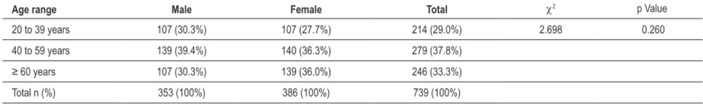
The distribution of individuals was uniform by gender and age range analyzed (Table 1). The means of systolic blood pressure (SBP), diastolic blood pressure (DBP), total cholesterol, triglycerides and creatinine significantly increased with the uric acid quintiles (Table 2). For HDL-c and for the eGFR, the opposite occurred: the higher means of these variables were observed together with the lowest quintiles of UA (Table 2). There was a prevalence of 48.3% of arterial hypertension, 33.6% of metabolic syndrome, 62.4% of overweight and obesity, 55.9% of hypercholesterolemia, 14.9% of altered fasting glycemia levels, 4.1% of diabetes, 24.9% of hypertriglyceridemia and 33.9% of low HDL levels in the total sample (Table 3).
The prevalences of arterial hypertension, overweight/obesity (Table 3) and metabolic syndrome (Figure 1) were significantly higher in the highest quintiles of uric acid (p < 0.0001).
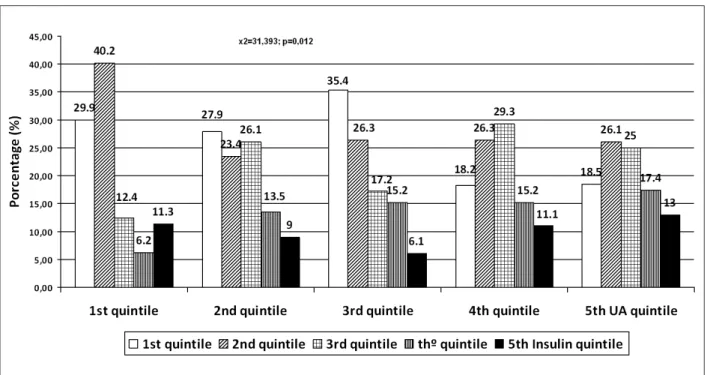
Regarding insulin and HOMA-IR, there was a higher predominance of the highest quintiles of insulin and HOMA-IR in the upper quintiles of uric acid with c² = 35.761 and p = 0.003 for HOMA-IR and c² = 31.393 and p = 0.012 for insulin (Figures 2 and 3).
Table 1 - Distribution of the population by sex and age ranges
Age range Male Female Total c2 p Value
20 to 39 years 107 (30.3%) 107 (27.7%) 214 (29.0%) 2.698 0.260
40 to 59 years 139 (39.4%) 140 (36.3%) 279 (37.8%)
≥ 60 years 107 (30.3%) 139 (36.0%) 246 (33.3%)
Table 2 - Means of blood pressure, anthropometric, metabolic and kidney function variables by UA quintiles
Variables G1 G2 G3 G4 G5 F p Comparison
2 by 2
SBP (mmHg) 130.80 ± 21.10 133.41 ± 20.60 133.52 ± 21.79 138.23 ± 20.48 144.29 ± 23.91 9.17 <0.0001 1<4; 1<5; 2<5; 3<5
DBP (mmHg) 82.32 ± 11.80 82.81 ± 12.00 85.29 ± 13.30 84.76 ± 11.60 89.15 ± 13.40 7.09 <0.0001 1<5; 2<5; 3<5
BMI (kg/m²) 25.54 ± 4.80 26.66 ± 4.60 26.94 ± 4.00 27.16 ± 4.10 27.89 ± 4.40 5.60 <0.0001 1<3; 1<4; 1<5;
AC (cm) 95.77 ± 13.70 98.47 ± 12.52 98.95 ± 9.99 99.45 ± 10.45 101.26 ± 11.05 4.34 0.002 1=2=3=4<5
Cholesterol (mg/dl) 201.8 ± 47.00 213.2 ± 56.60 209.6 ± 46.90 212.40 ± 45.40 229.8 ± 51.10 6.37 <0.0001 1<5; 2<5; 3<5; 4<5
Triglycerides (mg/dl) 99.61 ± 57.80 113.45 ± 62.30 121.96 ± 82.7 126.99 ± 63.0 141.64 ± 85.90 7.22 <0.0001 1<3; 1<4; 1<5; 2<5
HDL-c (mg/dl) 54.52 ± 14.60 51.76 ± 14.00 49.06 ± 14.40 48.48 ± 12.10 50.23 ± 14.50 4.10 0.003 1<3; 1<3; 1<4
LDL-c (mg/dl) 130.19 ± 43.63 137.35 ± 46.40 138.11 ± 41.07 139.12 ± 43.12 152.00 ± 47.59 4.44 0.002 1<5; 2<5
Creatinine (mg/dl) 0.79 ± 0.18 0.80 ± 0.20 0.83 ± 0.25 0.87 ± 0.20 0.92 ± 0.28 8.13 <0.0001 1<4; 1<5;2<5
GFR (ml/min) 112.16 ± 43.70 108.77 ± 39.40 103.25 ± 28.70 102.46 ± 33.50 94.62 ± 31.90 5.07 <0.0001
Glucose (mg/dl) 92.00 ± 29.02 88.31 ± 17.66 90.40 ± 20.30 91.87 ± 21.71 92.82 ± 18.50 1.01 0.39
Homa-IR 2.65 ± 5. 59 2.21 ± 2.02 2.29 ± 3.54 2.60 ± 2.91 2.67 ± 2.95 0.37 0.82
Insulin 12.11 ± 30.08 10.29 ± 7.74 10.66 ± 19.36 10.78 ± 6.33 11.33 ± 16.83 0.17 0.95
SBP - systolic blood pressure; DBP - diastolic blood pressure; BMI - body mass index; AC - abdominal circumference; GFR - glomerular iltration rate ; mmHg - millimeters of mercury; mg/dl - milligrams per deciliter; ml/min - milliliters per minute
Table 3 - Prevalence of cardiovascular risk factors in the total sample and by quintiles of uric acid
Prevalences Total G1 G2 G3 G4 G5 c² p
OW/O n (%) 466 (62.3) 68 (44.4) 95 (60.9) 99 (64.7) 92 (66.7) 112 (75.7) 33.67 <0.0001
SAH n (%) 365 (48.4) 58 (37.9) 67 (42.9) 68 (44.2) 73 (51.8) 99 (66.0) 28.95 <0.0001
Diabetes n (%) 31 (4.1) 8 (5.3) 6 (3.8) 4 (2.6) 6 (4.3) 7 (4.7) 15.92 0.44
Glucose intolerance n (%) 142 (18.9) 27 (17.8) 20 (12.8) 31 (20.4) 23 (16.3) 41 (27.5) 11.92 0.018
High cholesterol n (%) 420 (55.8) 72 (47.1) 79 50.6) 84 (54.5) 80 (56.7) 105 (70.5) 19.57 0.001
Triglycerides Alto n (%) 186 (24.9) 22 (14.4) 37 (23.9) 40 (26.5) 38 (27.0) 49 (33.1) 15.01 0.05
Low HDL-c n (%) 240 (34.0) 37 (26.8) 51 (34.5) 49 (34.0) 47 (36.2) 56 (38.4) 4.69 0.32
High LDL-c n (%) 376 (53.5) 61 (44.2) 71 (48) 74 (52.1) 74 (56.9) 96 (66.2) 16.745 0.002 OW/O - overweight and obesity; SAH - systemic arterial hypertension; n (%) - number and percentage.
Discussion
This study brought contributions to the better understanding of the behavior of uric acid in relation to the clinical, metabolic and renal function variables associated with a higher cardiovascular risk, especially as it included a significant number of non-hospitalized individuals from the Brazilian population, whose data were scarcely available up
to the present moment. Several studies have demonstrated that UA is an independent risk variable for cardiovascular risk factors2-5, 13-25.
The population studied in the present study is a convenience sample, with no sample size calculation, and thus, cannot reflect the characteristics of the general population. On the other hand, the fact that it has a Figure 2 -Insulin quintile according to uric acid (UA) quintile.
representative size and its non-hospital origin might have attenuated the possible selection bias.
Regarding the mean age of the individuals in this series (50.3 years), there are no significant differences in relation to most studies on the subject3,18, except for the studies carried
out in children and adolescents, such as the one by Bogalusa14
or the trial carried out by Forman et al26, in which mean age
was 61 years.
Regarding the presence of risk factors, an increased prevalence of systemic arterial hypertension was observed in the present study (48.3%), in contrast with the expected rate of 31.0% for the Brazilian southeast region. A possible explanation for this fact might be the age range of these individuals, as elderly patients (> 80 years) were included in the study, as well as the fact that 30.0% of the study population was older than 60 years, an age range at which the prevalence of arterial hypertension is known to be higher.
MS was observed in 34.5% of these individuals, higher than the prevalences described in several studies24,27. It is
noteworthy the fact that there are no national data on the subject for the purpose of comparison28. However, several
studies have shown prevalences of MS that vary from 8.0% to 70.7%, depending on the age range. It is possible that the rates observed in the present study are due not only to the oldest age range to which these individuals belong, but also because this population presented more than 60.0% of obesity prevalence.
Regarding diabetes, a similar prevalence was found to that observed in our country, which is 4.0%2, in large series, such
as the Framingham study’s2, which showed a prevalence of
2.7% of diabetes in the studied population.
From the hemodynamic point of view, it can be observed that there were higher means of BP at the highest quintiles of UA, which corresponds to the results found by Conen et al29.
As for the prevalence of arterial hypertension by quintiles of UA, it was clearly demonstrated that there was a significant association between these variables. This has been the result found in several publications, as it can be verified since 1994 with The Olivetti Heart Study23, where UA was
associated with cardiovascular risk factors such as arterial hypertension or, in the study carried out by Perlstein et al4
in 2,062 individuals, over a 21-year period, where arterial hypertension was also associated with higher quintiles of UA. Similarly the series by Mellen et al3, during a nine-year
follow-up period, showed that arterial hypertension was more prevalent in patients with higher levels of UA, in a sample of 9,104 bi-ethnic individuals of both sexes.
The biological explanation for this fact is supported by studies carried out with animal models with rats, which showed the development of arterial hypertension after the induction of hyperuricemia, caused by a probable decrease in nitric oxide in the renal macula densa and by direct stimulation of the renin-angiotensin system, with both mechanisms causing vasoconstriction and therefore, blood pressure increase6,7.
However, on the other hand, there is the possibility that the increase in UA might be present in clinical conditions known
to be pro-inflammatory, such as systemic arterial hypertension and MS, due to its antioxidant action, and therefore, represents only a biochemical defense mechanism30-32
against atherosclerosis progression. Thus, it would not be an independent risk factor for these syndromes, but just part of the clinical picture.
Regarding the metabolic variables, the prevalences of cholesterol, triglycerides and glucose intolerance were significantly higher in the higher quintiles of UA. The association with cholesterol and triglycerides was also found in the population studied by the Normative Aging Study. As for HDL-c, this study demonstrated an inverse association with the quintiles of UA and the same was verified in the 425 patients studied by Zocalli et al27.
At the analysis of distribution of insulin and HOMA-IR quintiles, in relation to the quintiles of UA, the present study showed a significant association between higher quintiles of UA and higher quintiles of both insulin and HOMA-IR, a finding that is consistent with the ones observed by Formam et al26. Regarding HOMA-IR, Ishizaka et al24 demonstrated
higher prevalences of this variable by quintiles of UA. As for the presence of diabetes, the present study showed that the prevalence of this pathology was significantly higher in the groups with higher levels of UA, a result consistent with large longitudinal studies, such as the Framingham study2 or
the cohort of 9,104 individuals from the ARIC study3. These
findings suggest that hyperuricemia is associated with insulin resistance, a condition underlying metabolic syndrome.
Regarding the MS, there was also a higher prevalence in the groups presenting higher levels of UA, and similarly, the literature demonstrates such association, as it can be observed in the study by Kawada et al25, where 981 Japanese workers
were assessed and which showed an association between MS and hyperuricemia.
The Ishizaka24 also demonstrated a significant association
between MS and UA levels, in a cross-sectional study of 8,144 individuals of both sexes, stratified by quartiles of UA. The highest prevalence of MS with increasing levels of UA was also demonstrated by a study carried out by Choi and Ford16, 17
using data from NHANES. In some other studies, such as the one by Coutinho et al33 and Desai et al15, it can be observed
that US was associated with components that constitute MS, and the higher the number of components of the syndrome, the higher the association.
Recently, in a study carried out in Brazil by Franco et al34,
in hypertensive patients from Cuiaba, demonstrated a higher prevalence of MS in patients with hyperuricemia34.
Conclusions and perspectives
The present study indicates the possibility that the uric acid is associated with cardiovascular risk variables and metabolic syndrome and that it can be useful in the individual cardiovascular risk assessment.
The existence of a physiopathological rational basis to explain the association between the uric acid and cardiovascular risk factor, together with the fact that uric acid is easy to measure and the existence of the appropriate therapy to treat it, reinforce the need for new researches to better understand its participation in the cardiovascular disease scenario.
Potential Conflict of Interest
No potential conflict of interest relevant to this article was reported.
Sources of Funding
There were no external funding sources for this study.
Study Association
This article is part of the thesis of master submitted by Monica Cristina Campos Barbosa, from Universidade do Estado do Rio de Janeiro.
References
1. Mohamed F. On chronic Bright’s disease, and its essential symptoms. Lancet. 1879; 1: 399-401. apud: Heinig M, Johnson RJ. Role of uric acid in hypertension, renal disease and metabolic syndrome. Cleve Clin J Med. 2006; 73 (12): 1059-63.
2. Sunstrom J, Sullivan L, D’agostinho R, Levy D, Kannnel W, Vasan R. Relations of serum uric acid to longitudinal blood pressure tracking and hypertension incidence. Hypertension. 2005; 45 (1): 28-33.
3. Mellen PB, Bleyer AJ, Erlinger TP, Evans G, Nieto FJ, Wagenknecht L, et al. Serum acid uric predicts incident hypertension in a biethnic cohort: the atherosclerosis risk in communities study. Hypertension. 2006; 48 (6):1037-42.
4. Perlstein TS, Gumieniak O, Williams GH, Sparrow D, Litonjua AA, Vokonas P, et al. Uric acid and the development of hypertension: the Normative Aging Study. Hypertension. 2006; 48 (6): 1031-6.
5. Krishnan E, Kwoh CK, Schumacher HR, Kuller L. Hyperuricemia and incidence of hypertension among men without metabolic syndrome. Hypertension. 2007; 49 (2): 298-303.
6. Mazzali M, Hughes J, Kim Y, Jefferson JA, Kang DH, Gordon KL, et al. Elevated uric acid increases blood pressure in the rat by a novel crystal-independent mechanism. Hypertension. 2001; 38 (5): 1101-6.
7. Mazzali M, Kanellis J, Han L, Feng L, Xia YY, Chen Q, et al. Hyperuricemia induces a primary renal arteriopathy in rats by a blood pressure-independent mechanism. Am J Physiol Renal. 2002; 282 (6): F991- F7.
8. Nakagawa T, Hu H, Zhariov S, Tuttle KR, Short RA, Glushakova O, et al. A causal role for uric acid in fructose e-induced metabolic syndrome. Am J Physiol Renal Physiol. 2006; 290 (3): F625-31.
9. Heinig M, Johnson RJ. Role of uric acid in hypertension, renal disease and metabolic syndrome. Cleve Clin J Med. 2006; 73 (12): 1059-63.
10. Matthews DR, Hosker JP, Rudenki AS, Naylor BA, Treacher DF, Turner RC. Homeostasis model assessment Insulin resistance and beta-cell function from fasting plasma glucose and insulin concentrations in man. Diabetologia. 1985; 28 (7): 412-9.
11. Brandão AP, Nogueira AR, Oliveira JE, Guimarães JI, Suplicy H, Brandão AA. (orgs.). / Sociedade Brasileira de Cardiologia. I Diretriz brasileira de diagnóstico e tratamento da síndrome metabólica. Arq Bras Cardiol. 2005; 84 (supl 1): 3-28.
12. Mion Jr D, Kohlmann Jr O, Machado CA, Amodeo C, Gomes MAM, Praxedes JN, et al. / Sociedade Brasileira de Cardiologia. V Diretrizes brasileiras de hipertensão arterial. Arq Bras Cardiol. 2007; 89 (3): e24-e79.
13. Viazzi F, Parodi D, Leoncini G, Parodi A, Falqui V, Ratto E, et al. Serum uric acid and target organ damage in primary hypertension. Hypertension. 2005; 45 (5): 991-6.
14. Alper AB, Chen W, Yau L, Srinivans SR, Berenson G, Hamm LL. Childhood uric acid predicts adult blood pressure: the Bogalusa Heart Study. Hypertension. 2005; 45 (1): 34-8.
15. Desai M, Santos R, Dalal D, Carvalho JA, Marten DR, Flynn JA, et al. Relation of serum uric acid with metabolic risk factors in asymptomatic middle-aged Brazilian men. J Am Coll Cardiol. 2005; 95 (7): 865-8.
16. Choi HK, Ford ES. Prevalence of metabolic syndrome in individuals with hyperuricemia. Am J Med. 2007; 120 (5): 442-7.
17. Ford ES, Li C, Cook S, Choi HK. Serum concentrations of uric acid and the metabolic syndrome among US children and adolescents. Circulation. 2007; 115 (19): 2526-32.
18. Niskanen LK, Laaksonen DE, Nyyssonen K, Alfthan G, Lakka HM, Lakka TA, et al. Uric acid as a risk factor for cardiovascular and all cause mortality in middle-ages men. Arch Intern Med. 2004; 164 (14): 1546-51.
19. Liese AD, Hense HW, Lowel H, Doring A, Tietze M, Keil U. Association of serum uric acid with all-cause and cardiovascular disease mortality and incident myocardial infarction in the MONICA Augsburg cohort. World Health Organization Monitoring Trends and Determinants in Cardiovascular Disease. Epidemiology. 1999; 10 (4): 391-7.
20. Culleton BF, Larson MG, Kannel WB, Levy D. Serum uric acid and risk for cardiovascular disease and death: the Framingham Heart Study. Ann Intern Med. 1999; 131 (1): 7-13.
21. Bengtsson C, Lapidus L, Stendhal C, Waldestrom J. Hyperuricemia and risk of cardiovascular disease and overall death: a 12-year follow-up of participants in the population study of women in Gothemburg, Sweden. Acta Med Scand. 1988; 224 (6): 549-55.
22. Taniguchi Y, Hayashi T, Tsumara K, Endo G, FujiiS, Okada K. Serum uric acid and the risk for hypertension and type 2 diabetes in Japanese men: the Osaka Health Survey. Hypertension. 2001; 19 (7): 1209-15.
23. Jossa F, Farinaro E, Panico S, Krogh V, Celentano E, Galasso R, et al. Serum acid uric and hypertension: the Olivetti Heart Study. J Hum Hypertens. 1994; 8 (9): 677-81.
24. Ishizaka N, Ishizaka I, Toda E, Nagai R, Yamakado M. Association between serum uric acid, metabolic syndrome, and carotid atherosclerosis in Japanese individuals. Arterioscler Thromb Vasc Biol. 2005; 25 (5): 1038-44.
25. Kawada T, Otsuka T, Katsumata M, Suzuki H. Serum uric acid is significantly related to the components of the metabolic syndrome in Japanese working men. J Cardiometab Syndr. 2007; 2 (3): 158-62.
26. Forman JP, Choi H, Curhan GC. Plasma uric acid level and risk for incident hypertension among men. J Am Soc Nephrol 2007; 18 (1): 287-92.
27. Zoccali C, Maio R, Mallamaci F, Sesti G, Perticone F. Uric acid and endothelial dysfunction in essential hypertension. J Am Soc Nephrol. 2006; 17 (5): 1466-71.
29. Conen D, Wietlisbach V, Bovet P, Shamaye C, Riesen W, Paccand F, et al. Prevalence of hyperuricemia and relation of serum uric acid with cardiovascular risk factors in developing country. BMC Public Health. 2004 Mar 25; 4-9.
30. Nieto FJ, Iribarren C, Gross MD, Comstock GW, Cutler RG. Uric acid and serum antioxidant capacity: a reaction to atherosclerosis? Atherosclerosis. 2000; 148 (1): 131-9.
31. Davies KJ, Sevanian A, Muakkassah-Kelly SF, Hochstein P. Uric acid-iron complexes: a new aspect of the antioxidant functions of uric acid. Biochemist J. 1986; 235 (3): 747-54.
32. Ames BN, Cathcart R, Schwiers E, Hochstein P. Uric acid provides an antioxidant defense in humans against oxidant- and radical- caused aging and cancer: a hypothesis. Proc Natl Acad Sci USA. 1981; 78 (11): 6858-62.
33. Coutinho TA, Turner ST, Peyser PA, Bielak LF, Sheedy PF, Kullo IJ. Association of serum uric acid with markers of inflammation, metabolic syndrome and subclinical coronary atherosclerosis. Am J Hypertens. 2007; 20 (1): 83-9.