CytogenetiCanalysisofmisCarriagematerial
681
Rev Assoc Med Bras 2010; 56(6): 681-3
IntroductIon
Miscarriage is the spontaneous or induced interruption of pregnancy until 20 complete weeks. Miscarriages occur in approximately 15% of diagnosed pregnancies1 and, although they are common, most women who have miscarriages give birth to a healthy child later in life.
The probability for a couple to have two consecutive miscar-riages ranges from 2.2 to 4%. Among the main causes for miscarriage are chromosome anomalies (whether numerical or structural)2,3, mostly represented by trisomies, by polyploidies and by the monosomy of sex-determining chromosome X.
Most miscarriages occur in the first trimester, generally between eight and 12 weeks, and half of these are caused by chromosome anomalies4. On the other hand, approximately 99% of pregnancies with chromosome anomalies evolve to miscarriage5. Some ultrasound signals, such as irregularities of the gestational sac, early restrictions in embryonic growth, increased diameter of yolk sac (larger than 8 mm), heart rate under 100 bpm and subchorionic hematoma are regarded as predictors of a bad prognosis6, 7.
Both the embryonic and fetal materials can be retrieved and cultivated for the research of chromosome anomalies. The most frequently observed aberrations are trisomy of chromosome 16
and monosomy of sex-defining chromosome X. It is known that trisomies are associated with greater maternal age2, 5, 8.
Causal investigation is formally indicated in cases of habitual abortion, defined as three or more consecutive miscar-riages, but some authors have been recommending cytogenetic analysis even in the first episode5, 9.
With the purpose of describing the abnormalities observed in our field, we presented a retrospective compilation of the karyotype analysis in miscarriage material sent for study.
Methods
This descriptive study has been approved by the Research Ethics Committee of Grupo Fleury and involved a retrospective analysis of the results of cytogenetic examinations of miscar-riage material sent to the Cytogenetics department of Grupo Fleury Medicina e Saúde after uterine curettage or manual vacuum aspiration (MVA) in cases of miscarriage in the first trimester (up to 12 weeks), coming from several hospitals in the state of São Paulo, between January, 2000 and June, 2009. All miscarriage material was sent in sealed containers with formal-dehyde and, occasionally, placed in transportation containers. The samples sent were carefully assessed using a stereo-scopic microscope by an experienced cytogeneticist for separa-tion of the embryonic or fetal material (chorionic villus) that was
*Correspondence:
Rua Cincinato Braga, 282 - Bela Vista
São Paulo – SP, Brazil CEP: 01333-910
AbstrAct
objectIve. To describe chromosome abnormalities in miscarriage material.
Methods. A retrospective compilation of karyotype analysis of slides stained with Band G was carried
out by optical microscopy with 428 miscarriage materials referred for study.
results. There were 145 normal results (33.9%) and 237 abnormal results (55.4%). In 46 samples there
was no cell growth (10.7%). Numerical abnormalities were the most frequent, especially trisomy 16 (41 cases), triploidy (27 cases), monosomy X (26 cases), tetraploidy (13 cases) and trisomy 15 (13 cases). conclusIon. Cytogenetic alterations are an important cause of miscarriages, and their detection is helpful
for the couple’s genetic counseling. Trisomy 16 is the most frequently found alteration.
Key Words: Cytogenetics. Miscarriage. Chromosomal alterations.
cytogenetIc
AnAlysIs
of
MIscArrIAge
MAterIAl
dAnIel lorber rolnIk1*, MárIo henrIque burlAcchInIde cArvAlho2, AnA lúcIA PereIrA MonteIro cAtelAnI3, AnA PAulA AlMeIdA rochA PInto4, julIAnA brAnco gonçAlves lIrA5, neusA kIyoMI kusAgArI5, PAulA bellIne3, MArIAde lourdes chAuffAIlle6
Study conducted at Grupo Fleury Medicina & Saúde, São Paulo, SP, Brazil
1- Ex-fellow em Medicina Fetal do Grupo Fleury Medicina & Saúde, São Paulo, SP
2- Doutorado em Obstetrícia – Assessor médico do setor de Medicina Fetal do Grupo Fleury Medicina & Saúde, São Paulo, SP 3- Doutorado em biologia - Bióloga do setor de Citogenética do Grupo Fleury Medicina & Saúde, São Paulo, SP
4- Bióloga do setor de Citogenética do Grupo Fleury Medicina & Saúde, São Paulo, SP 5- Biomédicas do setor de Citogenética do Grupo Fleury Medicina & Saúde, São Paulo, SP
rolnik Dl etal.
682
Rev Assoc Med Bras 2010; 56(6): 681-3Table 2 – Other abnormalities:
1 +13,+15
2 +21,+22
3 +13,+21
4 +9,+13
5 +18,+21
6 +9,+14
7 +16,+21
8 +15,+16
9 Der(14;14) 10 Der(13;14) 11 Der(13;14) 12 Der(10)t(10;16)(q25;q13) 13 R(14)(p11.2q32) 14 Der(6)t(6;X)(q11;q13) 15 48,XXY,+18 16 Add(17)(p11.2) 17 91,XXYY,+5,-21,-21 18 T(3;4)(q13;q35) 19 70,XXX,+22 20 68,XXX,-21 21 Der(18)(?) 22 68,XXY,-17 23 70,XXX,+18 24 -22,t(9;22) 25 T(3;13)(q27;q14) 26 Dup(9)(q21q34) 27 -15,der(15)add(15)(p11)
28 Mar
29 Add(8)(p21)
30 Inv(Y)
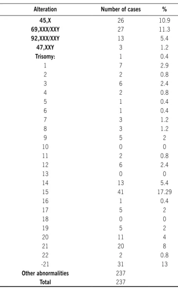
Table 1- List of numerical alterations
Alteration Number of cases %
45,X 26 10.9
69,XXX/XXY 27 11.3
92,XXX/XXY 13 5.4
47,XXY 3 1.2
Trisomy: 1 0.4
1 7 2.9
2 2 0.8
3 6 2.4
4 2 0.8
5 1 0.4
6 1 0.4
7 3 1.2
8 3 1.2
9 5 2
10 0 0
11 2 0.8
12 6 2.4
13 0 0
14 13 5.4
15 41 17.29
16 1 0.4
17 5 2
18 0 0
19 5 2
20 11 4
21 20 8
22 2 0.8
-21 31 13
Other abnormalities 237
Total 237
placed in culture, and the maternal material was discarded. The sample was divided for direct method and extended culture. Samples that contained no fetal matter or chorionic villus were refused. Cultures were processed as usual and, in summary, the direct method was conducted by placing the sample in Amniomax ® medium (5 ml with 7.0 pH) and left in incuba-tion for 24 to 48 hours in a carbon dioxide (CO2) hothouse at 37oC. Cultures were prepared by placing the sample in two Leighton tubes, one containing a half Chang D ® and another containing Amniomax ® (2.5 mL com 7.0 pH) which were then placed incubation in a CO2 hothouse at 37oC. When there was adequate growth, 60 uL colchicine were added for every 5mL of culture, for 45 minutes. The growth medium was then removed and 1% sodium citrate was added for 10 minutes at room temperature. This step was repeated twice and then the plates were prepared as usual and stained by G band.
Analysis was conducted with optic microscope (Olympus BX60 ®), counting 20 cells in metaphase stage, with capturing
by a computerized system (Applied Imaging ®) and karyotyping. The results were described according to the 2009 International System for Human Cytogenetic Nomenclature (ISCN)10.
results
In the assessed period, 428 karyotype analyses were performed in miscarriage material. The mean age of patients was 33 years (ranging from 13 to 46). We observed 145 normal results (33.9%) and 237 abnormal results (55.4%). In 46 samples there was no cell growth (10.7%). Numerical abnormalities were the most frequent, particularly trisomy 16 (41 cases), triplody (27 cases), monosomy X (26 cases), tetra-ploidy (13 cases) and trisomy 15 (13 cases) - Tables 1 and 2.
dIscussIon
CytogenetiCanalysisofmisCarriagematerial
683
Rev Assoc Med Bras 2010; 56(6): 681-3
professionals in these areas an a source of anxiety for parents, especially in cases of recurring miscarriages. A great part of first trimester miscarriages are due to chromosomopathies11. Chromosome abnormalities were observed in 55.4% of miscar-riage materials assessed in this study, thus confirming previous data from the literature, which has shown that 45% to 70% of all first trimester miscarriages are caused by chromosome abnormalities12-15.
The most frequent chromosomopathy in this study was that of chromosome 16, which concurs with data from the literature12-14. The occurrence of trisomies is associated with older mothers, which currently is a problem in the light of the increasing maternal age in most countries12. Some studies suggest that miscarriages without karyotype alterations, which could, therefore, have other causes (endocrine, immune, anatomical, etc.), are more frequent in women under 35 years old1.
Polyploidies and monosomy X are also common causes of miscarriage1,14, which was confirmed by this study. These abnormalities, however, do not have an evident association with maternal age12. The scarcity of data regarding the frequency of the different types of alterations in our field motivated the present study.
The detection of approximately 55% of abnormalities in miscarriage material, without previous selection, is in accor-dance with data from the literature4, 11. In a study conducted in China, Zhang et al.4 assessed 115 miscarriage materials and concluded that 61% presented chromosome abnormali-ties, 90% of which were numerical and 10% of which were structural, underscoring that polymerase chain reaction and comparative genomic hybridization are useful techniques for cases in which there is no cell growth in growth medium or when there is contamination by maternal cells11. The rate of cell growth failure mentioned in the literature ranges from 10% to 15%, which is in accordance with the rate found in this study (10.7%).
Although an isolated episode of miscarriage is a random event in the vast majority of cases and a full etiological investi-gation is not formally indicated, some authors1,5,8 suggest that, in these cases, the cytogenetic analysis could be comforting from a psychological perspective and influence the decision of attempting a new pregnancy, especially for patients aged 36 or older1.
In a similar study in which 420 miscarriage materials were assessed, stratified by maternal age, Stephenson et al.1 found 46% of chromosome abnormalities. The authors suggest researching other etiological factors after two early miscarriages with normal karyotype, which would lead to early diagnosis and to the introduction of more effective therapeutic measures1. Cytogenetic assessment in isolated cases would enable identifi-cation of alterations caused by random errors in meiosis. Finding a chromosome abnormality would render other investigations unnecessary, thus leading to cost reduction1.
The medical literature, however, is in need of studies that assess the recommendation of cytogenetic analysis in all cases of first trimester miscarriage. There is no data regarding the cost-effectiveness ratio of this practice.
conclusIon
Cytogenetic alterations, mainly aneuploidies, are an impor-tant cause of miscarriages and their detection is helpful to the couple’s genetic counseling. Among over 50 described altera-tions, trisomy 16 is the most frequently found, followed by other trisomies, polyploidies and monosomy X.
Cytogenetic analysis must be conducted as routine in cases of habitual abortion, but their use in everyday practice for isolated miscarriage episodes, in spite of being theoretically beneficial, should be further evaluated in future studies.
Conflict of interest: No conflicts of interest declared concer-ning the publication of this article.
referêncIAs
1. Stephenson MD, Awartani KA, Robinson WP. Cytogenetic analysis of miscar-riages from couples with recurrent miscarriage: a case-control study. Hum Reprod. 2002;17:446-51.
2. Gardo S. [Spontaneous abortion and genetic natural selection]. Orv Hetil. 1993;134:1459-64.
3. Gardo S, Bajnoczky K. [Chromosome analysis in spontaneous abortion using direct preparation of chorionic villi]. Orv Hetil. 1991;132:2727-9. 4. Ljunger E, Cnattingius S, Lundin C, Anneren G. Chromosomal anomalies in
first-trimester miscarriages. Acta Obstet Gynecol Scand. 2005;84:1103-7. 5. Morton NE, Chiu D, Holland C, Jacobs PA, Pettay D. Chromosome anomalies
as predictors of recurrence risk for spontaneous abortion. Am J Med Genet. 1987;28:353-60.
6. Choong S, Rombauts L, Ugoni A, Meagher S. Ultrasound prediction of risk of spontaneous miscarriage in live embryos from assisted conceptions. Ultra-sound Obstet Gynecol. 2003;22:571-7.
7. Varelas FK, Prapas NM, Liang RI, Prapas IM, Makedos GA. Yolk sac size and embryonic heart rate as prognostic factors of first trimester pregnancy outcome. Eur J Obstet Gynecol Reprod Biol. 2008;138:10-3.
8. Eiben B, Bartels I, Bahr-Porsch S, et al. Cytogenetic analysis of 750 sponta-neous abortions with the direct-preparation method of chorionic villi and its implications for studying genetic causes of pregnancy wastage. Am J Hum Genet. 1990;47:656-63.
9. Warburton D. Chromosomal causes of fetal death. Clin Obstet Gynecol. 1987;30:268-77.
10. Shaffer LG, Slovak ML, Campbell LJ. ISCN - An International System for Huma Cytogenetic Nomenclature. S Karger AG; 2009.
11. Zhang YX, Zhang YP, Gu Y, et al. Genetic analysis of first-trimester miscarriages with a combination of cytogenetic karyotyping, microsatellite genotyping and arrayCGH. Clin Genet. 2009;75:133-40.
12. Be C, Velasquez P, Youlton R. Spontaneous abortion: cytogenetic study of 609 cases. Rev Med Chil. 1997;125:317-22.
13. Ford JH, Wilkin HZ, Thomas P, McCarthy C. A 13-year cytogenetic study of spontaneous abortion: clinical applications of testing. Aust N Z J Obstet Gynaecol. 1996;36:314-8.
14. Philipp T, Kalousek DK. Generalized abnormal embryonic development in missed abortion: embryoscopic and cytogenetic findings. Am J Med Genet. 2002;111:43-7.
15. Goud TM, Mohammed Al Harassi S, Khalfan Al Salmani K, Mohammed Al Busaidy S, et al. A cytogenetic studies in couples with recurrent miscarriage in the Sultanate of Oman. Reprod Biomed Online. 2009;18:424-9.