(1) Escola Técnica de Artes -ETA, Universidade Federal de Alagoas-UFAL, Maceió, Alagoas, Brasil.
(2) Departamento de Fonoaudiologia, Universidade Federal de Pernambuco, Recife, Pernambuco, Brasil. (3) Departamento de Fonoaudiologia,
Universidade Federal da Paraíba, João Pessoa, Paraíba, Brasil.
(4) Hospital do Servidores do Estado de Pernambuco (HSE), Recife, Pernambuco, Brasil.
Source of support: MCTI/CNPQ/Universal 14/2014 - Faixa A
Conlict of interest: non-existent
Contributions of neuroimaging in singing voice studies:
a systematic review
Contribuições da neuroimagem no estudo da voz cantada:
revisão sistemática
Geová Oliveira de Amorim(1)
Lucas Carvalho Aragão Albuquerque(2)
Leandro de Araujo Pernambuco(3)
Patricia Maria Mendes Balata(4)
Brunna Thaís Luckwü-Lucena(3)
Hilton Justino da Silva(2)
Received on: March 06, 2017 Accepted on: July 08, 2017 Mailing address: Geová de Oliveira Amorim
ABSTRACT
It is assumed that singing is a highly complex activity, which requires the activation and interconnection of sensorimotor areas. The aim of the current research was to present the evidence from neuroimaging stu-dies in the performance of the motor and sensory system in the process of singing. Research articles on the characteristics of human singing analyzed by neuroimaging, which were published between 1990 and 2016, and indexed and listed in databases such as PubMed, BIREME, Lilacs, Web of Science, Scopus, and EBSCO were chosen for this systematic review. A total of 9 articles, employing magnetoencephalo-graphy, functional magnetic resonance imaging, positron emission tomomagnetoencephalo-graphy, and electrocorticography were chosen. These neuroimaging approaches enabled the identiication of a neural network interconnec -ting the spoken and singing voice, to identify, modulate, and correct pitch. This network changed with the singer’s training, variations in melodic structure and harmonized singing, amusia, and the relationship among the brain areas that are responsible for speech, singing, and the persistence of musicality. Since knowledge of the neural networks that control singing is still scarce, the use of neuroimaging methods to elucidate these pathways should be a focus of future research.
Keywords: Voice; Neuroimaging; Music
RESUMO
Admite-se que o canto seja uma atividade de alta complexidade pois requer ativação e interconexão de áreas sensório-motoras. Esta pesquisa teve como objetivo apresentar as evidências originadas por estu-dos de neuroimagem sobre a atuação do sistema motor e sensitivo na produção do canto. Na construção da revisão sistemática, foram premissas o período de publicação entre 1990 e 2016, artigos publicados em periódicos indexados e constantes nas bases de dados PubMed, BIREME, Lilacs, Web of Science,
INTRODUCTION
Singing is a specialized vocal behavior, which is only present in a very limited range of animals, including man and diverse species of birds. The production of a singing voice is mediated by a specialized cerebral
system that is constituted of speciic interconnected
areas of the brain1.
Singing animals can be differentiated into the following two groups on the basis of song learning: those that learn only for a period, and those that learn
their whole lives1. By comparing these two groups,
along with non-singing birds, song learning has emerged as a new evolutionary characteristic, which depends on the formation of new neural centers of control2.
Dissimilarities between the vocal behavior of man and their closest genetic relatives (chimpanzees and baboons) highlight that humans’ singing ability might not be derived from an ancestral learning of hominids species. The most likely hypothesis is that the system of human singing is a new neural specialization, which is analogous to the singing system of birds1,2. This specialization derives, among other factors, from man’s ability to exercise volitional control of vocal fundamental frequency, especially when singing without words, such as an arpeggio, which is crucially dependent on the movements of the vocal folds3.
Studies referring to the neurological aspects of song production in the literature are scarce, as researchers usually do not investigate the musical ability of non-singing subjects for means of comparison.
Within the last two decades, neuroimaging studies that aimed to identify the activation of brain’s senso-rimotor areas during repetitive or sustained singing of one note, the emission of a sung word in different rhythms, and pieces of popular music or Italian arias.
Harmonized singing, which is deined as the simulta -neous production of two or more sounds, has also been
studied. These indings add to the understanding of the
processes of perception and production in singing, which can help train singers and voice professionals, in addition to people with disorders of speech or song production, as both use the same the neural connec-tions2,4. This context is socially relevant when one considers the function of singing in social cohesion, motivation, and the structuring of group identity.
To contribute to this area of knowledge, the objective of the current study was to present original evidence, based on neuroimaging studies with a focus on the activation of the sensorimotor system in the production of song.
METHODS
A systematic review was performed, separately, by three trained researchers (GOA, HJS, and PMMB). The inclusion criteria for articles were as follows: containing one or more of the chosen descriptors (i.e., <neuroim-aging>, <voice>, <vocal training>, or <singers>); published in Portuguese, English, or Spanish, between 1990 and 2016, in indexed journals and present in one of the following databases: PubMed, BIREME, Lilacs, Web of Science, Scopus, or EBSCO.
The exclusion criteria were as follows: conference abstracts, book chapters, master’s theses, doctoral dissertations, or articles that involved research relating solely to the spoken voice.
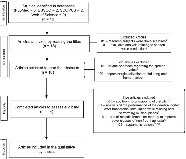
In total, 21 articles were found from the reference databases and analyzed according to the above-mentioned criteria. During the beginning of the analysis, two articles were excluded; one article was excluded because the research subjects were dove-like birds4, while the other referred exclusively to issues
relating to aspects concerning the spoken voice5.
After reading the abstracts, two more articles were excluded, since one presented a unique approach regarding the spoken voice6 and the other referred to the dopaminergic activation of bird song7. When the original version of the remaining articles were read in their entirety, the researchers independently excluded
ive more articles because of the following reasons:
a focus on the auditory-motor mapping of pitch8
control, an analysis of the performance of the cerebral cortex after transcranial stimulation while reading and
performing musical pieces9, a focus on the use of
melodic intonation therapy to improve severe cases
of non-luent aphasia10, as well as two other studies
because these were systematic reviews11,12. During
LITERATURE REVIEW
A total of 9 articles were included in this review. The articles were grouped according to the following central themes: alternation of the melodic structure in singing, harmonic singing, amusia, relationship between cerebral areas responsible for speech and song, and the persistence of musicality.
Perry et al.3 published one of the irst studies
regarding the identiication of cerebral regions involved
in singing simple songs (where only one note or pitch was maintained, which is known as the fundamental frequency of voice). Brown et al.1 analyzed the alter-ation of the melodic structure and harmonizalter-ation of
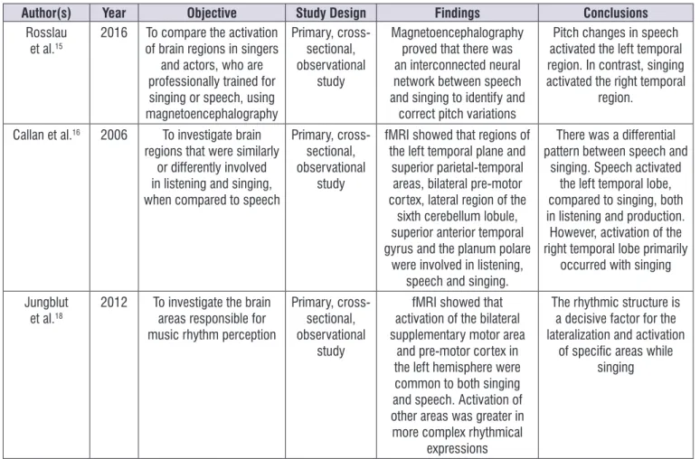
Wilson et al.14; Zarate, Wood and Zatorre4; Rosslau et al.15; Callan et al.16; Roux et al.17; and Jungblut et al.18, which investigated areas of the brain responsible for the production and perception of rhythm during singing (Figure 2).
Neuroimaging studies demonstrated that the learning and production of song (song control system) depend on the action of diverse areas of the brain,
acting in a speciic neural network, to grant meaning
to musicality, conceptualized as the ability to generate
meaning through making expressive music14. Prior
research on song production has conducted with a focus on the alteration of its melodic structure, harmo-nization, amusia, and the persistence of song and
Studies identified in databases (PubMed = 5, EBSCO = 2, SCOPUS = 2,
Web of Science = 9) (n = 18)
Articles analyzed by reading the titles (n = 18)
Articles selected to read the abstracts (n = 16)
Id
en
tif
ica
tio
n
Two articles excluded
01 –unique approach regarding the spoken voice6
01 –dopaminergic activation of bird song and human voice7
Excluded Articles:
01 – research subjects were dove-like birds4
01 – exclusive analysis relating to spoken voice production5
S
e
le
ct
io
n
Completed articles to assess eligibility (n = 14)
Five articles excluded
01 – auditory-motor mapping of the pitch8
01 – analysis of the performance of the cerebral cortex after transcranial stimulation while reading and
performing musical pieces9
01 – use of melodic intonation therapy to improve severe cases of non-fluent aphasia10
02 – systematic reviews11,12
Articles included in the qualitative synthesis
El
ig
ib
ilit
y
In
cl
usi
on
Author(s) Year Objective Study Design Findings Conclusions
Perry et al.3 1999 To investigate the cerebral
blood low during simple singing when compared to passive listening of complex-tones, using positron emission tomography (PET) Primary, cross-sectional, observational study
Cerebral blood low increases in brain areas related to motor control, as
during the speech, but the changes in the Heschl’s gyruswere related to the perception of fundamental
frequency
Singing and the emission of a vowel in a single pitch
seem to activate similar brain areas as speech; yet, in some regions an asymmetric activation, which was exclusively linked to singing was
observed. Brown et al. 1 2004 To investigate the
multifactorial vocal system using PET, to
assess the listening and response system
of non-professional singers, during repeated
singing, harmonizing new melodies, or singing
monotonically.
Primary, cross-sectional, observational
study
In general, there was a greater increase in blood
low to the primary and secondary auditory cortex, primary motor cortex, frontal
operculum, insula, posterior cerebellum and posterior
Brodmann area 22
The three tasks of listening to a response activated the frontal operculum (Broca’s area), which was involved in producing a sequence
and motor cognitive imitation, implied in musical
imitation and vocal learning
Terao et al.13 2006 To describe
psychophysical aspects of vocal amusia in a
professional tango singer, following a stroke
that affected the upper temporal cortex of the
right hemisphere
Case report Magnetic resonance imaging demonstrated damage in the right parietal cortex, which
affected pitch perception
The damage in the cortex of the right brain hemisphere
caused perceptive and expressive music loss
Roux et al.17 2009 To identify the brain areas
involved in singing and speech regions
Case report Stimulation electrocorticography during surgery for tumor removal demonstrated the
dissociation that exists between speech and singing, outside the primary
sensorimotor brain area
Identifying the dissociation indicated that these two
functions used distinct neural connections
Wilson et al.14 2011 To investigate the
relationship between musical and speech functions and their interaction with lyric
singing tasks Primary, cross-sectional, observational study Functional magnetic resonance imaging (fMRI) demonstrated that singing and speech were elicited by contiguous brain areas.
The interrelation between these areas was reduced as the singer became more
specialized
The specialization of lyrical singers caused less
interdependence of the cerebral areas devoted to singing and speech, demonstrating a more reined and less dependent
speech process Zarate, Wood,
Zatorre4
2010 To analyze the voluntary and involuntary pitch
regulation and its correlation with the neural
connections
Primary, cross-sectional, observational
study
fMRI proved that lower pitch adjustments are under less
voluntary control than the large variations
Author(s) Year Objective Study Design Findings Conclusions
Rosslau et al.15
2016 To compare the activation of brain regions in singers
and actors, who are professionally trained for singing or speech, using magnetoencephalography
Primary, cross-sectional, observational
study
Magnetoencephalography proved that there was an interconnected neural network between speech and singing to identify and
correct pitch variations
Pitch changes in speech activated the left temporal region. In contrast, singing activated the right temporal
region. Callan et al.16 2006 To investigate brain
regions that were similarly or differently involved in listening and singing, when compared to speech
Primary, cross-sectional, observational
study
fMRI showed that regions of the left temporal plane and
superior parietal-temporal areas, bilateral pre-motor cortex, lateral region of the
sixth cerebellum lobule, superior anterior temporal gyrus and the planum polare
were involved in listening, speech and singing.
There was a differential pattern between speech and
singing. Speech activated the left temporal lobe, compared to singing, both in listening and production. However, activation of the right temporal lobe primarily
occurred with singing Jungblut
et al.18
2012 To investigate the brain areas responsible for music rhythm perception
Primary, cross-sectional, observational
study
fMRI showed that activation of the bilateral supplementary motor area
and pre-motor cortex in the left hemisphere were common to both singing and speech. Activation of other areas was greater in more complex rhythmical
expressions
The rhythmic structure is a decisive factor for the lateralization and activation
of speciic areas while singing
Figure 2. Characteristics of the articles included in the systematic review
emission tomography (PET) to determine the blood
low of 13 volunteers who repetitively vocalized one
pitch or listened to complex tones of varying frequency such as singing, with the aim of comparing areas of the brain that were activated during both tasks. The authors based their study on the results of direct electric brain stimulation that were characterized by the production of sounds. They demonstrated that sound production
was associated with an increased blood low to cortical
regions such as the pre-central gyrus, supplementary motor region, and anterior cingulate cortex.
The localization of the supplementary motor region in repetitive singing was fundamentally identical with that of speech in the cingulate sulcus and cerebellum, yet with a peak corresponding to the lowest level of
vocal motor control. The authors also identiied inter -actions between the anterior cingulate and auditory cortices, which indicated that auditory cortical areas can perform decoding functions to offer feedback for
concentrating attention on the song itself, which is
deined as igure-background3.
These indings consequently led to other studies
with a focus on the cerebral areas involved in singing. Brown et al.1, performed a cross-sectional, observa-tional, interventional study, on song harmonization involving male and female participants who were all amateur singers. The study was conducted using PET, while the participants performed complete repetition of melodies, harmonic singing, vocalizing isochronic and monotonic sequences, or rested with closed eyes.
The authors identiied that harmonized singing,
where the individual produces two or more sounds simultaneously, resembled monophonic singing, in that the melody of the voice is lacking any accompaniment, such as the Gregorian chants. Both involve the creation
of a single melody line, making it dificult to parse the
attributed to singing harmonization. The authors
attributed their inding to a specialization for harmony
in the auditory area of the left hemisphere. Alternatively, an acoustic effect due to the presence of a greater number of notes and musical texture under harmonic conditions was also considered1.
Extending their study, Brown et al.1 also conirmed that the human singing system involved in imitation, repetition, and the adaptation of pitch depended on cerebral areas that can be hierarchically grouped into primary and secondary vocal and auditory cortex regions and high-level cognitive areas. The primary auditory cortex (BA 41) and motor cortex, which controls the mouth region (BA 4), were activated in all the study’s tasks. The tasks also activated BA 42 in the auditory cortex, BA 22 in the motor region (i.e., BA 6), the frontal operculum (BA 44/6), and the left insula. Thus, the researchers hypothesized that the upper part of the bilateral temporal lobe could comprise a third specialized auditory level for the processing of melodies with high-level pitch.
Amusia has been another focus of studies on
studying, based on neuroimaging indings. Amusia is the partial or total dificulty in perceiving melodic
sounds or rhythms, due to a dysfunction in the neural processing of music14. Terao et al.9 related a case of amusia in a professional tango singer following a cerebrovascular event. Magnetic resonance imaging (MRI) was used to identify a lesion in the upper part of the temporal cortex of the right hemisphere, which they concluded caused the alterations in the patient’s musical perception and recognition, related to pitch, timbre, and musicality. The singer’s perception of tempo and rhythm, however, were preserved because the left hemisphere was not affected. As the lesion was present in the right posterior portion of the insula, the
authors realized that the deicits in the patient’s singing
ability were related more to the motor performance of vocalization than to the auditory feedback. This
conirmed the possibility that amusia was a result of the impairment of pitch processing involving speciic
connections between the motor and auditory cortical areas, which destabilized the effective transformation of the auditory mechanism or the memory of intentional vocal emission19.
Analogous to the study by Terao et al.13 ive clinical cases based on amateur singers who underwent brain tumor removal surgery, in addition to speaking and singing tests, supported the hypothesis that there are common connections between the brain
regions associated with speech and singing. Electrocortigraphical data generated by cerebral stimu-lation during the brain surgery to remove the tumors demonstrated that singing was always affected when the pre-central gyrus was stimulated, independent of the handedness of the patient. The stimulation of facial areas, the tongue, and vocal folds also altered the singing, since this function requires bilaterality. However, the stimulation of the medial frontal gyrus and right inferior gyrus only provided interference in the patient’s singing. The authors concluded that the different alterations in singing and speech indicated that these functions activate different areas of the brain, at least at some stage, enabling better compre-hension in cases of amusia in patients without speech impairments.
Wilson et al.14 further investigated the hypothesis that distinct areas are used in the processing of speech and singing, such that secondary and tertiary auditory areas are linked to pitch, musicality, monophonic singing, melodic vocalization, and harmonic singing, which is different from speech1. However, to do this, they subjected high-performing opera singers to functional MRI (fMRI).
Wilson et al.14 demonstrated that non-high-perfor-mance singers used more cerebral areas of speech for singing, since there is an interconnecting neural network between these two areas. This behavior differ-entiated singers according to the complexity of their performance, in such a way that high-performance singers employed BA 6 with less intensity. Therefore, this study demonstrated that the professional singers’ training did not solely include high-performance vocals and pitch adjustments, but rather changed areas of the brain required for this process, making the acts of singing and speaking more independent from one
another. Briely, training processes directed toward the
development of singing ability are required to work on activities that explore various neural mechanisms.
participant had to read and sing the song presented on a screen, while their brain activity was recorded using
fMRI. Among the most important indings was the obser -vation that there was an overlap in the cerebral regions involved in the perception and production of song and speech, denoting the existence of an essential identity between lyrical song and speech. This suggested a mirrored neuronal system, capable of being activated by silently listening to music and producing a song.
According to the authors, the most important inding
was the increased activity in the right planum temporale during singing, compared to speaking, both for passive auditory perception and for production of songs, indicating that this brain region responded through the representative transformation between the auditory
and motor domains. Another important inding of this
study was regarding laterality, and statistical analysis of active voxels gave the conclusion more credibility. The
authors identiied more activity in the left temporal lobe
during speech than during singing, which was compa-rable during listening or production of singing. Further, there was greater activity in the right lobe during singing than during speech.
Zarate, Wood, and Zatorre4 proceeded with a study employing fMRI to study professional opera singers, with the intention of identifying brain regions used for voluntary and involuntary correction of the pitch by means of vocal motor integration. Initially the authors emphasized the importance of considering the constel-lation of neural structures involved in adjusting the pitch while singing. This complex network included motor/ pre-motor cortical networks (including the primary motor cortex, supplementary motor area, and anterior cingulate cortex), subcortical regions (such as the basal ganglia and thalamus), as well as structures in the brain stem, including the periaqueductal gray matter, substantia nigra, the reticular formation, and the band of motor neurons. This entire network of structures and their interconnections were involved in the production of correct pitch, as seen in loud environments where the interlocutors augmented or reduced the intensity of their speech to facilitate communication without losing
the emotion of the message. The authors4 compared
11 healthy subjects with no hearing problems and no history of singing with 13 professional singers, who were also healthy, with no hearing problems. The participants listened to their vocalizations with
of the anterior portion of the rostral cingulate zone was required for the discrete correction of pitch. For tasks where no pitch correction was needed, activation of the posterior superior temporal sulcus was observed. However, this correction was not present in the opera singers. This suggested that large pitch corrections are voluntary, while smaller corrections are involuntary and occur due to an interaction between two cerebral areas, preceding the voluntary correction mechanism. These data revealed the extent of the complexity of the process of frequency modulation and the diversity in the areas of the brain that are involved in this complex vocal activity.
Zarate et al.2,4 demonstrated the existence of an organization of neural networks for the perception of music and speech, which suggested the modulation of musical and speaking abilities. The basis of their study was the assumption that professional singers and actors have intensively trained voices, which they use in different semantic, syntactic, and emotional processing contexts. Rosslau et al.15 was also a pioneer
in analyzing the neural modiications required for
singing and speech processing, induced by training. They used magnetoencephalography to evaluate brain activity, due to its high sensitivity in the evaluation of tempos and medium-to-high accuracy in deter-mining the sources of brain activity during tasks. The researchers evaluated 15 singers (of whom 8 were women) and 15 actors (9 of whom were women), with a mean age of 29.2 years and 32.4 years, respectively. All the participants had more than 4 years of professional experience and practiced for at least 4 h daily.
Each participant had to judge the accuracy of the semantic congruity and the pitch of the last word of a song or spoken stimulus, and press a button to indicate the correctness or incorrectness of the stimulus. The participants underwent magnetoencephalography while completing the tasks. At the end of the exper-iment, each participant responded to a semi-structured interview to evaluate how fatigued they became when
judging words and pitch. The authors identiied the
existence of a global syntactic system, which governed melodic and prosodic aspects. The right hemisphere was dominant if the factors involved violation of pitch. Comparatively, the right temporal area were dominant in the presence of musical alterations that required concentrated attention to analyze the frequency.
This was attributed to more intense mental machinery and a strong command of musical cognition, which could be attributed to extensive training or is a mere prerequisite for this professional activity.
In addition to studies on pitch and harmony, a study on the neural basis of rhythm was also conducted by Jungblut et al.18. A total of 30 non-musical and healthy subjects were analyzed in the study and they underwent fMRI while rhythmically repeating vowels sung in monotone. The results demonstrated that the same areas involved in speech (i.e., the bilateral supplementary motor area, cingulate gyrus, and pre-motor cortex in the left hemisphere) were activated in the rhythmic emission of vowels. However, the bilateral pars orbitalis and left cingulate gyrus were also responsible for rhythmic complexity.
Some studies have also attempted to functionally investigate aspects, and the respective cerebral areas, which are related to emotions in the voice, to access intentions and feelings that are imprinted in the dynamics of communication,20,21 which can differ from the emotion elicited through song. However, further research is required on this topic.
CONCLUSION
Following a variable rhythm, harmonizing voices, and controlling fundamental frequency makes singing a unique human ability. It is a complex cerebral activity that combines the emission of speech and musicality, since it blends linguistic and acoustic components in a variety of ways.
Analyzing the areas of the brain involved in singing is challenging given the interconnection of cerebral areas and superimposition required to elicit speech and singing. Thus, the use of neuroimaging is of
fundamental importance in enabling the identiication
of the brain areas and hemispheric lateralization that is activated by singing. Although there has been an
advancement in this ield over the past two decades,
there is still a large knowledge gap, which still needs to
be illed.
REFERENCES
1. Brown S, Martinez MJ, Hodges DA, Fox PT,
Parsons LM. The song system of the human brain. Cogn. Brain Res. 2004;20(3):363-75.
2. Zarate JM. The neural control of singing. Front. Hum. Neurosci. 2013;7:237.
3. Perry DW, Zatorre RJ, Petrides M, Alivisatos B, Meyer E, Evans AC. Localization of cerebral activity during simple singing. Neuroreport. 1999;10(18):3979-84.
4. Zarate JM, Wood S, Zatorre RJ. Neural
networks involved in voluntary and involuntary vocal pitch regulation in experienced singers. Neuropsychologia. 2010;48(2):607-18.
5. Watson R, Latinus M, Charest I, Crabbe F, Belin P. People-selectivity, audiovisual integration and heteromodality in the superior temporal sulcus. Cortex. 2014;50(100):125-36.
6. Ozdemir E, Norton A, Schlaug G. Shared and distinct neural correlates of singing and speaking. Neuroimage. 2006;33(2):628-35.
7. Simonyan K, Horwitz B, Jarvis ED. Dopamine regulation of human speech and bird song: A critical review. Brain Lang. 2012;122(3):142-50. 8. Jones JA, Keough D. Auditory-motor mapping for
pitch control in singers and nonsingers. Exp. Brain Res. 2008;190(3):279-87.
9. Lo YL, Zhang HH, Wang CC, Chin ZY, Fook-Chong S, Gabriel C et al. Correlation of near-infrared spectroscopy and transcranial magnetic stimulation of the motor cortex in overt reading and musical tasks. Motor Control. 2009;13(1):84-99.
10. Schlaug G, Norton A, Marchina S, Zipse L, Wan CY. From singing to speaking: facilitating recovery
from nonluent aphasia. Future neurology.
2010;5(5):657-65.
11. Preti MG, Bolton TA, Ville DV. The dynamic functional connectome: State-of-the-art and perspectives. Neuroimage. iIn press. Doi: 10.1016/j. neuroimage.2016.12.061
12. Eippert F, Kong Y, Jenkinson M, Tracey I, Brooks JC. Denoising spinal cord fMRI data: Approaches to acquisition and analysis. Neuroimage. In press. Doi: 10.1016/j.neuroimage.2016.09.065.
13. Terao Y, Mizuno T, Shindoh M, Sakurai Y, Ugawa Y, Kobayashi S et al. Vocal amusia in a professional tango singer due to a right superior temporal cortex infarction. Neuropsychologia. 2006;44(3):479-88.
14. Wilson SJ, Abbott DF, Lusher D, Gentle EC,
Jackson GD. Finding your voice: A singing lesson from functional imaging. Hum. Brain Mapp. 2011;32(12):2115-30.
15. Rosslau K, Herholz SC, Knief A, Ortmann M,
16. Callan DE, Tsytsarev V, Hanakawa T, Callan AM, Katsuhara M, Fukuyama H et al. Song and speech: Brain regions involved with perception and covert production. Neuroimage. 2006;31(3):1327-42. 17. Roux FE, Borsa S, Démonet JF. The mute who
can sing: a cortical stimulation study on singing. J Neurosurg. 2009;110(2):282-8.
18. Jungblut M, Huber W, Pustelniak M, Schnitker R. The impact of rhythm complexity on brain activation during simple singing: An event-related fMRI study. Restor. Neurol. Neurosci. 2012;30(1):39-53.
19. Cuervo L da C, Mafioletti L de A. Musicalidade e
Amusia : interfaces de um mesmo ser musical. In: Anais do XI Simpósio Internacional de Cognição e Artes Musicais. Goiânia: Associação Brasileira de Cognição e Artes Musicais; 2015. p. 1-9.
20. Brück C, Kreifelts B, Wildgruber D. Emotional voices in context: A neurobiological model of multimodal affective information processing. Phys. Life Rev. 2011;8(4):383-403.
21. Frühholz S, Trost W, Grandjean D. The role of