Altered Spontaneous Brain Activity in
Patients with Acute Spinal Cord Injury
Revealed by Resting-State Functional MRI
Ling Zhu1, Guangyao Wu1*, Xin Zhou2, Jielan Li1, Zhi Wen1, Fuchun Lin2*
1Department of Magnetic Resonance Imaging, Zhongnan Hospital of Wuhan University, Wuhan, Hubei, China,2Wuhan Center for Magnetic Resonance, State Key Laboratory of Magnetic Resonance and Atomic and Molecular Physics, Wuhan Institute of Physics and Mathematics, Chinese Academy of Sciences, Wuhan, Hubei, China
*[email protected](GW);[email protected](FL)
Abstract
Background
Previous neuroimaging studies have provided evidence of structural and functional reorga-nization of brain in patients with chronic spinal cord injury (SCI). However, it remains un-known whether the spontaneous brain activity changes in acute SCI. In this study, we investigated intrinsic brain activity in acute SCI patients using a regional homogeneity (ReHo) analysis based on resting-state functional magnetic resonance imaging.
Methods
A total of 15 patients with acute SCI and 16 healthy controls participated in the study. The ReHo value was used to evaluate spontaneous brain activity, and voxel-wise comparisons of ReHo were performed to identify brain regions with altered spontaneous brain activity be-tween groups. We also assessed the associations bebe-tween ReHo and the clinical scores in brain regions showing changed spontaneous brain activity.
Results
Compared with the controls, the acute SCI patients showed decreased ReHo in the bilateral primary motor cortex/primary somatosensory cortex, bilateral supplementary motor area/ dorsal lateral prefrontal cortex, right inferior frontal gyrus, bilateral dorsal anterior cingulate cortex and bilateral caudate; and increased ReHo in bilateral precuneus, the left inferior pa-rietal lobe, the left brainstem/hippocampus, the left cingulate motor area, bilateral insula, bi-lateral thalamus and bibi-lateral cerebellum. The average ReHo values of the left thalamus and right insula were negatively correlated with the international standards for the neurologi-cal classification of spinal cord injury motor scores.
OPEN ACCESS
Citation:Zhu L, Wu G, Zhou X, Li J, Wen Z, Lin F (2015) Altered Spontaneous Brain Activity in Patients with Acute Spinal Cord Injury Revealed by Resting-State Functional MRI. PLoS ONE 10(3): e0118816. doi:10.1371/journal.pone.0118816
Academic Editor:Yong He, Beijing Normal University, Beijing, CHINA
Received:July 30, 2014
Accepted:January 6, 2015
Published:March 13, 2015
Copyright:© 2015 Zhu et al. This is an open access article distributed under the terms of theCreative Commons Attribution License, which permits unrestricted use, distribution, and reproduction in any medium, provided the original author and source are credited.
Data Availability Statement:Data are available upon request due to ethical restrictions protecting human participants’privacy. Data are available from the Medical Ethical Committee of the Zhongnan Hospital of Wuhan University for researchers who meet the criteria for access to confidential data. E-mail:[email protected]. TEL: +86 27 67813044. Fax: +86 27 67812787. URL:http://www.znhospital. cn/.
Conclusion
Our findings indicate that acute distant neuronal damage has an immediate impact on spon-taneous brain activity. In acute SCI patients, the ReHo was prominently altered in brain re-gions involved in motor execution and cognitive control, default mode network, and which are associated with sensorimotor compensatory reorganization. Abnormal ReHo values in the left thalamus and right insula could serve as potential biomarkers for assessment of neuronal damage and the prediction of clinical outcomes in acute SCI.
Introduction
Spinal cord injury (SCI) usually leads to a loss of motor and sensory function below the site of in-jury [1], owing to the disconnection of efferent motor and afferent sensory pathways between the lower body parts and the cortical and subcortical structures [2]. Approximately 50% of patients with SCI are diagnosed with tetraplegia and experience paralysis of all four limbs, whereas the re-mainder are diagnosed with paraplegia affecting the lower limbs [3]. Cerebral plasticity, the dy-namic potential of the brain to reorganize following damage, has been widely explored in the past decade since the development of various neuroimaging and neurophysiological techniques[4].
SCI is known to be associated with widespread structural and functional abnormality in the brain. Patients with SCI have been shown to have lower gray matter volume in the primary motor cortex (M1), primary somatosensory cortex (S1), medial prefrontal cortex and adjacent anterior cingulate cortex (ACC) as well as structural abnormalities in the same areas with re-duced gray matter volume, corticospinal and corticopontine tracts [5–8]. PET and fMRI have been used during motor task, revealing increased activation or novel activation of motor areas in both cortical and subcortical areas [9–12]. Almost all previous structural and functional in-vestigations have been conducted in the chronic stage post-SCI. Recently, one anatomical study has detected that atrophic and microstructural changes of corticospinal axons and senso-rimotor cortical areas occur within the first month in patients with SCI [13]. To our knowledge, as supported by a literature search, no resting-state functional magnetic resonance imaging (rs-fMRI) study has been performed in patients with acute SCI. Thus, it remains unclear whether the spontaneous brain activity changes in acute SCI patients.
Rs-fMRI based on the blood oxygenation level-dependent (BOLD) technique can detect spontaneous brain activity and endogenous neurophysiological processes of the human brain. Regional homogeneity (ReHo) [14], reflecting the temporal homogeneity of the BOLD signal, is commonly used to detect the spontaneous brain activity [15,16]. Considering the results of previous studies, it is likely that acute SCI alters spontaneous brain activity in the resting state. Therefore, we first employed ReHo to measure spontaneous brain activity and then performed a voxel-wise analysis to detect brain regions with affected intrinsic brain activity in patients with acute SCI. The associations between spontaneous brain activity and clinical scores were also investigated. Using these approaches, we sought to explore the effects of acute SCI on in-trinsic brain activity.
Materials and Methods
Subjects
We enrolled 15 patients with acute SCI who were admitted to the Zhongnan Hospital of Wuhan University. Patients with acute SCI satisfied the following inclusion criteria: (i) collection and analysis, decision to publish, or
preparation of the manuscript.
tetraplegia or paraplegia due to trauma, (ii) acute SCI (within the past 30 days), (iii) right-handedness (assessed using the Edinburgh Handedness Inventory [17]), and (iv) ability to give informed consent. The exclusion criteria as follows: (i) post-traumatic brain injury, (ii) history of seizure, and (iii) contraindications to MRI unless known to be safe in a magnetic environ-ment. A total of 16 gender- and age-matched healthy controls were recruited from the commu-nity through local advertisements. They were considered to be healthy, without prior history of neurological illness, and satisfied no exclusion criteria. The data of three SCI patients were ex-cluded because of excessive head motion (see theData Analysissection). As a result, 12 SCI pa-tients (mean age: 46.67±12.12 years; age range: 28–62 years) and 16 healthy controls (mean age: 46.06±9.44 years; age range: 28–58 years) were included in this study.
The study was approved by the Medical Ethical Committee of the Zhongnan Hospital of Wuhan University (approval number: 2011058). All participants or their relatives provided written informed consent after a complete description of the study was given to them.
ISNCSCI Assessment
Motor function was assessed using the international standards for the neurological classifica-tion of spinal cord injury (ISNCSCI), a revision of the ASIA (American Spinal Injury Associa-tion) classification [18]. This assessment provides the level of injury and muscle strength in the key muscles of the upper limbs (C5-C8 and T1 myotomes) and lower limbs (L2-L5 and S1 myotomes). The scales range from 0 to 100, with 100 indicating no impairment and 0 indicat-ing complete impairment.
Data Acquisition
All subjects were examined using a 3.0 Tesla MRI scanner (Magnetom Trio; Siemens Health-care, Erlangen, Germany) with an 8-channel phased-array head coil. The rs-fMRI data were ac-quired as follows: repetition time = 2000 ms, echo time = 30 ms, flip angle = 90°, acquisition matrix = 64×64, field of view = 240×240 mm2, and slice thickness = 4.5 mm with no gap. Each brain volume consisted of 30 slices, and each run contained 210 volumes. During the rs-fMRI scanning, subjects were instructed to keep quietly awake with their eyes closed.
Data Analysis
Data preprocessing was performed using the statistical parametric mapping (SPM8,http:// www.fil.ion.ucl.ac.uk/spm). For each subject, the first 10 volumes were discarded to allow for magnetization equilibration and the adaption of the subjects to the circumstance. The remain-ing volumes were slice-time corrected for the acquisition time and realigned to correct for head motion. Subjects with maximum translation exceeded 3.0 mm or maximum rotation exceeded 3.0° were excluded from this study. Based on this criterion, three SCI patients were excluded from the study. The realigned images were then spatially normalized to the Montreal Neuro-logical Institute space and resampled to 3×3×3 mm3. Finally, the linear trend was removed, and a band-pass filter (0.01–0.08 Hz) was applied to reduce the effects of physiological noise.
ReHo analysis was performed using the Resting State fMRI Data Analysis Toolkit (http:// restfmri.net/forum/REST). For each voxel, the ReHo value was defined as the Kendall’s coeffi-cient of concordance (KCC) of the time series of this voxel with its nearest 26 neighboring vox-els. Each standardized ReHo map was obtained by dividing the raw ReHo map by the global mean ReHo. Finally, the standardized ReHo maps were smoothed using a Gaussian kernel with 6 mm full width at half maximum (FWHM) and were used for the following
Statistical Analysis
To determine the brain regions with ReHo values significantly larger than the global mean ReHo, one-samplet-test with AlphaSim multiple comparison corrections was performed to ob-tain the group-specific ReHo map for each group.
The differences of translational and rotational head motion were also assessed between pa-tients and controls. To detect differences in ReHo between groups, a voxel-wise two-sample t-test was performed within the whole brain mask. The statistical map was set at a combined threshold ofp<0.005 for each voxel with a minimum cluster size of 26 voxels (702 mm3),
re-sulting in a corrected threshold ofpalpha<0.05 as determined via Monte Carlo simulation
(AlphaSim with the following parameters: single-voxelp= 0.005, FWHM = 6 mm, and cluster connection radius r = 5 mm, using the whole brain mask). It should be noted that, the stan-dardized ReHo maps rather than the raw ReHo maps were used in the two-samplet-test.
Subsequently, the brain regions with altered ReHo compared with healthy controls were ex-tracted as region-of-interest (ROI) masks and these ROI masks were then projected onto the ReHo maps of each subject, and the mean ReHo values within the ROIs were then calculated for post-hoc Pearson correlation analyses. Pearson correlation analyses were performed to de-tect correlations between the ISNCSCI scores and the mean ReHo values within the brain re-gions with altered ReHo compared with healthy controls. Apvalue of 0.05 (uncorrected) was used as threshold for significance. Statistical analysis was performed using Statistical Product and Service Solutions Statistics, Version 20.0 for Windows (IBM SPSS Statistics-win64).
Results
The subjects who participated in this study were all male. No significant difference was found between the two groups in age (p= 0.88). The mean post-injury duration was 16.83±4.34 days (range: 9–24 days). There were six paraplegic and six tetraplegic patients. Four patients had suffered complete SCI whereas the remaining eight had suffered incomplete SCI based on the ISNCSCI classification. More detailed information of patients with acute SCI is presented in
Table 1. There was no significant difference in head motion between the two groups (two-samplet-test,p= 0.51 for translational motion andp= 0.33 for rotational motion).
The results of one-samplet-test on ReHo maps of the SCI patients and the healthy controls are presented inFig. 1. Based on visual inspection, the ReHo maps of the two groups appeared to be similar. For both groups, extensive gray matter regions exhibited significantly larger than global mean ReHo values. These regions included the default mode network (DMN, including the precuneus, posterior cingulate cortex, bilateral inferior lateral parietal lobule and medial prefrontal cortex). In addition, we also observed other brain regions to exhibited higher ReHo values, including the visual areas, sensorimotor areas, prefrontal cortex, middle temporal cor-tex, striatum, thalamus, medial and lateral parietal corcor-tex, cerebellum and execution networks have higher ReHo values.
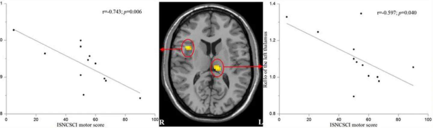
We found that the mean ReHo values of the left thalamus and the right insula were nega-tively correlated with the ISNCSCI motor scores in the SCI patients (r = -0.597,p= 0.040 and r = -0.743,p= 0.006, respectively) (Fig. 3).
Discussion
Widespread structural and functional abnormality in the brain has been reported in patients with SCI. In humans, similar to findings from experimental SCI, gray matter becomes atrophic and white matter integrity is reduced [5]. A spinal cord lesion affects primary sensorimotor areas connected to the lesioned area and can result in the reorganization of these and Table 1. Demographic and clinical information of patients with acute spinal cord injury.
NO Age at injury (years) Type of Injury Time of MRI after injury (days) AIS NLI ISNCSCI motor score
1 62 Fall 24 D T3 90
2 36 RTA 22 A T6 50
3 45 Fall 15 B L2 60
4 66 Fall 15 A C5 26
5 57 RTA 9 D C5 66
6 39 Fall 12 A C4 5
7 28 Fall 16 D C4 67
8 56 RTA 14 A T10 50
9 32 Fall 16 A T9 50
10 47 Fall 21 B T11 52
11 52 RTA 18 B C6 55
12 40 RTA 18 B C4 56
RTA, road traffic accident; AIS, American Spinal Injury Association Impairment Scale; grade A, complete, no motor or sensory function is preserved in the sacral segments S4 and S5; grade B, incomplete, sensory but not motor function is preserved below the neurological level and extends through the sacral segment S4-S5; grade C, incomplete, motor function is preserved below the neurological level, and more than half of key muscles below the neurological level have a muscle grade less than 3; grade D, incomplete, motor function is preserved below the neurological level and at least half of key muscles below the neurological level have a muscle grade of 3 or more; NLI, neurological level of injury; ISNCSCI, international standards for the neurological classification of spinal cord injury.
doi:10.1371/journal.pone.0118816.t001
Fig 1. Results of one-samplet-test on ReHo maps for healthy controls (CON, uppper) and patients with acute SCI (SCI, lower).Threshold was set to
p<0.05 with AlphaSim correction. The left side of the image corresponds to the right hemisphere of the brain. The underlying structure image is Ch2 image.
surrounding regions to compensate for sensorimotor loss [19,20]. However, almost all previ-ous studies have been performed in the chronic stage post-SCI. In the current study, ReHo was first employed to explore the changes in spontaneous brain activity and a voxel-wise analysis was then performed to detect brain regions with altered intrinsic brain activity in acute SCI pa-tients. The results indeed demonstrated that acute distant neuronal damage has an immediate impact on spontaneous brain activity.
ReHo measures the similarity or coherence of low frequency fluctuations (LFFs) within a given area based on hemodynamics. The LFF BOLD signal has been suggested to reflect spon-taneous neuronal activity [21–23]. Therefore, altered ReHo is most likely relevant to changes in the temporal aspects of regional spontaneous neural activity. Higher ReHo is thought to in-dicate greater temporal synchrony, whereas lower values are thought to represent decreased local coherence [24]. Results of one-samplet-test on ReHo maps of the two groups observed in Fig 2. Brain areas with altered ReHo compared with healthy controls (Alphasim corrected,palpha<0.05).The blue areas showed decreased ReHo in
acute SCI patients relative to healthy controls. The regions are the bilateral primary motor cortex/primary somatosensory cortex, bilateral supplementary motor area/ dorsal lateral prefrontal cortex, right inferior frontal gyrus, bilateral dorsal anterior cingulate cortex and bilateral caudate. The red areas showed increased ReHo in acute SCI patients. The regions include bilateral precuneus, the left inferior parietal lobe, the left brainstem/hippocampus, the left cingulate motor area, bilateral insula, bilateral thalamus and bilateral cerebellum. The left side of the image corresponds to the right hemisphere of the brain. The underlying structure image is Ch2 image.
the present study were consistent with previous studies [25–27], whereas abnormal ReHo was observed in cortical and subcortical brain regions in patients with acute SCI.
Compared with the controls, the acute SCI patients showed decreased ReHo prominently in brain regions involved in motor execution and cognitive control. First, brain regions with sig-nificantly decreased ReHo included the bilateral M1, SMA, right IFG and bilateral S1. M1, SMA and IFG are known to be critical in motor execution. The M1 is not only an executive motor area but also an area that contributes to movement sequence preparation [28] and motor control [29]. The SMA is thought to play a role in higher order activities related to movement, such as selection, preparation and sequencing of movements, as well as in move-ment execution [30]. The IFG programmes the sequential ordering of motor execution and is especially active in motor tasks of great difficulty or tasks which demand on selective attention [31]. Because sensorimotor function comprising motor function and sensory feedback from the spinal cord to the brain are expected to be greatly impaired or even absent in patients with Table 2. Brain areas with altered ReHo compared with healthy controls (Alphasim corrected,palpha<0.05).
Brain areas Hemisphere MNI coordinates (cluster
maxima, mm)
Peak T values Cluster size (voxels)
X Y Z
SCI>CON
Insula Left -33 3 15 6.03 90
Insula Right 39 9 15 4.88 36
Orbital middle frontal gyrus Right 33 42 -9 4.82 31
Inferior parietal lobule Left -42 -36 24 4.76 38
Superior temporal gyrus Right 72 -24 9 4.65 29
Cingulate motor area Left -12 3 45 4.39 44
Cerebellum/ lobe V and VI Bilateral 12 -63 -15 4.30 166
Thalamus Right 6 -12 15 4.21 77
Precuneus Left -21 -60 30 4.00 29
Thalamus Left -9 -24 9 3.96 43
Putamen Right 27 12 9 3.90 28
Precuneus Right 24 -54 42 3.86 28
Brainstem/hippocampus Left -21 -6 -9 3.81 69
Lingual gyrus Left -30 -54 -3 3.79 54
SCI<CON
Superior frontal gyrus/SMA/DLPFC Bilateral 15 42 51 5.92 783
Inferior frontal gyrus Right 48 30 -9 5.65 106
Inferior/middle temporal gyrus Right 48 -12 -24 5.17 73
Caudate Right 6 15 -3 5.15 110
Caudate Left -12 18 0 4.65 35
Precentral/postcentral gyrus/M1/S1 Left -27 -24 69 4.28 79
Inferior/middle occipital gyrus Left -24 -99 -9 4.25 86
Precentral/postcentral gyrus/M1/S1 Right 24 -27 69 4.24 62
Dorsal anterior cingulate cortex Bilateral -6 30 18 4.15 70
Inferior/middle occipital gyrus Right 24 -102 -9 4.14 33
Middle frontal gyrus Right 39 42 39 3.96 34
MNI: Montreal Neurological Institute; SCI, patients with acute SCI; CON, healthy controls; SMA, supplementary motor area; DLPFC, dorsal lateral prefrontal cortex; M1, primary motor cortex; S1, primary somatosensory cortex.
acute SCI, the decreased ReHo observed in the M1, SMA, IFG and S1 might reflect the motor execution deficits and a state of sensory deafferentation of these patients. Voxel-based mor-phometry (VBM) studies have found SCI patients with reduced gray matter volume in M1 and S1 [5,7,8], which might be the structural basis of the altered spontaneous brain activity in pa-tients with acute SCI. In addition, diffusion tensor imaging (DTI) has been used to evaluate the white matter microstructural changes following SCI. DTI analysis revealed structural abnor-malities in the brain regions with reduced gray matter volume as well as the corticospinal and corticopontine tracts of SCI subjects [5,6]. A rest SPECT study has also revealed regional blood flow reduction in M1, SMA, other movement-cortical areas and S1 in patients with SCI [32]. The decreased ReHo in S1 observed in our study is in line with neurophysiological evi-dence obtained from animal experiments: immediately (within minutes) after thoracic transec-tion of the spinal cord, the S1 cortical spontaneous activity at rest becomes strikingly slower and overall more silent [33].
Moreover, the patients with acute SCI showed decreased ReHo in other brain regions asso-ciated with cognitive control, i.e., the bilateral dACC, DLPFC and caudate. Cognitive control supports flexible behavior by selecting actions that are consistent with our goals and appropri-ate to our environment [34]. There is ample evidence that the control of any voluntary move-ment relies upon both higher-level cognitive and lower-level movemove-ment mechanisms [35]. Studies using functional neuroimaging techniques have related cognitive control to activity in the ACC and DLPFC [36]. Various functions have been ascribed to the dACC, including the modulation of attention or executive functions, complex motor control and the anticipation of cognitively demanding tasks [37]. Changes in DLPFC activity are often associated with the modulation of ACC activity and can be explained using several computational models that de-fine the lateral PFC, ACC and parietal cortex as the core components involved in executive control [38]. The caudate is consider to be involved in cognitive functions [39] playing a critical role in supporting the planning and execution of strategies and behavior required for complex goals [40]. The decreased spontaneous brain activity observed in regions within the cognitive control network may indicate a reduced level of cognitive control capability in patients with acute SCI, which may be an additional cause for the hypoactivation of the motor
execution network.
Precuneus, IPL and hippocampus are components of DMN [41]. The increased ReHo in these brain regions might suggest that the DMN is abnormal in patients with acute SCI. The other brain regions with increased ReHo in the acute SCI patients included the left CMA, bilat-eral insula, bilatbilat-eral thalamus and cerebellum, may be associated with the sensorimotor Fig 3. Correlations between the mean ReHo values of the affected regions and ISNCSCI motor scores in patients with acute SCI.Mean ReHo values of the left thalamus and the right insula were negatively correlated with the ISNCSCI motor scores.
compensatory reorganization related to both motor execution deficits and sensory deafferenta-tion. The CMA has been suggested to play a pivotal role in processing the information neces-sary to select voluntary actions in accordance with the subject’s internal and external requirements [42]. The human insula has been revealed map to the sensorimotor network of the brain. This area has been repeatedly demonstrated to be involved in various somato- and viscerosensory stimuli. In addition to this sensory processing, movement was elicited by electri-cal stimulation of this region in humans, which indicates that this region plays a role in sensori-motor processing [43]. The thalamus is a relay center subserving both sensory and motor mechanisms [44]. Clinical studies suggest that cerebellum lobes V and VI are principally en-gaged in motor control and somatosensory functions [45]. Strong additional activation of the thalamus and cerebellum has been shown using PET in patients with SCI during the perfor-mance of motor task [10]. The authors of this study assumed that when afferent input from the spinal cord is reduced, more complex processing of the remaining inputs leads to stronger acti-vation, or possibly to disinhibition, of the neuronal centers involved, i.e., the thalamus
and cerebellum.
Furthermore, the altered spontaneous brain activity of the left thalamus and right insula may reflect clinical outcomes. We found negative correlations between the ReHo values of the left thalamus and right insula and the ISNCSCI motor scores in the patients with acute SCI. As mentioned above, both the insula and thalamus play critical roles in integrating sensorimotor processing. The explanation of these negative correlations might be that more severe motor im-pairment leads to more evident compensatory reorganization. However, it is difficult to explain why there is a lateralization in these two brain regions.
Several limitations of our study should be mentioned. Because of the relatively low inci-dence, acuteness and severity of this disorder, only 12 male patients in the acute stage were in-cluded in the present study. Studies have demonstrated that the post-injury brain
reorganization may follow a dynamic time course [46,47]. Thus, another limitation was that we did not conduct this work as a longitudinal study because of low compliance. Finally, fur-ther analysis of function connectivity of the involved brain regions in the resting state should be conducted in future work.
In conclusion, the abnormal ReHo observed after remote spinal lesions demonstrated that even acute distant neuronal damage has an immediate impact on spontaneous brain activity. The spontaneous brain activity in brain regions associated with sensorimotor, cognitive control and DMN have changed in patients with acute SCI. Abnormal ReHo values in the left thalamus and right insula could serve as potential biomarkers for the assessment of the neuronal damage and the prediction of clinical outcomes in acute SCI.
Author Contributions
Conceived and designed the experiments: LZ GW FL. Performed the experiments: LZ GW. An-alyzed the data: FL. Contributed reagents/materials/analysis tools: LZ GW FL XZ JL ZW. Wrote the paper: LZ GW FL.
References
1. Dietz V, Curt A. Neurological aspects of spinal-cord repair: promises and challenges. Lancet Neurol. 2006; 5:688–694. PMID:16857574
2. Curt A, Alkadhi H, Crelier GR, Boendermaker SH, Hepp-Reymond MC, Kollias SS. Changes of non-af-fected upper limb cortical representation in paraplegic patients as assessed by fMRI. Brain. 2002; 125:2567–2578. PMID:12390981
4. Nardone R, Holler Y, Brigo F, Seidl M, Christova M, Bergmann J, et al. Functional brain reorganization after spinal cord injury: systematic review of animal and human studies. Brain Res. 2013; 1504:58–73.
doi:10.1016/j.brainres.2012.12.034PMID:23396112
5. Wrigley PJ, Gustin SM, Macey PM, Nash PG, Gandevia SC, Macefield VG, et al. Anatomical changes in human motor cortex and motor pathways following complete thoracic spinal cord injury. Cereb Cor-tex. 2009; 19:224–232. doi:10.1093/cercor/bhn072PMID:18483004
6. Guleria S, Gupta RK, Saksena S, Chandra A, Srivastava RN, Husain M, et al. Retrograde Wallerian de-generation of cranial corticospinal tracts in cervical spinal cord injury patients using diffusion tensor im-aging. J Neurosci Res. 2008; 86:2271–2280. doi:10.1002/jnr.21664PMID:18335542
7. Jurkiewicz MT, Crawley AP, Verrier MC, Fehlings MG, Mikulis DJ. Somatosensory cortical atrophy after spinal cord injury: a voxel-based morphometry study. Neurology. 2006; 66:762–764. PMID: 16534122
8. Henderson LA, Gustin SM, Macey PM, Wrigley PJ, Siddall PJ. Functional reorganization of the brain in humans following spinal cord injury: evidence for underlying changes in cortical anatomy. J Neurosci. 2011; 31:2630–2637. doi:10.1523/JNEUROSCI.2717-10.2011PMID:21325531
9. Alkadhi H, Brugger P, Boendermaker SH, Crelier G, Curt A, Hepp-Reymond MC, et al. What disconnec-tion tells about motor imagery: evidence from paraplegic patients. Cereb Cortex. 2005; 15:131–140.
PMID:15238440
10. Bruehlmeier M, Dietz V, Leenders KL, Roelcke U, Missimer J, Curt A. How does the human brain deal with a spinal cord injury? Eur J Neurosci. 1998; 10:3918–3922. PMID:9875370
11. Curt A, Bruehlmeier M, Leenders KL, Roelcke U, Dietz V. Differential effect of spinal cord injury and functional impairment on human brain activation. J Neurotrauma. 2002; 19:43–51. PMID:11852977 12. Hotz-Boendermaker S, Hepp-Reymond MC, Curt A, Kollias SS. Movement observation activates lower
limb motor networks in chronic complete paraplegia. Neurorehabil Neural Repair. 2011; 25:469–476.
doi:10.1177/1545968310389184PMID:21343526
13. Freund P, Weiskopf N, Ashburner J, Wolf K, Sutter R, Altmann DR, et al. MRI investigation of the sen-sorimotor cortex and the corticospinal tract after acute spinal cord injury: a prospective longitudinal study. Lancet Neurol. 2013; 12:873–881. doi:10.1016/S1474-4422(13)70146-7PMID:23827394 14. Zang Y, Jiang T, Lu Y, He Y, Tian L. Regional homogeneity approach to fMRI data analysis.
Neuro-image. 2004; 22:394–400. PMID:15110032
15. Wu T, Zang Y, Wang L, Long X, Li K, Chan P. Normal aging decreases regional homogeneity of the motor areas in the resting state. Neurosci Lett. 2007; 423:189–193. PMID:17709202
16. Liu H, Liu Z, Liang M, Hao Y, Tan L, Kuang F, et al. Decreased regional homogeneity in schizophrenia: a resting state functional magnetic resonance imaging study. Neuroreport. 2006; 17:19–22. PMID: 16361943
17. Oldfield RC. The assessment and analysis of handedness: the Edinburgh inventory. Neuropsycholo-gia. 1971; 9:97–113. PMID:5146491
18. Kirshblum SC, Waring W, Biering-Sorensen F, Burns SP, Johansen M, Schmidt-Read M, et al. Refer-ence for the 2011 revision of the International Standards for Neurological Classification of Spinal Cord Injury. J Spinal Cord Med. 2011; 34:547–554. doi:10.1179/107902611X13186000420242PMID: 22330109
19. Donoghue JP, Suner S, Sanes JN. Dynamic organization of primary motor cortex output to target mus-cles in adult rats. II. Rapid reorganization following motor nerve lesions. Exp Brain Res. 1990; 79:492–503. PMID:2340869
20. Jain N, Catania KC, Kaas JH. Deactivation and reactivation of somatosensory cortex after dorsal spinal cord injury. Nature. 1997; 386:495–498. PMID:9087408
21. Logothetis NK, Pauls J, Augath M, Trinath T, Oeltermann A. Neurophysiological investigation of the basis of the fMRI signal. Nature. 2001; 412:150–157. PMID:11449264
22. Pelled G, Goelman G. Different physiological MRI noise between cortical layers. Magn Reson Med. 2004; 52:913–916. PMID:15389942
23. Lu H, Zuo Y, Gu H, Waltz JA, Zhan W, Scholl CA, et al. Synchronized delta oscillations correlate with the resting-state functional MRI signal. Proc Natl Acad Sci U S A. 2007; 104:18265–18269. PMID: 17991778
24. Philip NS, Kuras YI, Valentine TR, Sweet LH, Tyrka AR, Price LH, et al. Regional homogeneity and resting state functional connectivity: associations with exposure to early life stress. Psychiatry Res. 2013; 214:247–253. doi:10.1016/j.pscychresns.2013.07.013PMID:24090510
26. Fang W, Lv F, Luo T, Cheng O, Liao W, Sheng K, et al. Abnormal regional homogeneity in patients with essential tremor revealed by resting-state functional MRI. PLoS One. 2013; 8:e69199. doi:10.1371/ journal.pone.0069199PMID:23869236
27. Song Y, Mu K, Wang J, Lin F, Chen Z, Yan X, et al. Altered spontaneous brain activity in primary open angle glaucoma: a resting-state functional magnetic resonance imaging study. PLoS One. 2014; 9: e89493. doi:10.1371/journal.pone.0089493PMID:24586822
28. Gerloff C, Corwell B, Chen R, Hallett M, Cohen LG. The role of the human motor cortex in the control of complex and simple finger movement sequences. Brain. 1998; 121 (Pt 9):1695–1709. PMID:9762958 29. Binkofski F, Fink GR, Geyer S, Buccino G, Gruber O, Shah NJ, et al. Neural activity in human primary
motor cortex areas 4a and 4p is modulated differentially by attention to action. J Neurophysiol. 2002; 88:514–519. PMID:12091573
30. Hiroshima S, Anei R, Murakami N, Kamada K. Functional localization of the supplementary motor area. Neurol Med Chir (Tokyo). 2014; 54:511–520. PMID:24305024
31. Liakakis G, Nickel J, Seitz RJ. Diversity of the inferior frontal gyrus—a meta-analysis of neuroimaging
studies. Behav Brain Res. 2011; 225:341–347. doi:10.1016/j.bbr.2011.06.022PMID:21729721 32. Cermik TF, Tuna H, Kaya M, Tuna F, Gultekin A, Yigitbasi ON, et al. Assessment of regional blood flow
in cerebral motor and sensory areas in patients with spinal cord injury. Brain Res. 2006; 1109:54–59.
PMID:16859656
33. Aguilar J, Humanes-Valera D, Alonso-Calvino E, Yague JG, Moxon KA, Oliviero A, et al. Spinal cord in-jury immediately changes the state of the brain. J Neurosci. 2010; 30:7528–7537. doi:10.1523/ JNEUROSCI.0379-10.2010PMID:20519527
34. Badre D. Cognitive control, hierarchy, and the rostro-caudal organization of the frontal lobes. Trends Cogn Sci. 2008; 12:193–200. doi:10.1016/j.tics.2008.02.004PMID:18403252
35. Mutha PK, Haaland KY. Cognitive aspects of motor control. Cortex. 2014; 57:299–300; discussion
306–298. doi:10.1016/j.cortex.2014.03.001PMID:24726068
36. MacDonald AW 3rd, Cohen JD, Stenger VA, Carter CS. Dissociating the role of the dorsolateral pre-frontal and anterior cingulate cortex in cognitive control. Science. 2000; 288:1835–1838. PMID: 10846167
37. Bush G, Luu P, Posner MI. Cognitive and emotional influences in anterior cingulate cortex. Trends Cogn Sci. 2000; 4:215–222. PMID:10827444
38. Procyk E, Goldman-Rakic PS. Modulation of dorsolateral prefrontal delay activity during self-organized behavior. J Neurosci. 2006; 26:11313–11323. PMID:17079659
39. Mestres-Misse A, Turner R, Friederici AD. An anterior-posterior gradient of cognitive control within the dorsomedial striatum. Neuroimage. 2012; 62:41–47. doi:10.1016/j.neuroimage.2012.05.021PMID: 22617829
40. Grahn JA, Parkinson JA, Owen AM. The cognitive functions of the caudate nucleus. Prog Neurobiol. 2008; 86:141–155. doi:10.1016/j.pneurobio.2008.09.004PMID:18824075
41. Long XY, Zuo XN, Kiviniemi V, Yang Y, Zou QH, Zhu CZ, et al. Default mode network as revealed with multiple methods for resting-state functional MRI analysis. J Neurosci Methods. 2008; 171:349–355.
doi:10.1016/j.jneumeth.2008.03.021PMID:18486233
42. Picard N, Strick PL. Imaging the premotor areas. Curr Opin Neurobiol. 2001; 11:663–672. PMID: 11741015
43. Kurth F, Zilles K, Fox PT, Laird AR, Eickhoff SB. A link between the systems: functional differentiation and integration within the human insula revealed by meta-analysis. Brain Struct Funct. 2010; 214:519–534. doi:10.1007/s00429-010-0255-zPMID:20512376
44. Herrero MT, Barcia C, Navarro JM. Functional anatomy of thalamus and basal ganglia. Childs Nerv Syst. 2002; 18:386–404. PMID:12192499
45. Stoodley CJ, Schmahmann JD. Functional topography in the human cerebellum: a meta-analysis of neuroimaging studies. Neuroimage. 2009; 44:489–501. doi:10.1016/j.neuroimage.2008.08.039PMID: 18835452
46. Jurkiewicz MT, Mikulis DJ, McIlroy WE, Fehlings MG, Verrier MC. Sensorimotor cortical plasticity dur-ing recovery followdur-ing spinal cord injury: a longitudinal fMRI study. Neurorehabil Neural Repair. 2007; 21:527–538. PMID:17507643