Rev. Col. Bras. Cir. 2016; 43(3):
INTRODUCTION
P
rostate cancer is the most common malignancy inmen. A large portion of the male population is sub-jected to screening tests, which makes early diagnosis in-creasingly frequent. Many of these patients are currently treated with laparoscopic radical prostatectomy (LRP) as a primary surgical approach aimed at cure1,2.
The vesicourethral anastomosis between the bladder neck and the membranous urethra for recon-struction of the lower urinary tract after removal of the prostate is a crucial point of LRP. Leakage of urine be-tween the anastomosis stitches in the postoperative peri-od is common, but is usually of low output and self-limit-ed, during two or three days3.
Persistent vesicourethral anastomotic leaks (PVAL) can be defined as significant urinary losses through the drain after the third postoperative day, usu-ally above 100 or 200 ml. It is a rare event, about which there is little published literature. However, its occurrence is of difficult control for the medical staff and patients,
prolonging hospital stay, and bringing risks of potentially serious complications.
The objective of this study is to analyze the results of a endoscopic, minimally invasive approach to control PVAL when conservative treatment fails, thus avoiding more invasive surgical procedures, such as repair by conventional open surgery or nephrostomy, options traditionally used as the last resort in such cases.
METHODS
A total of 620 patients with adenocarcinoma of the prostate, clinical stage T1c, and a mean age of 61 years, underwent transperitoneal laparoscopic radical prostatectomy (LRP). The vesicourethral anastomosis was made with wire 3-0 Monocryl, as described by Van Veltho-ven et al., without bladder neck plasty prior to the anas-tomosis3,4. Ten patients had persistent vesicourethral
anas-tomotic leaks (PVAL), with urine output by the perivesical drain of 100-400 ml in 24 hours, reaching 400-1100 ml on the second day after surgery. The fluids collected from the
DOI: 10.1590/0100-69912016003011
Artigo Original
Minimally invasive treatment of vesicourethral leak after
laparoscopic radical prostatectomy
Tratamento minimamente invasivo para fístula vesicouretral após prostatectomia
radical videolaparoscópica
Tiago Rivello elmoR1; mauRicio RubinsTein2; guilheRme lima3; anTonio cesaR cRuz3; Clovis
F
RagaT
enóRioP
eReiRa3;
iRineu RubinsTein2.1 - Escola de Medicina e Cirurgia da Universidade Federal do Estado do Rio de Janeiro (EMC-UNIRIO); 2 - Universidade Federal do Estado do Rio de Janeiro (UNIRIO), Rio de Janeiro, RJ, Brasil; 3 - Instituto de Medicina Integral Professor Fernando Figueira (IMIP), Recife, PE, Brasil.
185-188
A B S T R A C T
Objective: to describe our experience with a minimally invasive approach for persistent vesicourethral anastomotic leak (PVAL) after Laparoscopic Radical Prostatectomy (LRP). Methods: from 2004 to 2011, two surgeons performed LRP in 620 patients. Ten patients had PVAL, with initially indicated conservative treatment, to no avail. These patients underwent a minimally invasive operation, consisting of an endoscopically insertion of two ureteral catheters to direct urine flow, fixed to a new urethral catheter. We maintained the ureteral catheters for seven days on average to complete resolution of urine leakage. The urethral catheter was removed after three weeks of surgery. Results: the correction of urine leakage occurred within a range of one to three days, in all ten patients, without complications.
There were no stenosis of the bladder neck and urinary incontinence on long-term follow-up. Conclusion: the study showed that PVAL after laparoscopic radical prostatectomy can be treated endoscopically with safety and excellent results.
186
Rev. Col. Bras. Cir. 2016; 43(3):
tubes were consistent with urine after laboratory results. All patients underwent total abdomen computed tomog-raphy, which showed collection of fluid within the pelvis. The ureters were preserved and the bladder Folley cathe-ters were correctly positioned in the bladder. A retrograde cystogram was also carried out by urinary catheters in all ten patients, clearly showing contrast leak through the vesicourethral anastomosis (Figure 1).
Initially, conservative techniques had been used, such as traction and attachment of the bladder catheter to the patient’s thigh so that the catheter balloon occluded the urine leakage site, associated with a lower fluid intake. After the failure of these initial measures, these ten patients un-derwent endoscopic intervention for treatment of persistent urinary fistulas. The time interval between the LRP and the endoscopic reintervention ranged from three to nine days.
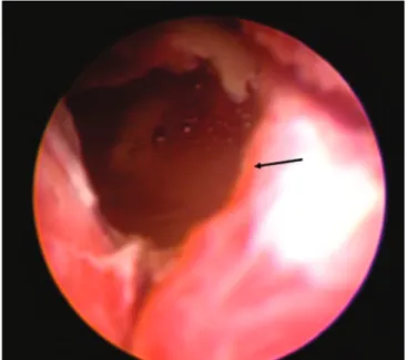
The procedure consisted of bilaterally placing ureteral catheters exteriorized alongside a new Folley catheter, to direct the urine out via the urethra and to re-duce the leakage through the fistula, allowing its closure. Initially, we carried out a urethrocystoscopy with a rigid, 19Fr cystoscope, under sedation and local anesthetic gel, allowing the exact identification of the fistula opening and its location relative to the ureteral ostia (Figure 2). Then 6Fr ureteral catheters were inserted bilaterally over a hydrophilic guidewire under radioscopic control and
externalized through the urethra, along with a new, 18 Folley bladder catheter, also placed in the bladder with the aid of a guidewire.
All patients underwent a control retrograde cystogram to verify the complete resolution of the urine leakage before removal of the ureteral catheters, which took place after seven days. The bladder catheter was re-moved three weeks after prostatectomy.
RESULTS
The resolution of the persistent vesicourethral anastomosis leak (PVAL) occurred within a range of one to three days for all ten patients. There were no periop-erative or immediate postopperiop-erative complications of the reintervention. The drains were removed after leakage become less than 50 ml per day (Table 1). There were no bladder neck stenosis or urinary incontinence after a mean follow up of 12 months (6-18 months).
DISCUSSION
Laparoscopic radical prostatectomy (LRP) is a procedure that requires great skill in making the vesicoure-thral anastomosis. Leakage of urine by the anastomosis is very common, but the persistent one is a rare event. The incidence of persistent vesicourethral anastomotic leaks (PVAL) after radical prostatectomy has been estimated at 0.9% to 2.5%. It is generally treated with ineffective con-servative measures, and when it requires a therapeutic ap-proach, procedures are very invasive for the patient, such as conventional open surgery or nephrostomy5.
To confirm the PVAL diagnosis there are several available exams. Conventional cystogram remains a very useful tool to this day, and allows to record the contrast ex-travasation with simple radiographic images after injection through the bladder catheter. However, images obtained by computed tomography can provide more information, especially with three-dimensional volume estimate, which allows to better define the conduct in cases where the flow rate through the fistula is not very high. Lee et al. found a statistically significant difference between the urinary leak detection rate by cystography using tomography and
con-Elmor
Minimally invasive treatment of vesicourethral leak after laparoscopic radical prostatectomy
185-188
187
Rev. Col. Bras. Cir. 2016; 43(3):
ventional cystography (80.4% vs. 54.3%). Therefore, even when conventional cystogram shows normal results, the leak can be detected by tomography6,7.
After the PVAL diagnosis, it is in the form of treatment that lies the difficulty for urologists. This is a complication that can indefinitely prolong patients’ hos-pitalization and presents risks of adverse developments, such as secondary infection by resistant germs.
Moinzadeh et al. previously presented a con-servative technique of continuous suction by the Folley catheter to help in these cases. However, all conservative techniques, including continuous suction catheter, have failed to correct the intraperitoneal fistula8.
In another study, the same group described the use of a percutaneous nefroureteral suction catheter hav-ing multiple fenestrae along its length and which allows the suction of urine, both from the bladder and from the renal pelvis. Thus, it forms a proximal urinary diversion, which allows the closure of the fistula. This minimally in-vasive technique avoids conducting bilateral nephrosto-mies or other reconstructive procedures, but also pres-ents complexity compared to the endoscopic procedure9.
Yossepowitch et al., from Urology Institute of the University of Tel Aviv, described their experience in treating PVAL after open radical prostatectomy, using the same endoscopic approach of our study. They treated 1,480 patients with open radical prostatectomy between 1996 and 2007. Seven patients had PVAL and underwent a rigid cystoscopy with a 19Fr cystoscope, followed by the bilateral placement of 5Fr ureteral catheters over a hydro-philic guidewire under fluoroscopic control. The average time between the intervention and removal of the pelvic drain (drain <50ml per day) was two days. The catheters were maintained for nine days on average. There was res-olution of the urinary fistulas in all seven patients, which was confirmed by control cystography10.
Our results were similar to the Yossepowitch group ones, with the same surgical technique. We be-lieve, therefore, that the persistent vesicourethral anas-tomotic leaks (PVAL) can be treated by endoscopically draining the urinary system, with ease and security. The procedure is an alternative, less aggressive approach than any other surgical treatment, with excellent results.
Table 1. Relationship between the high output of urine through the drain in patients with PVAL after LRP and the time of resolution of fistulas after endoscopic rapprochement.
Patient Interval between procedures
(with PVAL)
Fistula output (mean 24h)
Resolution after rapprochement (output <50ml)
1 9 days 400ml 24 hours
2 6 days 720ml 24 hours
3 3 days 950ml 48 hours
4 5 days 650ml 48 hours
5 6 days 450ml 48 hours
6 4 days 800 ml 72 hours
7 4 days 850ml 72 hours
8 7 days 560ml 24 hours
9 3 days 1100ml 72 hours
10 6 days 480ml 48 hours
PVAL: Persistent vesicourethral anastomotic leak.
Elmor
Minimally invasive treatment of vesicourethral leak after laparoscopic radical prostatectomy
185-188
188
Rev. Col. Bras. Cir. 2016; 43(3):
REFERENCES
1. Sociedade Brasileira de Urologia. Doenças da próstata: vença o tabu. Rio de Janeiro: Elsevier; 2003.
2. Brasil. Ministério da Saúde. Secretaria de Assistência à Saúde. Instituto Nacional de Câncer. Programa nacio-nal de controle do câncer da próstata: documento de consenso. Rio de Janeiro: INCA; 2002.
3. Van Velthoven RF, Ahlering TE, Peltier A, Skarecky DW, Clayman RV. Technique for laparoscopic running urethrovesical anastomosis: the single knot method. Urology. 2003;61(4):699-702.
4. van Velthoven RF. Laparoscopic radical prostatec-tomy: transperitoneal versus retroperitoneal approa-ch: is there an advantage for the patient? Curr Opin Urol. 2005;15(2):83-8.
5. Castillo OA, Alston C, Sanchez-Salas R. Persistent ve-sicourethral anastomotic leak after laparoscopic ra-dical prostatectomy: laparoscopic solution. Urology. 2009;73(1):124-6.
6. Lee HJ, Shin CI, Hwang SI, Jung SI, Kim SH, Lee SE, et al. MDCT cystography for detection of vesicoure-thral leak after prostatectomy. AJR Am J Roentgenol. 2008;191(6):1847-51.
7. Schoeppler GM, Buchner A, Zaak D, Khoder W, Staehler M, Stief CG, et al. Detection of urinary leakage after radical retropubic prostatectomy by contrast enhanced ultrasound - do we still need conventional retrograde cystography? BJU Int. 2010;106(11):1632-7.
8. Moinzadeh A, Abouassaly R, Gill IS, Libertino JA. Continuous needle vented foley catheter suction for urinary leak after radical prostatectomy. J Urol. 2004;171(6 Pt 1):2366-7.
9. Shah G, Vogel F, Moinzadeh A. Nephroureteral stent on suction for urethrovesical anastomotic leak after robot-assisted laparoscopic radical prostatectomy. Urology. 2009;73(6):1375-6.
10. Yossepowitch O, Baniel J. Persistent vesicourethral anastomotic leak after radical prostatectomy: a no-vel endoscopic solution. J Urol. 2010;184(6):2452-5.
Recebido em: 27/02/2016
Aceito para publicação em: 05/05/2016 Conflito de interesse: nenhum.
Fonte de financiamento: nenhuma.
Endereço para correspondência:
Tiago Rivello Elmor
E-mail: [email protected]
185-188
Elmor
Minimally invasive treatment of vesicourethral leak after laparoscopic radical prostatectomy
R E S U M O
Objetivo: descrever nossa experiência com uma abordagem minimamente invasiva para fístula de anastomose vesicouretral persistente (FAVP) após prostatectomia radical laparoscópica (PRL). Métodos: de 2004 a 2011, 620 pacientes foram submetidos à prostatectomia rad-ical laparoscópica realizada por dois cirurgiões. Dez pacientes apresentaram FAVP e o tratamento conservador foi inicialmente indicado sem sucesso. Esses pacientes foram submetidos a uma reoperação minimamente invasiva, por via endoscópica, com inserção de dois cateteres ureterais para direcionar o fluxo urinário, fixados a um novo cateter uretral. Os cateteres ureterais foram mantidos por sete dias, em média, até a completa resolução do vazamento de urina. O cateter uretral foi removido após três semanas da cirurgia. Resultados: a correção do vazamento de urina ocorreu dentro de um intervalo de um a três dias em todos os dez pacientes, sem complicações. Não foram observadas estenose de colo vesical ou incontinência urinária após acompanhamento em longo prazo. Conclusão: o estudo mostrou que a FAVP após a prostatectomia radical laparoscópica pode ser tratada por via endoscópica com segurança e excelentes resultados.