Comparative analysis of endoscopic and histopathological
features of superficial elevated lesions resected by endoscopic
mucosal resection in the distal and proximal colon
Análise comparativa dos aspectos endoscópicos e histopatológicos das lesões
superficialmente elevadas ressecadas por mucosectomias no cólon distal e proximal
Artur Adolfo PArAdA1,2; CArmen AustrAliA PArede mArCondesribAs1
, filAdelfio euClydes VenCo3; José Celso Ardengh2; mAriAnA
AmArAl reis2; mAtheus degioVAni1,2; miguel reynAldo VArCA-neto2; nildede rodrigues diger1,2; roberto el ibrAhim3, KAssiA
fernAn-dA CordoVA1, mAríliA dA Cruz fAgundes1, hAmilton moreirA1, luiz fernAndo Kubrusly1
1 - Programa de Pós-Graduação em Princípios da Cirurgia, Faculdade Evangélica do Paraná/Hospital Universitário Evangélico de Curitiba/ Instituto de Pesquisas Médicas, Curitiba, PR, Brasil; 2 - Serviço de Endoscopia Gastrointestinal do Hospital Nove de Julho, São Paulo, SP, Brasil; 3 - Laboratório Diagnóstika Patologia Cirúrgica e Citologia, São Paulo, SP, Brasil.
INTRODUCTION
C
olorectal cancer is one of the major medical problems throughout the world1-3. The proportion of proximal carcinomas has increased relative to the distal ones4 and the protection afforded by colonoscopy in the proximal colon is lower than in the distal5. Many studies suggest that the interval carcinomas, which are diagnosed few years after colonoscopy, are more proximal, and whose diagnosis was missed, among various factors, due to the development from superficial lesions5,6. At the same time, endoscopists started to increasingly diagnose non-polypoid or superficial lesions, and Laterally Spreading Tumor (LST) lesions7.In recent years the serrated lesions, which are often superficially elevated lesions, have been subject of much discussion, but there are still some disagreements
and difficulties in their diagnosis and characterization by endoscopists and pathologists. Even so, they are now considered important, representing 7.5% to 30% of all colorectal carcinomas according to several authors8.
This work, emphasizing the histogenesis of col-orectal cancer, aimed to study mucosectomy specimens of superficially elevated lesions of 1 cm or more in diam-eter, comparing their endoscopic and pathologic features in the distal and proximal colon.
METHODS
The study was retrospective, cross-section-al, observationcross-section-al, in which we evaluated the specimens from patients undergoing colonoscopies with endoscop-ic mucosectomies of superfendoscop-icially elevated lesions with A B S T R A C T
Objective: to compare endoscopic and histopathologic features of superficial, elevated lesions with one or more centimeters in diame-ter, diagnosed by videocolonoscopy on the distal and proximal colon, and subjected to mucosal resection.Methods: we conducted a retrospective, cross-sectional, observational study involving 8,075 videocolonoscopies. From this total, we evaluated 166 mucosectomies in 145 patients with superficial, elevated lesions with a diameter equal to or greater than 1cm. Results: the lesion prevalence was lower in G1 than in G2 (34.9% vs. 65%). The mean age, gender distribution and size (1.9cm in G1 versus 2.0cm in G2, p=0.921) were similar. There was no difference of mucosal surfaces in relation to the location (p=0.575). Considering Intraepithelial neoplasias, both the low grade, high grade (including carcinomas) and hyperplasic ones showedd no difference (p=0.527), nor did the neoplastic lesions when divided into serrated and non-serrated (p=0.124). Excluding 13 hyperplastic lesions and two carcinomas, 124 (82.1%) were non-serra-ted and 27 (17.9%), serranon-serra-ted. Conclusion: were found no significant differences between endoscopic and histopathological aspects of superficial, elevated lesions of 1cm or more in diameter in distal colon compared with the proximal, when resected by mucosectomy. Although not significant, there was a tendency of association between the location of the lesion and the presence of serrated features.
more than 1 cm in diameter, in the period from 2011 to 2014 at the Hospital Nove de Julho, São Paulo, SP, Brazil. The examinations were performed with sedation controlled by an anesthesiologist and the lesions were resected by the mucosectomy technique. We considered both the 0-LST and 0-IIa lesions (classification of Paris) as superficially elevated lesions. We classified their surfaces as granular, nodular and smooth after chromoendoscopy with indigo carmine 0.4%. Lesions 2-2.5 cm in diameter were resected en bloc and with more than 2.5-3 cm by fragment (piecemeal) mucosectomy.
We stretched the specimens in cardboard with needles and fixed them in 10% formalin. Subsequently, we cut every 2mm, and microscopically examined them with hematoxylin and eosin. We divided the invasion of the submucosa into three levels: sm1, sm2 and sm3. We histologically classified lesions by the Vienna classifi-cation. Lesions with cellular atypia and cytoarchitecture were subdivided in serrated and not serrated, keeping hyperplastic polyps as a separate group.
Finally, evaluations of serrated lesions were re-considered in accordance with the guidance of the World Health Organization (WHO), including hyperplastic polyps with 1cm or more in diameter as serrated lesions8-10. These, when with atypia (sessile serrated adenomas/polyps – SSA/ Ps) were considered low-grade or high-grade intraepithe-lial neoplasias, serrated type (IN-LG-S or IN-HG-S). The adenomatous lesions were considered as low-grade or high-grade intraepithelial neoplasias, or as non-serrated low-grade or high-grade intraepithelial neoplasia.
The splenic flexure is considered proximal by some authors11 and distal by others12. In this work, we considered the splenic flexure, descending and sigmoid colon as distal (G1), and the cecum, ascending and trans-verse colon as proximal (G2).
We described the results of the variables evalu-ated in the study as frequencies and percentages (qualita-tive variables). For the age of the patients, we present the mean values and standard deviation. For the comparison of lesions’ locations (distal and proximal) with the qualitative variables, we used the Fisher exact test or chi-square test. W considered p values < 0.05 as statistically significant. Data were analyzed with the software IBM SPSS Statistics v.20.
RESULTS
We carried out 166 mucosectomies (2% of to-tal colonoscopies) in 145 patients. Of these, 52 (35.9%) had 58 lesions in G1. The mean age was 64.2 years (+/- 12.3 years, 33-89); 25 (48.1%) were men and 27 (51.9%), women. In G2, 100 individuals (69%) had 108 lesions, with a mean age of 65.4 years (+/- 10.2 years, 38-89); 45 (45%) were men and 55 (55%), women.
Table 1 shows the frequencies and percentage of lesions according to ranges in size at each location.
When comparing the size of lesions in the dis-tal colon with the proximal one, there was no significant difference (p=0.921). We also show the frequencies and percentage of lesions according to the surface’s charac-teristics at each location. There was no significant differ-ence between the locations of the injury and the surface’s characteristics (p=0.575).
For the statistical test, we considered low grade, high-grade and hyperplastic intraepithelial neopla-sia. The two cases of carcinoma were grouped with the high-grade intraepithelial neoplasias (Table 2).
When comparing G1 with G2 lesions, there was no significant difference (p=0.527). Table 3 shows the comparison between non-serrated intraepithelial neoplasias and serrated ones, excluding the hyperplastic polyps (n=13) and carcinomas (n=2).
In Table 3 we divided these 151 lesions in two groups, considering them as serrated and non-ser-rated and showed the results restricted to lesions with low-grade and high-grade intraepithelial neoplasia. In all analyzes, there were no significant differences be-tween the types of lesions and their locations bebe-tween G1 or G2.
Tables 4 and 5 present the frequencies and percentages according to the surface and size, with the histopathology, at each location.
DISCUSSION
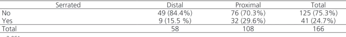
The sessile serrated adenomas / polyps (SSA/Ps) predominate in the right colon13. They tend to be flat in the proximal colon (75%), 64% being bigger than 5mm, and 17% bigger than 10mm. The proximal hyperplastic polyps with more than 5mm could be classified as ser-rated8, while most non-serrated or adenomatous lesions would occur in the left colon14. Authors state that the proximal hyperplastic polyps, greater than 10mm may be considered sessile serrated adenomas. With these crite-ria, we would have had 41 serrated lesions, nine (22%) in G1 and 32 (78%) in G2. In G1, they correspond to 15.5% of 58, and in G2, including the sm1 serrated car-cinoma, 29.6% of 108. Of the 125 non-serrated lesions, 49 (39.2%) occurred in G1 and 76 (60.8 %), in G2. The statistical test (0.061) was not significant, but showed a tendency to the association between the location and the presence of serrated lesions.
In an American study of 100 serrated lesions, 88 were located in the colon proximal to the splenic flex-ure. The vast majority were superficially elevated lesions15. This paper presents similar data, ie, of the 41 serrated
(including hyperplastic polyps), 32 (78%) were located in the G2 and nine (21.9%), in G1.
A Japanese multicenter study analyzed 154 hy-perplastic polyps with 1cm or more in diameter. Most sessile serrated adenomas with atypia (SSA/Ps), 90 of 107 (84.1%), and those who were not sessile serrated adenomas (non-SSA/Ps, thus without atypia), 33 of 47 (70.2%) were in the proximal colon16, as observed in this study, where 77.7% of serrated lesions with atypia (21 of 27 lesions) and 76.9% of serrated lesions without atypia (10 of 13 hyperplastic le-sions) were located in the proximal colon (G2).
A very large series of a Korean group of 28,544 colonoscopies diagnosed 143 sessile serrated adenomas / polyps (SSA/Ps) (0.5%). Of these, 123 (86%) were proxi-mal to the splenic flexure and nine (6.3%) had more than 1cm in diameter17. In the literature, the average size of sessile serrated adenomas was 8.1mm16. We diagnosed 27 sessile serrated adenomas with 1cm or more in diam-eter, six in G1 (22.2%) and 21 in G2 (77.7%).
In this series, with these criteria, four of 41 serrated lesions (9.7%) and 31 of 125 non-serrated (24.8%) had high-grade IN or sm1carcinomas. In G1, of the 49 non-serrated le-sions, 11 had high-grade IN or sm1 carcinomas (22.4%) and
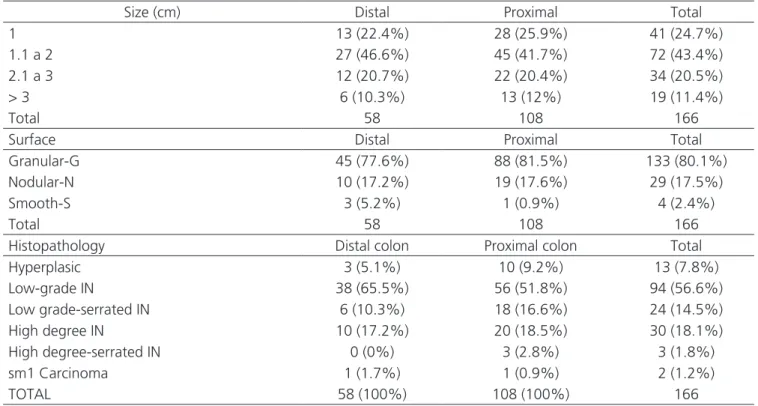
Table 1. Frequencies and percentages of lesions according to ranges of sizes, surfaces, and histopathology in the distal and proximal colon.
Size (cm) Distal Proximal Total
1 13 (22.4%) 28 (25.9%) 41 (24.7%)
1.1 a 2 27 (46.6%) 45 (41.7%) 72 (43.4%)
2.1 a 3 12 (20.7%) 22 (20.4%) 34 (20.5%)
> 3 6 (10.3%) 13 (12%) 19 (11.4%)
Total 58 108 166
Surface Distal Proximal Total
Granular-G 45 (77.6%) 88 (81.5%) 133 (80.1%)
Nodular-N 10 (17.2%) 19 (17.6%) 29 (17.5%)
Smooth-S 3 (5.2%) 1 (0.9%) 4 (2.4%)
Total 58 108 166
Histopathology Distal colon Proximal colon Total
Hyperplasic 3 (5.1%) 10 (9.2%) 13 (7.8%)
Low-grade IN 38 (65.5%) 56 (51.8%) 94 (56.6%)
Low grade-serrated IN 6 (10.3%) 18 (16.6%) 24 (14.5%)
High degree IN 10 (17.2%) 20 (18.5%) 30 (18.1%)
High degree-serrated IN 0 (0%) 3 (2.8%) 3 (1.8%)
sm1 Carcinoma 1 (1.7%) 1 (0.9%) 2 (1.2%)
TOTAL 58 (100%) 108 (100%) 166
none in the serrated lesions. In G2, 20 of the 76 non-serrated lesions (26.3%) and four serrated (4/32 = 16.7%) were high-grade intraepithelial neoplasia or sm1carcinomas.
In a Brazilian publication, it was shown that le-sions larger than 1 cm tend to be pedunculated, with ad-enomatous component, and patients over 50 years of age are more likely to present sessile polyps in the proximal colon18. In an American study with 2400 patients, 10% of diagnosed polyps were serrated. The right colon lesions, when compared by size, were more likely to be dysplas-tic19. In this study we diagnosed 41 serrated lesions in 166 mucosectomies’ specimens (24.7%), 55 being lesions with atypia in G1 (94.8% ) and 98 in G2 (90.4%), with no sta-tistical difference between the two groups.
In a Korean study of 47 proximal serrated lesions, 43 were slightly elevated lesions, and of these, nine were at high risk, two with dysplasia and seven with diameter greater than 10mm. The average size was 6mm20. In this study, 32 were slightly elevated lesions in G2, all with 1 cm or more in
diameter, and four were high-grade IN or sm1 carcinomas. Recent publications of few cases series (n=12)21 demonstrated that even small serrated lesions may have invasive carcinoma, with sizes between 8.5 and 11.3 mm, suggesting malignant transformations are rare, but fast. This rapid progression aspect was not confirmed in an-other study, in which the average age of patients with sessile serrated adenomas was 61, of the sessile serrated adenomas with high-grade atypia, 72, and of the cancer related to sessile serrated adenomas, 7622.
A Japanese research evaluated 141 serrated lesions, 107 being slightly elevated lesions, preferably in the right colon (81.8%), with an average size of 13 mm, with intramucosal carcinoma in 13.6% (3/22 SSA/Ps)23. In this study, considering the high degree IN as intramucosal carcinoma, we found three lesions of 27 sessile serrat-ed adenomas (11.1%) and one sm1 carcinoma, which would total four carcinomas in 28 SSA/Ps (14 2%), with an average size of 14 mm, all in G2.
Table 2. Histopathological aspects of the sample (n = 166).
Histopathology Distal Proximal Total
Low-grade IN 44 (75.9%) 74 (68.5%) 118 (71.1%)
High-grade IN and carcinoma* 11 (19%) 24 (22.2%) 35 (21.1%)
Hyperplastic polyp 3 (5.2%) 10 (9.3%) 13 (7.8%)
Total 58 108 166
* Two cases of adenocarcinoma (one distal non-serrated on and one proximal, serrated)
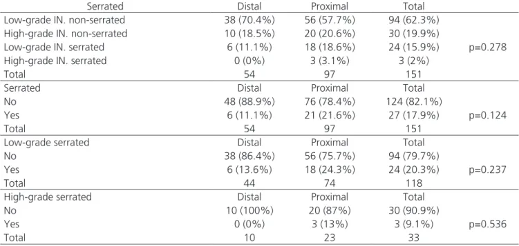
Table 3. Serrated and non-serrated lesions and intraepithelial neoplasia.
Serrated Distal Proximal Total
Low-grade IN. non-serrated 38 (70.4%) 56 (57.7%) 94 (62.3%)
p=0.278
High-grade IN. non-serrated 10 (18.5%) 20 (20.6%) 30 (19.9%)
Low-grade IN. serrated 6 (11.1%) 18 (18.6%) 24 (15.9%)
High-grade IN. serrated 0 (0%) 3 (3.1%) 3 (2%)
Total 54 97 151
Serrated Distal Proximal Total
No 48 (88.9%) 76 (78.4%) 124 (82.1%)
p=0.124
Yes 6 (11.1%) 21 (21.6%) 27 (17.9%)
Total 54 97 151
Low-grade serrated Distal Proximal Total
No 38 (86.4%) 56 (75.7%) 94 (79.7%)
p=0.237
Yes 6 (13.6%) 18 (24.3%) 24 (20.3%)
Total 44 74 118
High-grade serrated Distal Proximal Total
No 10 (100%) 20 (87%) 30 (90.9%)
p=0.536
Yes 0 (0%) 3 (13%) 3 (9.1%)
Total 10 23 33
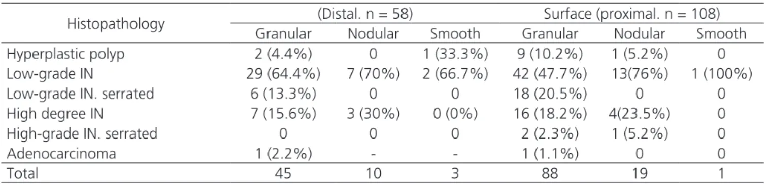
The submucosal invasion index for lateral spread-ing lesions with homogeneous surfaces is very low (< 2%), even in large lesions, while in those with mixed surfaces, with larger nodules, this ratio is higher (up to 7%)24. The two cases of carcinoma in this series occurred lesions with granular sur-face (1.5% of 133), one being serrated with 1 cm in G2, and the other non-serrated, with a 2.5 cm diameter in G1.
Sessile serrated adenomas and traditional ses-sile adenomas (TSA) have been considered precancerous neoplastic lesions and the hyperplastic polyp, void of malignant potential. However, one author considers the hyperplastic polyp with more than 1 cm also with ma-lignant potential24. Sessile serrated adenomas with obvi-ous dysplasia present, according to some authors, with an estimated higher probability to evolve to cancer than conventional adenomas (5.3% versus 2.2%).
The progression of sessile serrated adenoma to cancer would be faster than that of conventional ade-nomas. Progress to invasive carcinoma has already been shown to take place in eight months. The data suggest that the serrated sessile adenomas may be present for many years with few changes; however, they may rapidly progress to invasive carcinomas, even without dysplasia and with less than 10mm in diameter25.26.
New technologies can help to better distinguish hyperplastic lesions from the serrated and not serrated le-sions and to determine the most adequate procedure to adopt in each case during colonoscopy27.28.
In recent years, serrated lesions were also in-cluded in the colonoscopy follow-up recommendation. Nevertheless, it is not yet clear whether the size of 10mm – used to define conventional adenomas as advanced –
Table 5. Histopathology and lesion size in the distal and proximal colon.
Histopathology
Lesion size (cm) (Distal. n = 58)
Lesion size (cm) (Proximal. n = 108)
1 1.1 a 2 2.1 a 3 > 3 1 1.1 a 2 2.1 a 3 > 3
Hyperplastic polyp 2
(15.4%) 1 (3.7%)
1 (3.6%)
8 (17.8%)
1 (7.7%)
Low-grade IN 10
(76.9%)
19 (70.4%)
7 (58.3%)
2 (33.3%)
15 (53.6%)
21 (46.7%)
13 (59.1%)
7 (53.8%)
Low-grade IN. serrated 1
(7.7%)
2 (7.4%)
1 (8.3%)
2 (33.3%)
7 (25%)
9 (20%)
1 (4.5%)
1 (7.7%)
High-grade IN 5
(18.5%) 3 (25%)
2 (33.3%)
2 (7.1%)
7 (15.6%)
7 (31.8%)
4 (30.8%)
High-grade IN. serrated 2
(7.1%)
1 (4.5%)
Adenocarcinoma 1
(8.3%)
1 (3.6%)
Total 13 27 12 6 28 45 22 13
IN= intraepithelial neoplasia
Table 4. Histopathology and surface features of lesions in the distal and proximal colon
Histopathology (Distal. n = 58) Surface (proximal. n = 108)
Granular Nodular Smooth Granular Nodular Smooth
Hyperplastic polyp 2 (4.4%) 0 1 (33.3%) 9 (10.2%) 1 (5.2%) 0
Low-grade IN 29 (64.4%) 7 (70%) 2 (66.7%) 42 (47.7%) 13(76%) 1 (100%)
Low-grade IN. serrated 6 (13.3%) 0 0 18 (20.5%) 0 0
High degree IN 7 (15.6%) 3 (30%) 0 (0%) 16 (18.2%) 4(23.5%) 0
High-grade IN. serrated 0 0 0 2 (2.3%) 1 (5.2%) 0
Adenocarcinoma 1 (2.2%) - - 1 (1.1%) 0 0
Total 45 10 3 88 19 1
should also be applied to serrated sessile adenomas28. In conclusion, there were no significant differ-ences between the endoscopic and histopathological as-pects of superficially elevated lesions with more than 1cm
in diameter resected by mucosectomy from the distal colon compared with the proximal one. Although not significant, there is a tendency to the association between the location of the lesion and the presence of serrated features.
Tabela 6. Results according to the criteria of the World Health Organization (WHO).
Serrated Distal Proximal Total
No 49 (84.4%) 76 (70.3%) 125 (75.3%)
Yes 9 (15.5 %) 32 (29.6%) 41 (24.7%)
Total 58 108 166
p=0.061
REFERENCES
1. Snover DC. Update on the serrated pathway to col-orectal carcinoma. Hum Pathol. 2011;42(1):1-10. 2. Baxter NN, Goldwasser MA, Paszat LF, Saskin R, Urbach
DR, Rabeneck L. Association of colonoscopy and death from colorectal cancer. Ann Intern Med. 2009;150(1):1-8. 3. Nahas SC, Nahas CSR, Bustamante-Lopez LA, Pinto
RA, Marques CFS, Campos FG, et al. Prognostic factors of surgically-treated patients with cancer of the right colon: a ten years’ experience of a single universitary institution. ABCD, arq bras cir dig. 2015;28(1):3-7. 4. Caldarella A, Crocetti E, Messerini L, Paci E. Trends in
colorectal incidence by anatomic subsite from 1985 to 2005: a population-based study. Int J Colorectal Dis. 2013;28(5):637-41.
5. Brenner H, Chang-Claude J, Seiler CM, Rickert A, Hoffmeister M. Protection from colorectal cancer after colonoscopy: a population-based, case-control study. Ann Intern Med. 2011;154(1):22-30.
6. Laiyemo AO, Doubeni C, Sanderson AK 2nd, Pinky PF, Badurdeen DS, Doria-Rose VP, et al. Likelihood of missed and recurrent adenomas in the proxi-mal versus the distal colon. Gastrointest Endosc. 2011;74(2):253-61.
7. Lambert R, Tanaka S. Laterally spreading tumors in the colon and rectum. Eur J Gastroenterol Hepatol. 2012;24(10):1123-34.
8. Anderson JC. Pathogenesis and management of ser-rated polyps: current status and future directions. Gut Liver. 2014;8(6):582-9.
9. Rex DK, Ahnen DJ, Baron JA, Batts KP, Burke CA, Burt RW, et al. Serrated lesions of the colorectum: review and recommendations from an expert panel. Am J Gastroenterol. 2012;107(9):1315-29.
10. Snover DC, Ahnen DJ, Burt RW, Odze RD. Serrated polyps of the colon and rectum and serrated polyp-osis. In: Bosman FT, Carneiro F, Hruban RH, editors. WHO classification of tumours of the digestive sys-tem. Lyon, France: IARC; 2010. p.160-5.
R E S U M O
Objetivo: comparar aspectos endoscópicos e histopatológicos de lesões superficialmente elevadas, com um ou mais centímetros de diâmetro, diagnosticadas por videocolonoscopias e ressecadas por mucosectomias do cólon distal com as do cólon proximal. Métodos:
estudo foi retrospectivo, transversal, observacional, envolvendo 8075 videocolonoscopias. Avaliou-se 166 mucosectomias em 145 paci-entes com lesões superficialmente elevadas com diâmetro igual ou maior do que 1cm. Resultados: a prevalência de lesões foi menor no G1 do que no G2 (34,9% x 65%). A média de idade, a distribuição por sexo e o tamanho (1,9cm no G1 e 2cm no G2, p=0,921) foram semelhantes. Não houve diferenças das superfícies em relação à localização (p=0,575). Considerando neoplasia intraepitelial de baixo grau, neoplasia intraepitelial de alto grau (incluindo carcinomas) e hiperplásicas, não houve diferença (p=0,527), assim como quando foram divididas as lesões neoplásicas em serrilhadas e não serrilhadas (p=0,124). Excluindo-se 13 lesões hiperplásicas e duas com carcinomas, 124 (82,1%) foram não serrilhadas e 27 (17,9%) serrilhadas. Conclusão: não foram observadas diferenças significativas entre os aspectos endoscópicos e os histopatológicos das lesões superficialmente elevadas, com 1cm ou mais de diâmetro, ressecadas por mucosectomia do cólon distal em relação ao proximal. Embora não significante, há tendência à associação entre a localização da lesão e a presença de características serrilhadas.
11. Rustagi T, Rangasamy P, Myers M, Sanders M, Vazi-ri H, Wu GY, et al. Sessile serrated adenomas in the proximal colon are likely to be flat, large and occur in smokers. World J Gastroenterol. 2013;19(32):5271-7. 12. Benedix F. Kube R. Meyer F, Schmidt U, Gastinger
I, Lippert H; Colon/Rectum Carcinomas (Primary Tu-mor) Study Group. Comparison of 17,641 patients with right- and left-sided colon cancer: differences in epidemiology, perioperative course, histology, and survival. Dis Colon Rectum. 2010;53(1):57-64. 13. Abdeljawad K, Vemulapalli KC, Kahi CJ, Cummings
OW, Snover DC, Rex DK. Sessile serrated polyp prev-alence determined by a colonoscopist with a high le-sion detection rate and an experienced pathologist. Gastrointest Endosc. 2015;81(3):517-24.
14. Noffsinger AE. Serrated polyps and colorectal can-cer: new pathway to malignancy. Annu Rev Pathol. 2009;4:343-64.
15. Raju GS, Vadyala V, Slack R, Krishna SG, Ross WA, Lynch PM, et al. Adenoma detection in patients un-dergoing a comprehensive colonoscopy screening. Cancer Med. 2013;2(3):391-402.
16. Shida Y, Ichikawa K, Fujimori T, Fujimori Y, Tomita S, Fujii T, et al. Differentiation between sessile serrated adenoma/polyp and non-sessile serrated adenoma/ polyp in large hyper plastic polyp: a Japanese collab-orative study. Mol Clin Oncol. 2012;1(1):53-8. 17. Kim HY, Kim SM, Seo JH, Park EH, Kim N, Lee DH.
Age-specific prevalence of serrated lesions and their subtypes by screening colonoscopy: a retrospective study. BMC Gastroenterol. 2014;14:82.
18. Silva SM, Rosa VF, Santos ACN, Almeida RM, Oliveira PG, Sousa JB. Influência da idade do paciente e do tamanho dos pólipos colorretais nos achados histopa-tológicos. ABCD, arq bras cir dig. 2014;27(2):109-13. 19. Qumseya BJ, Coe S, Wallace MB. The effect of polyp lo-cation and patient gender on the presence of dysplasia in colonic polyps. Clin Transl Gastroenterol. 2012;3:e20. 20. Lee CK, Kim YM, Shim JJ, Jang JY. Prevalence of
proximal serrated polyps and conventional adeno-mas in an asymptomatic average-risk screening pop-ulation. Gut Liver. 2013;7(5):524-31.
21. Fujita K, Yamamoto H, Matsumoto T, Hirahashi M,
Gushima M, Kishimoto J, et al. Sessile serrated ad-enoma with early neoplastic progression: a clinico-pathologic and molecular study. Am J Surg Pathol. 2011;35(2):295-304.
22. Lash RH, Genta RM, Schuler CM. Sessile serrated ad-enomas: prevalence of dysplasia and carcinoma in 2139 patients. J Clin Pathol. 2010;63(8):681-6. 23. Hasegawa S, Mitsuyama K, Kawano H, Arita K,
Maeyama Y, Akagi Y, Watanabe Y, et al. Endoscop-ic discrimination of sessile serrated adenomas from other serrated lesions. Oncol Lett. 2011;2(5):785-9. 24. Hiraoka S, Kato J, Fujiki S, Kaji E, Morikawa T,
Murakami T, et al. The presence of large serrated polyps increases risk for colorectal cancer. Gastroen-terol. 2010;139(5):1503-10.
25. Menacho AM, Reimann A, Hirata LM, Ganzerella C, Ivano FH, Sugisawa R. Double-blind prospective ran-domized study comparing polyethylene glycol to lac-tulose for bowel preparation in colonoscopy. ABCD, arq bras cir dig. 2014;27(1):9-12.
26. Kagueyama FMN, Nicoli FM, Bonatto MW, Orso IRB. Importance of biopsies and histological evaluation in patients with chronic diarrhea and normal colonos-copies. ABCD, arq bras cir dig. 2014;27(3):184-7. 27. Saul C, Prolla JC, Silva VD, Teixeira CR, Parada AA.
Morphometric digital measurement of the luminal opening area of colonic crypts (pits) can differentiate the adenomas from other colonic lesions. Arq Gas-troenterol. 2009;46(2):107-10.
28. Moss A, Bourke MJ, Williams SJ, Hourigan LF, Brown G, Tam W, et al. Endoscopic mucosal resection out-comes and prediction of submucosal cancer from advanced colonic mucosal neoplasia. Gastroenterol-ogy. 2011;140(7):1909-18.
Recebido em: 27/02/2016
Aceito para publicação em: 28/04/2016 Conflito de interesse: nenhum.
Fonte de financiamento: nenhuma.
Endereço para correspondência: Artur Adolfo Parada