One Dose of
Staphylococcus aureus
4C-Staph
Vaccine Formulated with a Novel
TLR7-Dependent Adjuvant Rapidly Protects Mice
through Antibodies, Effector CD4
+
T Cells,
and IL-17A
Francesca Mancini1,2¤a, Elisabetta Monaci1¤a, Giuseppe Lofano1,3¤b, Antonina Torre1, Marta Bacconi1,4¤a, Simona Tavarini1¤a, Chiara Sammicheli1¤a, Letizia Arcidiacono1¤a, Bruno Galletti1¤a, Donatello Laera1¤a, Michele Pallaoro1¤a, Giovanna Tuscano1¤a, Maria Rita Fontana1¤a, Giuliano Bensi1¤a, Guido Grandi1¤c, Silvia Rossi-Paccani1¤a,
Sandra Nuti1¤a, Rino Rappuoli1¤a, Ennio De Gregorio1¤a, Fabio Bagnoli1¤a, Elisabetta Soldaini1¤a
*, Sylvie Bertholet1¤a
1Novartis Vaccines and Diagnostics, S.r.l., Research Center, Siena, Italy,2Department of Biomedical Sciences, University of Padua, Padua, Italy,3Department of Biology and Biotechnologies“Charles Darwin”, University of Rome“La Sapienza”, Rome, Italy,4Department of Biotechnology, Chemistry and Pharmacy, University of Siena, Siena, Italy
¤a Current address: GlaxoSmithKline Vaccines S.r.l., Siena, Italy
¤b Current address: Ragon Institute of MGH, MIT and Harvard, Cambridge, Massachusetts, United States of America
¤c Current address: Centre for Integrative Biology, University of Trento, Trento, Italy *[email protected]
Abstract
A rapidly acting, single dose vaccine againstStaphylococcus aureuswould be highly ben-eficial for patients scheduled for major surgeries or in intensive care units. Here we show that one immunization with a multicomponentS.aureuscandidate vaccine, 4C-Staph, for-mulated with a novel TLR7-dependent adjuvant, T7-alum, readily protected mice from death and from bacterial dissemination, both in kidney abscess and peritonitis models, outperforming alum-formulated vaccine. This increased efficacy was paralleled by higher vaccine-specific andα-hemolysin-neutralizing antibody titers and Th1/Th17 cell
responses. Antibodies played a crucial protective role, as shown by the lack of protection of 4C-Staph/T7-alum vaccine in B-cell-deficient mice and by serum transfer experiments. Depletion of effector CD4+T cells not only reduced survival but also increasedS.aureus
load in kidneys of mice immunized with 4C-Staph/T7-alum. The role of IL-17A in the con-trol of bacterial dissemination in 4C-Staph/T7-alum vaccinated mice was indicated byin vivoneutralization experiments. We conclude that single dose 4C-Staph/T7-alum vaccine promptly and efficiently protected mice againstS.aureusthrough the combined actions of antibodies, CD4+effector T cells, and IL-17A. These data suggest that inclusion of an
adjuvant that induces not only fast antibody responses but also IL-17-producing cell-OPEN ACCESS
Citation:Mancini F, Monaci E, Lofano G, Torre A, Bacconi M, Tavarini S, et al. (2016) One Dose of
Staphylococcus aureus4C-Staph Vaccine Formulated with a Novel TLR7-Dependent Adjuvant Rapidly Protects Mice through Antibodies, Effector CD4+T Cells, and IL-17A. PLoS ONE 11(1): e0147767. doi:10.1371/journal.pone.0147767
Editor:John S Tregoning, Imperial College London, UNITED KINGDOM
Received:October 12, 2015
Accepted:January 7, 2016
Published:January 26, 2016
Copyright:© 2016 Mancini et al. This is an open access article distributed under the terms of the
Creative Commons Attribution License, which permits unrestricted use, distribution, and reproduction in any medium, provided the original author and source are credited.
Data Availability Statement:All relevant data are within the paper and its Supporting Information files.
Funding:This work was supported by internal funding by Novartis Vaccines and Diagnostics S.r.l. The funder provided support in the form of salaries for all authors but did not have any additional role in the study design, data collection and analysis, decision to publish, or preparation of the manuscript.
mediated effector responses could efficaciously protect patients scheduled for major sur-geries or in intensive care units.
Introduction
Staphylococcus aureusinfections of the bloodstream or deep wound are a serious complication of major surgeries, including cardiothoracic and orthopedic surgery, resulting in significant morbidity and mortality [1,2].S.aureusis also the most commonly isolated microorganism from patients in intensive care units, which have mortality rates that reach 60% [3]. Due to methicillin-resistantS.aureusinfections, up to one third of patients diagnosed withS.aureus
bacteremia succumb even when treated with appropriate antibiotic therapy [4]. Therefore, a vaccine that provides rapid protection againstS.aureusduring the post-operative or intensive care period would address an important unmet medical need.
We recently developed a four-componentS.aureusvaccine (4C-Staph) consisting of HlaH35L, EsxAB, FhuD2, and Csa1A recombinant proteins [5]. HlaH35Lis a genetically
detoxi-fied mutant ofα-hemolysin (Hla) [6], a highly conserved exotoxin that plays a prominent role in early stages of invasive infections disrupting epithelial and endothelial barriers, contributing to pathogen-associated mortality [6,7]. Immunization with HlaH35Lpartially protected mice
against staphylococcal pneumonia, peritonitis, and skin infections inducing functional anti-bodies neutralizing the lytic activity of native Hla [8–10]. Remarkably, however, Hla neutraliza-tion was not sufficient to eradicateS.aureusinfection, suggesting that additional antigens are required for an efficacious vaccine [9]. EsxAB is a fusion of the two ESAT-6-like secreted viru-lence factors EsxA and EsxB associated to abscess formation, which may facilitate persistence and spread of the pathogen in the infected host [11–13]. FhuD2 is a lipoprotein involved in iron uptake and in early stages of invasiveS.aureusinfection [14–16], while Csa1A is a putative lipoprotein whose role in pathogenesis is under investigation [17]. We have recently shown that two doses of 4C-Staph vaccine formulated with alum protected against a panel of epidemi-ologically relevantS.aureusstrains in kidney abscess, peritonitis, skin, and pneumonia mouse models ofS.aureusinfection [5].
Adjuvants enhance and accelerate adaptive immune responses toward a co-administered antigen, while also directing the quality of the immune response [18,19]. Three major types of cell-mediated effector immunity meant to optimally respond to distinct threats have been iden-tified: type 1 protects against intracellular pathogens and comprises IFN-γ-producing cells (e.g. Th1 cells), type 2 protects against helminths and comprises IL-4/IL-13-producing cells (e.g. Th2), while type 3 protects against extracellular bacteria and fungi and comprises IL-17-pro-ducing cells (e.g. Th17) [20]. Aluminum salts-based adjuvants, which are included in many licensed vaccines, preferentially induce type 2 responses while agonists of Toll-like receptors (TLRs), a family of receptors that recognize pathogen-associated molecular patterns [21,22], induce mainly type 1 responses [18]. The potential of small molecule immune potentiators (SMIPs) targeting TLR7, an endosomal TLR that recognizes single-stranded RNAs, as vaccine adjuvants has been shown in pre-clinical settings, especially for Imiquimod. However, systemic activation induced by these SMIPs has posed safety issues [23]. The rational design of SMIP.7-10, a novel TLR7 agonist that can be stably adsorbed to alum (T7-alum), minimized systemic exposure and inflammation while retainedin vivopotency [24].
Therefore, in this study, we investigated whether single immunization with 4C-Staph/ T7-alum conferred prompt protection againstS.aureusinfection. We found that this was indeed the case and we elucidated the immune mechanisms involved.
Materials and Methods
Vaccine formulations and animal immunizations
Vaccine antigens, HlaH35L, EsxAB, FhuD2, and Csa1A, were amplified by PCR fromS.aureus
NCTC8325 strain and cloned as tagless constructs. Antigens were purified and adsorbed to alum by incubation with aluminum hydroxide (alum) with or without SMIP-7.10 (T7), at pH 6.5–7.0 and osmolality 0.308±0.060 Osm/Kg, with slow stirring for a few hours at room tem-perature (RT) [5]. One dose of vaccine (100μl) consisted in 10μg of each antigen adsorbed to
2 mg/ml alum alone (4C-Staph/alum) or together with 50μg SMIP-7.10 (4C-Staph/T7-alum).
Formulations were adjusted to physiological ranges of pH and isotonicity and had an endo-toxin content˂1.5 European U/ml.
Five-week old female BALB/c (Charles River Laboratories) or JH(Taconic) mice were used.
For active immunization, mice were immunized i.m. (50μl/hind leg quadriceps). Control mice
received equal amounts of alum, or T7-alum. For passive immunization, pools of sera were prepared from BALB/c mice immunized once with 4C-Staph/T7-alum (immune serum) or T7-alum (control serum) by 32 days. Five-week-old naïve BALB/c mice were injected with 150μl of immune or control serum into the tail vein the day before challenge withS.aureus.
Bacterial inoculum and challenge
S.aureusNewman strain (kindly provided by Professor Schneewind, University of Chicago) was grown in tryptic soy broth (Difco Laboratories) at 37°C shaking at 250 rpm to an optical density at 600 nm (OD600) of 2. After centrifugation at 3,320 x g for 10 min., bacteria were
washed with 50 ml of phosphate-buffered saline (PBS, pH 7.4; Invitrogen) and then diluted in PBS to obtain the required infectious dose/mouse. Inocula were verified experimentally by plat-ing on tryptic soy agar and enumeration of the colony formplat-ing units (CFU) the day after.
Kidney abscess model: mice were challenged i.v. with 100μl of a sublethal dose of bacteria
(~ 2×10⁷CFU). After 4 days, mice were euthanized; both kidneys were removed, homogenized in pool in 2 ml PBS, 2-fold serially diluted, and plated in duplicate for CFU counts.
Peritonitis model: mice were challenged i.p. with 100μl of a LD80of bacteria (~5 x 10⁸
CFU). Mice were monitored daily for 15 or 30 days, as indicated. Mice that survived were euthanized and CFU in kidneys were enumerated as described above.
Quantification of 4C-Staph-specific and Hla-neutralizing antibodies
Vaccine-specific antibody titers were measured by Luminex (Luminex1200 TM) at a fixed serum dilution. HlaH35L, FhuD2, EsxAB and Csa1A purified proteins were covalently
conju-gated to the free carboxyl groups of xMAP multi-analyte microspheres (Luminex Corpora-tion). Antigen-specific antibodies were revealed using PE-labeled secondary antibodies. For total IgG, median fluorescence intensity (MFI) values were converted into relative Luminex units (RLU)/ml using a mouse serum as standard.
SpectraMax1
340PC384 Absorbance Microplate Reader (Molecular Devices). Percent hemoly-sis was calculated using as denominator the OD540obtained following lysis of rabbit RBCs
without mouse serum. Hla neutralizing titers were expressed as the reciprocal of the serum dilution inhibiting 50% of Hla hemolysis (ED50) given by native Hla, calculated using a "log
(agonist) vs. normalized response" nonlinear regression curve (GraphPad Software, Inc.).
Intracellular cytokine staining
Splenocytes were isolated 12 days after vaccination, plated at 1-2x10⁶cells/well in 96-well plates, and stimulated with vaccine proteins (HlaH35L, EsxAB, FhuD2 and Csa1A, 10μg/ml
each) together with anti-CD28 and anti-CD49d mAb (2μg/ml each, BD Biosciences) at 37°C
for 16–18 h. Brefeldin A (5μg/ml) was added for the last 4 h. The cells were then stained with
Live/DeadYellow (Invitrogen), fixed and permeabilized with Cytofix/Cytoperm (BD Biosci-ences), washed in Perm/Wash buffer (BD BiosciBiosci-ences), incubated with anti-CD16/CD32 Fc block (BD Biosciences) for 20 min at RT, and stained with the following fluorochrome-conjugated mAbs anti: CD3-PerCP Cy5.5, CD4-V500, IFN-γ-PE, IL-2-APC, TNF-AF700, CD44-V450 (BD Pharmingen), CD8-PE Texas Red (Invitrogen), IL-17A-PE Cy7, IL-4-AF488 and IL-13-AF488 (eBioscience) in Perm/Wash buffer (BD Biosciences) for 20 min at RT, washed twice in Perm/Wash buffer, suspended in PBS. Samples were acquired on a LSRII spe-cial order flow cytometer (BD Biosciences) and T-cell responses were analyzed using FlowJo software (TreeStar) applying the gating strategy described inS1 Fig.
Depletion of effector CD4
+T cells
To deplete effector CD4+T cells, mice were vaccinated on day 0, injected twice i.p. with 100μg
of a rat anti-CD4 mAb (clone GK1.5; Areta International) or isotype-matched control antibody (isot. ctr., rat IgG2b; R&D System) on day 6 and 8, and challenged withS.aureuson day 10. CD4+cell depletion efficiency was evaluated by flow cytometry on heparinized blood taken the day before infection (day 9). For this purpose, RBCs were lysed with RBCs lysis buffer (Biole-gend). White blood cells were stained with Live/Dead Yellow (Invitrogen), incubated with anti-CD16/CD32 Fc block (BD Biosciences), and stained with: anti-CD3-PerCP Cy5.5, (BD Phar-mingen), anti-CD8-PE TexasRed (Invitrogen), and anti-CD4-Pacific Blue (Invitrogen) in PBS 0.1% BSA for 20 min at room temperature. Cells were fixed with Cytofix (BD Biosciences), sus-pended in PBS and analyzed by LSR II flow cytometer (BD Biosciences). No CD4+T cells were detected in the peripheral blood indicating that depletion occurred efficiently (S2 Fig).
In vivo
cytokine neutralization
To neutralize IL-17A and/or IFN-γin vivo, mice immunized with 4C-Staph/T7-alum or T7-alum were injected i.p. with 100μg of a neutralizing mouse anti-IL-17A mAb (clone 17F3;
BioXCell) and/or a neutralizing rat anti-IFN-γmAb (clone XMG 1.2; BioXCell) every other day starting from 3 h prior toS.aureuschallenge (day 12 after vaccination) until sacrifice. Control mice received the same amount of isot. ctr. antibodies (mouse IgG1 and/or rat IgG1; BioXCell).
Statistical analysis
CD4+T cells in response to 4C-Staph/alum or 4C-Staph/T7-alum vaccination were performed using a Student'sttest and a partial permutation test. For total IgG titers, Kruskall-Wallis and Dunn's multiple comparisons tests were used to evaluate differences in vaccine-specific anti-body titers. Statistical analyses were performed using GraphPad Prism 6 software (GraphPad Software, Inc.) and SPICE version 5.1 (http://exon.niaid.nih.gov).
Ethics statement
All animal studies were carried out in compliance with current Italian legislation on the care and use of animals in experimentation (Legislative Decree 26/2014, authorizations 185/2011-B and 249/2011-B approved by the Italian Ministry of Health) and with the Novartis Animal Welfare Policy and Standards (authorizations AWB 201105 and AWB 201106). After infection, mice were monitored daily using a dedicated clinical score system based on the following parameters: piloerection, hunched posture, decreased mobility, rolling/twisting movements and weight loss. Mice were euthanized by cervical dislocation when they exhibited either one of the following humane endpoints: a loss of 20% bodyweight and/or moderate to severe signs of rolling/twisting movements that were pre-established in agreement with Novartis Animal Welfare Policies. All animals were provided environmental enrichment and food and water were offeredad libitum.
Results
One dose of 4C-Staph/T7-alum vaccine confers rapid and efficacious
protection against
S
.
aureus
We tested the efficacy of one dose of adjuvanted 4C-Staph vaccines in the mouse kidney abscess and peritonitis models. For this purpose, BALB/c mice were vaccinated i.m. with 4C-Staph/T7-alum or 4C-Staph/alum vaccines. Control groups of mice received T7-alum or alum. Twelve days after immunization, mice were challenged withS.aureusNewman strain.
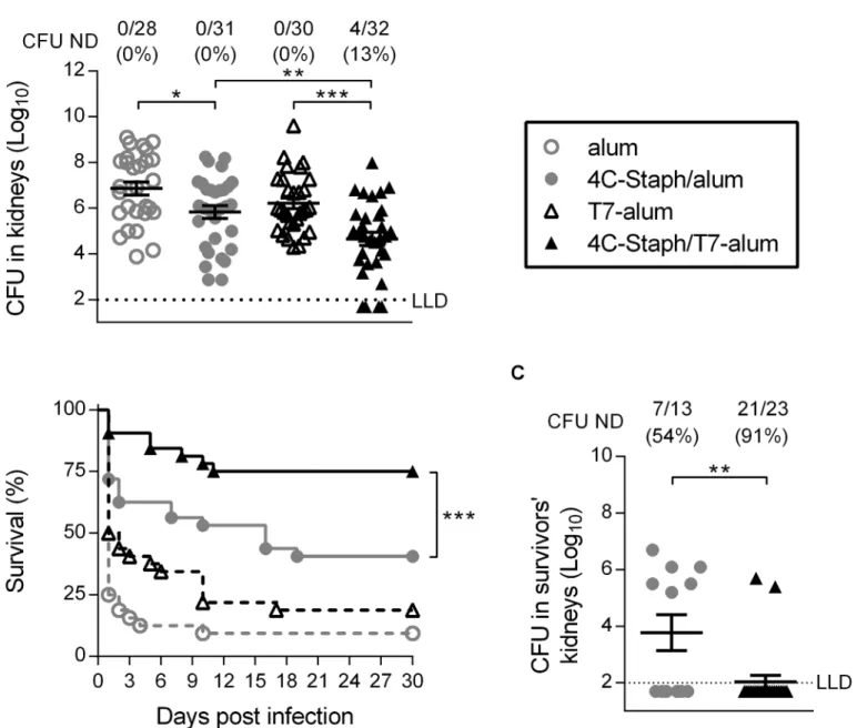
In the kidney abscess model, mice were challenged i.v. with a sublethal dose of bacteria (2x10⁷CFU). After 4 days mice were sacrificed and CFU in kidneys were enumerated. As shown inFig 1A, 4C-Staph/T7-alum-vaccinated mice showed a 1.55 log10reduction in CFU/
kidneys compared to T7-alum-treated control mice (4.66 ± 0.28 and 6.21 ± 0.23 log10,
mean ± SEM CFU/kidneys, respectively,p= 0.0003), and a 1.14 log10reduction in
CFU/kid-neys compared to 4C-Staph/alum-immunized animals (5.80 ± 0.28 log10CFU/kidneys,
p= 0.0080). Remarkably, 4 out of 32 mice vaccinated with 4C-Staph/T7-alum had no detect-able bacteria in kidneys compared to 0 out of 31 mice in the 4C-Staph/alum group.
In the peritonitis model, which can be used to study the spread of bacteria into the blood-stream [9], mice were challenged i.p. (5x10⁸CFU) with a lethal dose ofS.aureusand their sur-vival was monitored for 30 days. One month after infection, 75% of mice immunized with 4C-Staph/T7-alum survivedS.aureuschallenge compared to 41% of mice immunized with 4C-Staph/alum (p= 0.0058,Fig 1B). Furthermore, mice vaccinated with 4C-Staph/T7-alum that survived had significantly less bacteria in kidneys (1.7 log10reduction,p= 0.0091) and
91% of them had no detectable bacteria in kidneys as compared to survivors in the 4C-Staph/ alum group (54%,Fig 1C).
Fig 1. One dose of 4C-Staph/T7-alum vaccine protects better than 4C-Staph/alum in kidney abscess and peritonitis models ofS.aureusinfection. BALB/c mice were immunized once i.m. with 4C-Staph/T7-alum or 4C-Staph/alum. Control mice were injected with T7-alum or alum alone. After 12 days, mice were challenged withS.aureusNewman strain. (A) Kidney abscess model. Mice (n = 28–32) were injected i.v. with 2 x 10⁷CFU. Four days later, both kidneys of each mouse were homogenized in pool and CFU enumerated. Each symbol represents one mouse, and data are the merge of three independent experiments. Mean±SEM of each group are shown. The dotted line indicates the lower limit of detection (LLD).*p<0.05,**p<0.01 by one-way ANOVA and Sidak's multiple comparisons test. Number of survivors with non-detectable CFU (CFU ND) in kidneys/total number of survivors and corresponding percentages are reported above the graph. (B-C) Peritonitis model. Mice (n = 32) were injected i.p. with 5 x 10⁸CFU. Survival was monitored for 30 days after challenge. Data are the merge of three independent experiments.***p<0.001 by Log-rank test. (C) Thirty days afterS.aureusinfection, survivors were euthanized, both kidneys were homogenized and CFU enumerated. Each symbol represents one mouse. Mean±SEM of each group is shown.**p<0.01 by unpaired Studentttest, one-tailed. Number of survivors with CFU ND in kidneys/total number of survivors and corresponding percentages are reported above the graph.
4C-Staph/T7-alum vaccine rapidly induces functional and protective
antibodies
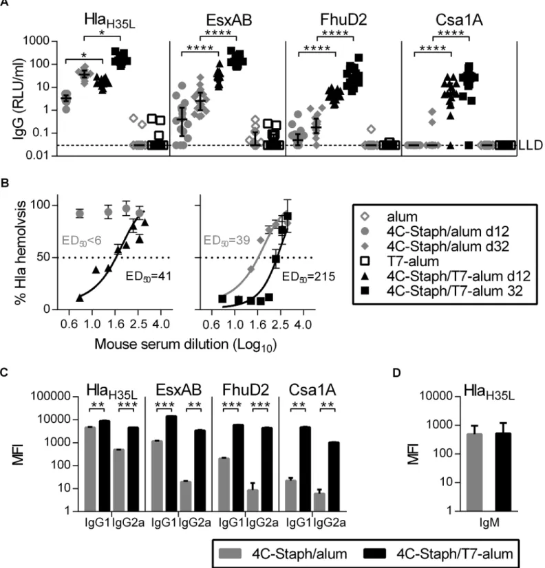
The induction of antibodies specific for each component of the vaccine by 4C-Staph/T7-alum and 4C-Staph/alum was evaluated 12 and 32 days after vaccination. IgG specific for HlaH35L,
EsxAB, FhuD2, and Csa1A were detected in sera from all mice vaccinated with 4C-Staph/ T7-alum by day 12 and further increased by day 32, with the exception of one mouse that had no detectable anti-Csa1A IgG (Fig 2A). In contrast, only HlaH35L-specific IgG were detected in
all sera from mice immunized with 4C-Staph/alum 12 days after vaccination, while EsxAB-and FhuD2-specific IgG were detected only in some sera, EsxAB-and Csa1A-specific IgG were not found. By day 32, HlaH35L- and EsxAB-specific IgG were found in sera from all 4C-Staph/alum
vaccinated mice, while FhuD2- and Csa1A-specific IgG were found only in some sera. Higher levels of IgG1 and IgG2a specific for each vaccine component were found in sera of mice vacci-nated with 4C-Staph/T7-alum as compared to sera of mice vaccivacci-nated with 4C-Staph/alum (Fig 2C), while no differences in vaccine-specific IgM levels were observed (Fig 2D). In addi-tion, Hla neutralizing titers were measurable in sera from mice vaccinated with 4C-Staph/ T7-alum by day 12 (ED50= 41) and increased by 5-fold at day 32 (ED50= 215), while
Hla-neutralizing activity was detected in sera from mice vaccinated with 4C-Staph/alum at d32 (ED50= 39) but not at day 12 (ED50<6,Fig 2B). Altogether, these data show that single dose
of 4C-Staph/T7-alum vaccine readily induced not only seroconversion of vaccinated mice against each 4C-Staph component but also Hla-neutralizing antibodies.
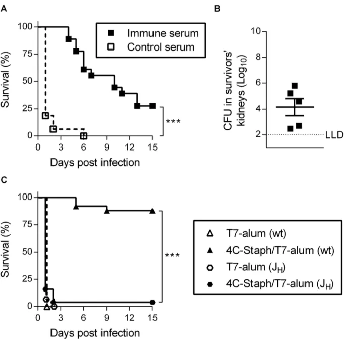
To determine if antibodies induced by 4C-Staph/T7-alum vaccination were protective against a lethal challenge withS.aureus, sera collected from mice 32 days after immunization with 4C-Staph/T7-alum (or T7-alum, as control) were passively transferred into naïve mice one day before i.p. infection. All mice that received immune serum were protected from death during the first 3 days after infection (Fig 3A). Thereafter, the survival gradually declined to become 28% at day 15, when all survivors had bacteria in kidneys (Fig 3B). In contrast, 100% of mice that received control sera died by day 6 after infection. Finally, we showed that 4C-Staph/T7-alum vaccination required B cells to protect mice againstS.aureusinfection by using JHmice, which lack mature B cells and antibodies [25]. Within two days after i.p.
chal-lenge, 96% of JHmice vaccinated with 4C-Staph/T7-alum succumbed to the infection while
88% of BALB/c mice were still alive 15 days after infection (p<0.0001,Fig 3C).
Overall these data suggest that antibodies induced by 4C-Staph/T7-alum vaccine are neces-sary and sufficient to protect against early death caused byS.aureusi.p. infection.
4C-Staph/T7-alum induces vaccine-specific Th1 and Th17 cells
4C-Staph/T7-alum (114.6 to 318.7) by 32 days indicates a difference significant withp<0.05. Bars represent SEM. (C) Vaccine-specific IgG1 and IgG2a. Columns represent median MFI with interquartile range of pooled sera from vaccinated mice (n = 16, same pools as in B) bled at d32.**p<0.01,
***p<0.001 by unpaired Studentttest, two-tailed. (D) HlaH35L-specific IgM. Columns represent median MFI with interquartile range of sera from vaccinated mice (n = 12) bled at d12. IgM specific for EsxAB, FhuD2 and Csa1A were at the limit of detection (data not shown). Data shown are the merge of two independent experiments.
doi:10.1371/journal.pone.0147767.g002
Fig 3. One dose of 4C-Staph/T7-alum induces protective antibodies.Sera from mice immunized with 4C-Staph/T7-alum (immune serum), or T7-alum as negative control (control serum), by 32 days were pooled and injected i.v. (150μl/mouse) in naïve BALB/c mice (n = 16) 24 h before i.p. challenge withS.
aureus.(A) Survival was monitored for 15 days post challenge. Data are the merge of two independent experiments.***p<0.001 by Log-rank test.(B) Fifteen days afterS.aureusinfection, survivors were euthanized, both kidneys were homogenized and CFU enumerated. Each symbol represents one mouse.(C) B cell/antibody-deficient JHmice, or BALB/c (wt) as control, were immunized with 4C-Staph/T7-alum or T7-alum alone. Twelve days after vaccination, mice (n = 25 for 4C-Staph/T7-alum; n = 15 for T7-alum) were challenged i.p. withS.aureusand their survival was monitored for 15 days. Data are the merge of four independent experiments.***p<0.001 by Log-rank test.
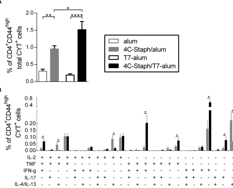
TNF+IFN-γ+) and Th17 cells (IL-17A+), but lower percentages of Th2 cells (IL-4+/IL-13+, IL-2+IL-4+/IL-13+and IL-2+TNF+IL-4+/IL-13+) than 4C-Staph/alum (Fig 4B). Interestingly, 4C-Staph/T7-alum also induced higher percentages of polyfunctional IL-2+TNF+IFN-γ+CD4 T cells.
These results demonstrated that one dose of 4C-Staph/T7-alum induced higher frequencies of vaccine-specific cytokine-producing CD4+T cells and polarized the CD4+T cells more towards a Th1/Th17 phenotype as compared to 4C-Staph/alum.
Fig 4. Magnitude and quality of vaccine-specific CD4+T-cell responses induced by 4C-Staph/T7-alum and 4C-Staph/alum.Splenocytes from single
mice (n = 16) vaccinated with 4C-Staph/T7-alum, 4C-Staph-alum, T7-alum or alum by 12 days were stimulated or not with vaccine antigensin vitro, stained and analyzed by intracellular cytokine staining. CD4+CD44highT cells producing IL-2, TNF, IL-4/IL-13,
IFN-γor IL-17A were identified (seeS1 Figfor gating strategy). The response of unstimulated cells was subtracted from that of stimulated cells. Data are the merge of four independent experiments. (A) Percentages of CD4+CD44highT cells producing any combination of IL-2, TNF, IL-4/IL-13,
IFN-γor IL-17A in response to vaccine protein stimulation (CD4+CD44hightotal CYT+cells) were calculated applying Boolean gates. Bars represent mean±SEM.
*p<0.05,**p<0.01,***p<0.001 by one-way ANOVA and Sidak post-test. (B) Percentages of CD4+CD44highT cells producing IL-2, TNF, IL-4/IL-13,
IFN-γand/or IL-17A in each of the possible combinations (CD4+CD44highCYT+) in response to vaccine proteins stimulation calculated applying Boolean gates. No cells expressing more than 3 cytokines at once were detected. Bars represent mean±SEM.*p<0.05 by unpaired Studentttest, two-tailed, and a partial permutation test.
CD4
+T cells induced by 4C-Staph/T7-alum contribute to the protection
against
S
.
aureus
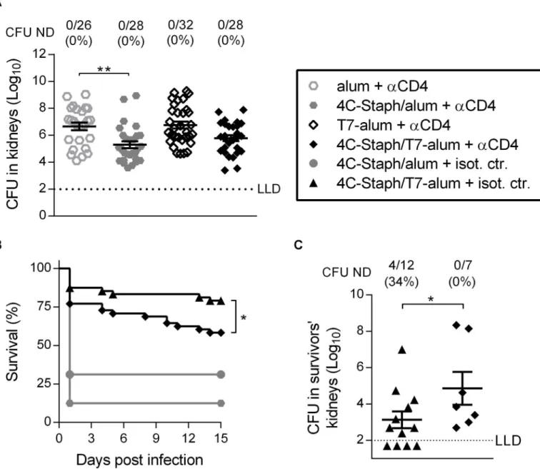
To assess if vaccine-specific CD4+T cells play a role in protection, mice were treated with an anti-CD4 mAb 6 and 8 days after immunization with 4C-Staph/alum or 4C-Staph/T7-alum to allow T cell-dependent antibody production, but to eliminate CD4+T cells with effector func-tion beforeS.aureuschallenge (S2 Fig). In the kidney abscess model, the depletion of CD4+T cells abolished the protection conferred by 4C-Staph/T7-alum, as evidenced by the numbers of CFU found in kidneys that was not different in the T7-alum vaccine group (p= 0.145,Fig 5A). In contrast, CD4+T cell depletion did not impact the efficacy of 4C-Staph/alum (statistically significant decrease in CFU in kidneys vs. mice vaccinated with alum,p= 0.002). In the perito-nitis model, depletion of CD4+T cells significantly decreased the survival of mice vaccinated with 4C-Staph/T7-alum (58% survival vs. 79% of mice treated with isotype control antibody,
p= 0.034), but not of mice vaccinated with 4C-Staph/alum (13% survival vs. 31% of mice treated with isotype control antibody,p= 0.207,Fig 5B). In addition, depletion of CD4+T cells in mice vaccinated with 4C-Staph/T7-alum caused an increase of 1.74 log10in the CFU/kidneys
of survivors as compared to isotype control antibody-treated mice (p= 0.0372,Fig 5C). Overall, these data show that effector CD4+T cells contribute to the protection conferred by one dose of 4C-Staph vaccine adjuvanted with T7-alum both in the kidney abscess and perito-nitis models, although other mechanisms of protection were involved, in agreement with the role of humoral immunity that we have shown inFig 3.
Neutralization of IL-17A increases the bacterial load in kidneys of mice
vaccinated with 4C-Staph/T7-alum upon i.p.
S
.
aureus
infection
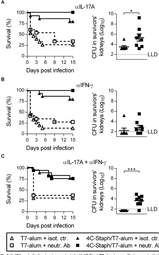
Depletion of effector CD4+T cells had no effect on protection conferred by vaccination with 4C-Staph/alum in either the kidney abscess or the peritonitis models (Fig 5A and 5B) indicat-ing that the quality of effector CD4+T cells induced by 4C-Staph/T7-alum vaccination is important. Since 4C-Staph/T7-alum induced a vaccine-specific CD4+T cell response more polarized towards Th1 and Th17 cells as compared to 4C-Staph/alum (Fig 4B), we neutralized IL-17A and IFN-γ, separately or together, immediately before and repeatedly after i.p. chal-lenge to block the effects of these cytokines produced in response toS.aureus. Treatment with neutralizing mAb specific for IL-17A and/or IFN-γhad no effect on survival compared to treat-ment with isotype-matched control antibody (Fig 6A–6C, left panels). However, neutralization of IL-17A alone or in combination with IFN-γcaused a 1.85 log10(p= 0.019) and a 1.66 log10
(p= 0.0002) CFU increase, respectively, in kidneys of mice that survived the infection com-pared to survivors injected with isotype-matched control antibody (Fig 6A and 6C, right panels). Neutralization of IFN-γalone did not cause a significant increase in CFU/kidneys (p= 0.253,Fig 6B, right panel). These data indicated that IL-17A produced in mice vaccinated with 4C-Staph/T7-alum in response toS.aureusi.p. infection is important to control dissemi-nation of staphylococci to organs distant from the infection site.
Discussion
We showed that 4C-Staph/T7-alum vaccine reduced by more than 100-fold the bacterial burden in kidneys of mice infected i.v. and protected roughly 80% of vaccinated mice from the lethal outcome associated with an i.p. challenge, outperforming 4C-Staph/alum formulation. Remarkably, 91% of 4C-Staph/T7-alum vaccinated mice that survived the i.p. infection had no detectable staphylococci in kidneys as compared to 54% of 4C-Staph/alum vaccinated mice.
Fig 5. One dose of 4C-Staph/T7-alum vaccine induces protective CD4+effector T cells.BALB/c mice injected once with 4C-Staph/alum, 4C-Staph/
T7-alum, alum or T7-alum, were injected i.p. with an anti-CD4 (αCD4) or isotype control (isot. ctr.) on day 6 and 8 after vaccination. (A) Ten days after vaccination, mice (n = 26–32) were challenged i.v. withS.aureus. Four days later, the kidneys of each mouse were homogenized in pool and CFU enumerated. Each data point represents a mouse, data are the merge of three independent experiments. Mean±SEM is reported. The dotted line indicates the lower limit of CFU detection.**p<0.01 by one-way ANOVA and Sidak's multiple comparisons test. (B-C) Ten days after vaccination mice were challenged i.p. withS.aureus. (B) Survival was monitored for 15 days after challenge (n = 48). The difference betweenαCD4 and isot. ctr. was statistically significant (*p<0.05) for mice vaccinated with 4C-Staph/T7-alum but not for mice vaccinated with 4C-Staph/alum by Log-rank test. (C)Fifteen days afterS.
aureusinfection, survivors were euthanized, both kidneys homogenized and bacteria enumerated as CFU. Each data point represents a mouse. Data from a representative experiment out of 4 (n = 16) are shown as mean±SEM.*p<0.01 by Studentttest, one-tailed unpaired. Number of survivors with CFU ND in kidneys/total number of survivors and corresponding percentages are reported above the graph.
These observations on one hand showed that 4C-Staph/T7-alum promoted a more efficient control of bacteria than 4C-Staph/alum, and on the other hand highlighted that considering just survival rates as an index ofS.aureusvaccine efficacy can lead to overestimated results. Indeed, the dichotomy between survival and bacterial burden has been recently highlighted by an elegant study by Maureret al. [28].
The immunologic correlates of protection induced by 4C-Staph/T7-alum vaccine consisted in faster: (i) seroconversion of nearly 100% of vaccinated mice; (ii) production of antibodies specific for each of the vaccine antigens; (iii) induction of Hla-neutralizing antibodies; (iv) induction of vaccine-specific CD4+T cells, a higher percentage of which produced IFN-γ, IL-17A, and TNF than those induced by 4C-Staph/alum.
Protection conferred by 4C-Staph/T7-alum was mediated by antibodies, as demonstrated by the lack of efficacy of the vaccine in B cell-deficient mice and the passive protection against
S.aureuschallenge achieved by transferring immune sera. Furthermore, depletion of effector CD4+T cells in mice vaccinated with 4C-Staph/T7-alum, but not 4C-Staph/alum, resulted in an increase in renal bacterial burden upon i.v. or i.p. challenge as well as in reduction of sur-vival rate upon i.p. challenge, indicating that effector CD4+T cells’polarization towards Th1 and Th17 induced by 4C-Staph/T7-alum (but not 4C-Staph/alum) vaccination is likely to be crucial. Indeed, neutralization of IL-17A, alone or together with IFN-γ(but not IFN-γalone) resulted in a 100-fold increase in bacterial load in kidneys of 4C-Staph/T7-alum-vaccinated mice. While not statistically significant, there was a trend for an increase in CFU in IFN-γ -neu-tralizing antibody-treated mice and a more marked increase in CFU in mice treated with the combination of IL-17A- and IFN-γ-neutralizing antibodies compared to IL-17A-treated mice, suggesting that IFN-γmight also play a role in protection. Neutralization of either cytokine, alone or in combination, had no effect on survival. This result together with the partial effect on survival of effector CD4+T cell depletion as opposed to the dramatic effect of the lack of B cells/antibodies in JHmice indicated that antibodies are required at the moment of
intraperito-neal infection to control bacterial growth and toxicity, giving time to effector CD4+T cells to expand and exert their protective role.
The importance of the humoral response in protection againstS.aureusinfection is shown by preclinical as well as clinical data (reviewed in [26]). Passive transfer of antibodies raised against different staphylococcal antigens conferred partial protection againstS.aureusin mouse models [8,14,29]. In humans, circulating antibodies to severalS.aureusantigens are commonly found, particularly in colonized subjects, which present milder disease outcomes to systemic infections as compared to non-colonized patients (reviewed in [26]). In this study, passive vaccination protected all animals from death in the first days after i.p. challenge, while survival rates decreased afterwards, suggesting that a continuous supply of functional antibod-ies is needed, and/or mechanisms other than antibodantibod-ies contribute to 4C-Staph/T7-alum-induced protection. The concept that antibodies alone are insufficient to protect againstS.
aureusinfection is supported by the lack of efficacy of several passive and active immunization strategies in phase 3 clinical trials, despite high antibody titers were achieved [26,30].
The role of cell-mediated immunity and in particular of IL-17-producing cells, belonging either to the innate immunity (e.g.γδT cells) or to the adaptive immunity (i.e. Th17), in
(A-C, left panels) Survival was monitored for 15 days after challenge. Data are the merge of two independent experiments. No statistically significant differences between mice vaccinated with 4C-Staph/T7-alum treated with neutr. Ab or isot. ctr. (Log-rank test). (A-C, right panels) Fifteen days afterS.aureusinoculation, survivors (n = 7–8) were euthanized, both kidneys homogenized and CFU enumerated. Each symbol represents a mouse. Mean±SEM. One representative experiment out of two is shown. The dotted line indicates the lower limit of CFU detection.*p<0.01;***p<0.001 by unpaired Studentttest, one-tailed.
protection againstS.aureus is well documented both in patients with genetic defects that affect the IL-23/IL-17 immune axis and mice deficient inil-17a,il-17foril-17ra[26,31–33]. IL-17A and IL-17F act on many non-immune cells, including epithelial cells, inducing: (i) production of chemokines that in turn mobilize and recruit neutrophils and macrophages to the site of infection; and (ii) production of anti-microbial peptides [34]. Both mechanisms promoteS.
aureusclearance (reviewed in [27]). In addition, human Th17 can kill bacteria, includingS.
aureus, through the anti-microbial action of IL-26 that they produce [35]. In mice, IL-17 pro-duced byγδT cells was protective againstS.aureusinfection [36–39], while IL-17 produced by Th17 in response to vaccination with Als3p, ClfA, and IsdB contributed to protection against
S.aureus[40–42], supporting our own observations with 4C-Staph/T7-alum.
In the peritonitis model, IL-17A neutralization and CD4+effector T cell-depletion equally increased the bacterial load in kidneys of mice vaccinated with 4C-Staph/T7-alum, suggesting that Th17 are the main source of IL-17A in these settings. The contribution of IL-17-producing cells other than Th17 deserves further investigation. The fact that effector CD4+T cell deple-tion, but not IL-17A (or IFN-γ) neutralization, affected also mice survival indicated that other cytokines produced by CD4+effector T cells might be important. IL-17F, which was produced by vaccine-specific CD4+T cells (data not shown) and was not neutralized by the anti-IL-17A antibody, could be such a cytokine. IL-17F contributes to controlS.aureusinfection since dou-bleil-17a-/-il-17f-/-deficient mice, but not singleil-17a-/-oril-17f-/-deficient mice, were more susceptible to opportunisticS.aureusinfections [43]. TNF could also be important since it can not only synergize with IL-17A and IL-17F, which on their own are poor activators of signaling [44,45], but also prime neutrophils, rendering them faster and more efficient against patho-gens [46,47].
In summary, we showed that one dose of 4C-Staph/T7-alum vaccine elicits a fast and effica-cious protection againstS.aureussystemic as well as peripheral infection through the induc-tion of vaccine-specific funcinduc-tional antibodies, CD4+effector T cells, and IL-17A. Cooperation between humoral and cell-mediated immunity is likely to be required to achieve efficacious protection against the broad spectrum of pathologies induced byS.aureusinfections.
Supporting Information
S1 Fig. Gating-tree for phenotypic and functional characterization of vaccine-specific CD4+T cells by polychromatic intracellular flow cytometry.Splenocytes from single mice immunized by 12 days were stimulated or not with vaccine antigens (10μg/ml each)in vitro.
Splenocytes were then stained and analyzed by intracellular cytokine staining. Live cells were identified based on Live/Dead staining. Lymphocytes were gated based on their forward side scatter (FSC) vs. side scatter (SSC) profile. Singlets were gated based on their SSC properties. CD4+CD44highT cells were identified based on CD3, CD4, and CD44 expression. Inside the CD4+CD44highT-cell population, cells producing IL-2, TNF, IL-4/IL-13, IFN-γor IL-17A were identified setting gates on non-stimulated cells (not shown). The dot plots refer to splenocytes of a representative mouse immunized with 4C-Staph/T7-alum stimulatedin vitrowith vaccine proteins.
(TIF)
Acknowledgments
We wish to thank the staff at Novartis Animal Research Center, particularly Marco Tortoli and Stefania Torricelli for skilled animal handling, and Benn Reeves for the dedicatedS.aureus
clinical score system; Sara Marchi, Claudia Facciotti, and Elena Cartocci for the supply of 4C-Staph proteins; Luisa Galli-Stampino for contributing to intracellular cytokine staining set up; Alessandra Anemona for statistical analysis support.
Author Contributions
Conceived and designed the experiments: FM EM MRF GB GG RR EDG FB ES SB. Performed the experiments: FM EM GL AT MB LA DL GT SR-P ES. Analyzed the data: FM EM AT GT MRF BG MP SR-P GB SN ES SB. Contributed reagents/materials/analysis tools: ST CS. Wrote the paper: FM FB ES SB.
References
1. Chen LF, Arduino JM, Sheng S, Muhlbaier LH, Kanafani ZA, Harris AD, et al. Epidemiology and out-come of major postoperative infections following cardiac surgery: Risk factors and impact of pathogen type. Am J Infect Control. 2012; 40(10):963–8. doi:10.1016/j.ajic.2012.01.012PMID:22609237
2. Kanafani ZA, Arduino JM, Muhlbaier LH, Kaye KS, Allen KB, Carmeli Y, et al. Incidence of and preoper-ative risk factors forStaphylococcus aureusbacteremia and chest wound infection after cardiac sur-gery. Infect Control Hosp Epidemiol. 2009; 30(3):242–8. Epub 2009/02/10. doi:10.1086/596015PMID:
19199534.
3. Vincent J, Rello J, Marshall J, Silva E, Anzueto A, Martin CD, et al. International study of the prevalence and outcomes of infection in intensive care units. JAMA. 2009; 302(21):2323–9. doi:10.1001/jama. 2009.1754PMID:19952319
4. van Hal SJ, Jensen SO, Vaska VL, Espedido BA, Paterson DL, Gosbell IB. Predictors of mortality in
Staphylococcus aureusBacteremia. Clin Microbiol Rev. 2012; 25(2):362–86. Epub 2012/04/12. doi:
10.1128/cmr.05022-11PMID:22491776; PubMed Central PMCID: PMCPmc3346297.
5. Bagnoli F, Fontana MR, Soldaini E, Mishra RPN, Fiaschi L, Cartocci E, et al. Vaccine composition for-mulated with a novel TLR7-dependent adjuvant induces high and broad protection against Staphylo-coccus aureus. Proc Natl Acad Sci U S A. 2015. doi:10.1073/pnas.1424924112
6. Menzies BE, Kernodle DS. Site-directed mutagenesis of the alpha-toxin gene ofStaphylococcus aureus: role of histidines in toxin activity in vitro and in a murine model. Infect Immun. 1994; 62 (5):1843–7. Epub 1994/05/01. PMID:8168947; PubMed Central PMCID: PMCPmc186423. 7. Berube BJ, Bubeck Wardenburg J.Staphylococcus aureusalpha-toxin: nearly a century of intrigue.
Toxins. 2013; 5(6):1140–66. Epub 2013/07/31. PMID:23888516; PubMed Central PMCID: PMCPmc3717774.
8. Bubeck Wardenburg J, Schneewind O. Vaccine protection againstStaphylococcus aureuspneumonia. J Exp Med. 2008; 205(2):287–94. Epub 2008/02/13. doi:10.1084/jem.20072208PMID:18268041; PubMed Central PMCID: PMCPmc2271014.
9. Rauch S, DeDent AC, Kim HK, Bubeck Wardenburg J, Missiakas DM, Schneewind O. Abscess Forma-tion and Alpha-Hemolysin Induced Toxicity in a Mouse Model ofStaphylococcus aureusPeritoneal Infection. Infect Immun. 2012; 80(10):3721–32. doi:10.1128/iai.00442-12PMID:22802349
10. Kennedy AD, Bubeck Wardenburg J, Gardner DJ, Long D, Whitney AR, Braughton KR, et al. Targeting of alpha-hemolysin by active or passive immunization decreases severity of USA300 skin infection in a mouse model. J Infect Dis. 2010; 202(7):1050–8. Epub 2010/08/24. doi:10.1086/656043PMID:
20726702; PubMed Central PMCID: PMCPmc2945289.
11. Burts ML, Williams WA, DeBord K, Missiakas DM. EsxA and EsxB are secreted by an ESAT-6-like sys-tem that is required for the pathogenesis ofStaphylococcus aureusinfections. Proc Natl Acad Sci U S A. 2005; 102(4):1169–74. doi:10.1073/pnas.0405620102PMID:15657139
12. Korea CG, Balsamo G, Pezzicoli A, Merakou C, Tavarini S, Bagnoli F, et al. Staphylococcal Esx pro-teins modulate apoptosis and release of intracellularStaphylococcus aureusduring infection in epithe-lial cells. Infect Immun. 2014; 82(10):4144–53. Epub 2014/07/23. doi:10.1128/iai.01576-14PMID:
25047846; PubMed Central PMCID: PMCPmc4187876.
Infect Immun. 2015; 83(1):339–45. Epub 2014/11/05. doi:10.1128/iai.02498-14PMID:25368117; PubMed Central PMCID: PMCPmc4288882.
14. Mishra RPN, Mariotti P, Fiaschi L, Nosari S, Maccari S, Liberatori S, et al.Staphylococcus aureus
FhuD2 is involved in the early phase of staphylococcal dissemination and generates protective immu-nity in mice. J Infect Dis. 2012; 206(7):1041–9. doi:10.1093/infdis/jis463PMID:22829645
15. Mariotti P, Malito E, Biancucci M, Lo Surdo P, Mishra RP, Nardi-Dei V, et al. Structural and functional characterization of theStaphylococcus aureusvirulence factor and vaccine candidate FhuD2. Biochem J. 2013; 449(3):683–93. Epub 2012/11/02. doi:10.1042/bj20121426PMID:23113737.
16. Sebulsky MT, Heinrichs DE. Identification and characterization of fhuD1 and fhuD2, two genes involved in iron-hydroxamate uptake inStaphylococcus aureus. J Bacteriol. 2001; 183(17):4994–5000. Epub 2001/08/08. PMID:11489851; PubMed Central PMCID: PMC95374.
17. Schluepen C, Malito E, Marongiu A, Schirle M, McWhinnie E, Lo Surdo P, et al. Mining the bacterial unknown proteome: identification and characterization of a novel family of highly conserved protective antigens inStaphylococcus aureus. Biochem J. 2013; 455(3):273–84. Epub 2013/07/31. doi:10.1042/ bj20130540PMID:23895222.
18. Reed SG, Orr MT, Fox CB. Key roles of adjuvants in modern vaccines. Nat Med. 2013; 19(12):1597– 608. Epub 2013/12/07. doi:10.1038/nm.3409PMID:24309663.
19. Maisonneuve C, Bertholet S, Philpott DJ, De Gregorio E. Unleashing the potential of NOD- and Toll-like agonists as vaccine adjuvants. Proc Natl Acad Sci U S A. 2014; 111(34):12294–9. Epub 2014/08/20. doi:10.1073/pnas.1400478111PMID:25136133; PubMed Central PMCID: PMCPmc4151741. 20. Annunziato F, Romagnani C, Romagnani S. The 3 major types of innate and adaptive cell-mediated
effector immunity. J Allergy Clin Immunol. 2015; 135(3):626–35. Epub 2014/12/22. doi:10.1016/j.jaci. 2014.11.001PMID:25528359.
21. Medzhitov R, Janeway CA Jr. Innate Immunity: The Virtues of a Nonclonal System of Recognition. Cell. 1997; 91(3):295–8. doi:10.1016/S0092-8674(00)80412-2PMID:9363937
22. Akira S, Takeda K. Toll-like receptor signalling. Nat Rev Immunol. 2004; 4(7):499–511. Epub 2004/07/ 02. doi:10.1038/nri1391PMID:15229469.
23. Vasilakos JP, Tomai MA. The use of Toll-like receptor 7/8 agonists as vaccine adjuvants. Expert Rev Vaccines. 2013; 12(7):809–19. doi:10.1586/14760584.2013.811208PMID:23885825
24. Wu TY, Singh M, Miller AT, De Gregorio E, Doro F, D'Oro U, et al. Rational design of small molecules as vaccine adjuvants. Sci Transl Med. 2014; 6(263):263ra160. Epub 2014/11/21. doi:10.1126/ scitranslmed.3009980PMID:25411473.
25. Chen J, Trounstine M, Alt FW, Young F, Kurahara C, Loring JF, et al. Immunoglobulin gene rearrange-ment in B cell deficient mice generated by targeted deletion of the JH locus. Int Immunol. 1993; 5 (6):647–56. Epub 1993/06/01. PMID:8347558.
26. Pozzi C, Lofano G, Mancini F, Soldaini E, Speziale P, De Gregorio E, et al. Phagocyte subsets and lym-phocyte clonal deletion behind ineffective immune response toStaphylococcus aureus. FEMS Micro-biol Rev. 2015. Epub 2015/05/23. doi:10.1093/femsre/fuv024PMID:25994610.
27. Proctor RA. Challenges for a universalStaphylococcus aureusvaccine. Clin Infect Dis. 2012; 54 (8):1179–86. Epub 2012/02/23. doi:10.1093/cid/cis033PMID:22354924.
28. Maurer K, Reyes-Robles T, Alonzo F 3rd, Durbin J, Torres VJ, Cadwell K. Autophagy mediates toler-ance toStaphylococcus aureusalpha-toxin. Cell Host Microbe. 2015; 17(4):429–40. Epub 2015/03/31. doi:10.1016/j.chom.2015.03.001PMID:25816775; PubMed Central PMCID: PMCPmc4392646. 29. Kim HK, DeDent A, Cheng AG, McAdow M, Bagnoli F, Missiakas DM, et al. IsdA and IsdB antibodies
protect mice againstStaphylococcus aureusabscess formation and lethal challenge. Vaccine. 2010; 28(38):6382–92. Epub 2010/03/17. doi:10.1016/j.vaccine.2010.02.097PMID:20226248; PubMed Central PMCID: PMCPmc3095377.
30. Fattom A, Matalon A, Buerkert J, Taylor K, Damaso S, Boutriau D. Efficacy profile of a bivalent Staphy-lococcus aureusglycoconjugated vaccine in adults on hemodialysis: Phase III randomized study. Hum Vaccin Immunother. 2015; 11(3):632–41. Epub 2014/12/09. doi:10.4161/hv.34414PMID:25483694. 31. Cypowyj S, Picard C, Marodi L, Casanova JL, Puel A. Immunity to infection in IL-17-deficient mice and humans. Eur J Immunol. 2012; 42(9):2246–54. Epub 2012/09/06. doi:10.1002/eji.201242605PMID:
22949323; PubMed Central PMCID: PMCPmc3720135.
32. Lanternier F, Cypowyj S, Picard C, Bustamante J, Lortholary O, Casanova JL, et al. Primary immunode-ficiencies underlying fungal infections. Curr Opin Pediatr. 2013; 25(6):736–47. Epub 2013/11/19. doi:
10.1097/mop.0000000000000031PMID:24240293; PubMed Central PMCID: PMCPmc4098727. 33. Gaffen SL, Jain R, Garg AV, Cua DJ. The IL-23-IL-17 immune axis: from mechanisms to therapeutic
testing. Nat Rev Immunol. 2014; 14(9):585–600. Epub 2014/08/26. doi:10.1038/nri3707PMID:
34. Ye P, Rodriguez FH, Kanaly S, Stocking KL, Schurr J, Schwarzenberger P, et al. Requirement of inter-leukin 17 receptor signaling for lung CXC chemokine and granulocyte colony-stimulating factor expres-sion, neutrophil recruitment, and host defense. J Exp Med. 2001; 194(4):519–27. Epub 2001/08/22. PMID:11514607; PubMed Central PMCID: PMCPmc2193502.
35. Meller S, Di Domizio J, Voo KS, Friedrich HC, Chamilos G, Ganguly D, et al. TH17 cells promote micro-bial killing and innate immune sensing of DNA via interleukin 26. Nat Immunol. 2015; 16(9):970–9. Epub 2015/07/15. doi:10.1038/ni.3211PMID:26168081.
36. Cho JS, Pietras EM, Garcia NC, Ramos RI, Farzam DM, Monroe HR, et al. IL-17 is essential for host defense against cutaneousStaphylococcus aureusinfection in mice. J Clin Invest. 2010; 120(5):1762– 73. Epub 2010/04/07. doi:10.1172/jci40891PMID:20364087; PubMed Central PMCID: PMC2860944. 37. Cheng AG, Kim HK, Burts ML, Krausz T, Schneewind O, Missiakas DM. Genetic requirements for
Staphylococcus aureusabscess formation and persistence in host tissues. FASEB J. 2009; 23 (10):3393–404. doi:10.1096/fj.09-135467PMID:19525403
38. Maher BM, Mulcahy ME, Murphy AG, Wilk M, O'Keeffe KM, Geoghegan JA, et al. Nlrp-3-driven interleu-kin 17 production by gammadeltaT cells controls infection outcomes duringStaphylococcus aureus
surgical site infection. Infect Immun. 2013; 81(12):4478–89. Epub 2013/10/02. doi: 10.1128/iai.01026-13PMID:24082072; PubMed Central PMCID: PMCPmc3837970.
39. Murphy AG, O'Keeffe KM, Lalor SJ, Maher BM, Mills KH, McLoughlin RM.Staphylococcus aureus
infection of mice expands a population of memory gammadelta T cells that are protective against sub-sequent infection. J Immunol. 2014; 192(8):3697–708. Epub 2014/03/14. doi:10.4049/jimmunol. 1303420PMID:24623128; PubMed Central PMCID: PMCPmc3979672.
40. Joshi A, Pancari G, Cope L, Bowman EP, Cua D, Proctor RA, et al. Immunization with Staphylococcus aureus iron regulated surface determinant B (IsdB) confers protection via Th17/IL17 pathway in a murine sepsis model. Hum Vaccin Immunother. 2012; 8(3):336–46. doi:10.4161/hv.18946PMID:
22327491
41. Lin L, Ibrahim AS, Xu X, Farber JM, Avanesian V, Baquir B, et al. Th1-Th17 cells mediate protective adaptive immunity againstStaphylococcus aureusandCandida albicansinfection in mice. PLoS Pathog. 2009; 5(12):e1000703. doi:10.1371/journal.ppat.1000703PMID:20041174
42. Narita K, Hu D-L, Mori F, Wakabayashi K, Iwakura Y, Nakane A. Role of Interleukin-17A in Cell-Medi-ated Protection againstStaphylococcus aureusInfection in Mice Immunized with the Fibrinogen-Bind-ing Domain of ClumpFibrinogen-Bind-ing Factor A. Infect Immun. 2010; 78(10):4234–42. doi:10.1128/iai.00447-10
PMID:20679443
43. Ishigame H, Kakuta S, Nagai T, Kadoki M, Nambu A, Komiyama Y, et al. Differential roles of interleu-kin-17A and -17F in host defense against mucoepithelial bacterial infection and allergic responses. Immunity. 2009; 30(1):108–19. Epub 2009/01/16. doi:10.1016/j.immuni.2008.11.009PMID:
19144317.
44. Qian Y, Liu C, Hartupee J, Altuntas CZ, Gulen MF, Jane-wit D, et al. The adaptor Act1 is required for interleukin 17-dependent signaling associated with autoimmune and inflammatory disease. Nat Immu-nol. 2007; 8(3):247–56. doi:http://www.nature.com/ni/journal/v8/n3/suppinfo/ni1439_S1.html. PMID:
17277779
45. Gaffen SL. Structure and signalling in the IL-17 receptor family. Nat Rev Immunol. 2009; 9(8):556–67. doi:10.1038/nri2586PMID:19575028
46. van Kessel KP, Bestebroer J, van Strijp JA. Neutrophil-Mediated Phagocytosis of Staphylococcus aureus. Front Immunol. 2014; 5:467. Epub 2014/10/14. doi:10.3389/fimmu.2014.00467PMID:
25309547; PubMed Central PMCID: PMCPmc4176147.