Regulate Development and Pathogenicity by Modulating
Intracellular Cyclic AMP Levels in
Magnaporthe oryzae
Haifeng Zhang1, Kaiyue Liu1, Xing Zhang1, Wei Tang1, Jiansheng Wang1, Min Guo1, Qian Zhao1, Xiaobo Zheng1, Ping Wang2, Zhengguang Zhang1*
1Department of Plant Pathology, College of Plant Protection, Nanjing Agricultural University, and Key Laboratory of Monitoring and Management of Crop Diseases and Pest Insects, Ministry of Agriculture, Nanjing, China,2Department of Pediatrics and the Research Institute for Children, Louisiana State University Health Sciences Center, New Orleans, Louisiana, United States of America
Abstract
Cyclic AMP (cAMP) signaling plays an important role in regulating multiple cellular responses, such as growth, morphogenesis, and/or pathogenicity of eukaryotic organisms such as fungi. As a second messenger, cAMP is important in the activation of downstream effector molecules. The balance of intracellular cAMP levels depends on biosynthesis by adenylyl cyclases (ACs) and hydrolysis by cAMP phosphodiesterases (PDEases). The rice blast fungusMagnaporthe oryzae
contains a high-affinity (PdeH/Pde2) and a low-affinity (PdeL/Pde1) PDEases, and a previous study showed that PdeH has a major role in asexual differentiation and pathogenicity. Here, we show that PdeL is required for asexual development and conidial morphology, and it also plays a minor role in regulating cAMP signaling. This is in contrast to PdeH whose mutation resulted in major defects in conidial morphology, cell wall integrity, and surface hydrophobicity, as well as a significant reduction in pathogenicity. Consistent with both PdeH and PdeL functioning in cAMP signaling, disruption ofPDEHonly partially rescued the mutant phenotype ofDmagBandDpka1. Further studies suggest that PdeH might function through a feedback mechanism to regulate the expression of pathogenicity factor Mpg1 during surface hydrophobicity and pathogenic development. Moreover, microarray data revealed new insights into the underlying cAMP regulatory mechanisms that may help to identify potential pathogenicity factors for the development of new disease management strategies.
Citation:Zhang H, Liu K, Zhang X, Tang W, Wang J, et al. (2011) Two Phosphodiesterase Genes,PDELandPDEH, Regulate Development and Pathogenicity by Modulating Intracellular Cyclic AMP Levels inMagnaporthe oryzae. PLoS ONE 6(2): e17241. doi:10.1371/journal.pone.0017241
Editor:Chaoyang Xue, University of Medicine & Dentistry of New Jersey - New Jersey Medical School, United States of America
ReceivedNovember 6, 2010;AcceptedJanuary 22, 2011;PublishedFebruary 2 , 2011
Copyright:ß2011 Zhang et al. This is an open-access article distributed under the terms of the Creative Commons Attribution License, which permits unrestricted use, distribution, and reproduction in any medium, provided the original author and source are credited.
Funding:This work was supported by Special Fund for Agro-scientific Research in the Public Interest (200803008 to ZG Zhang), the Natural Science Foundations of China (30771394 and 30971890 to XB Zheng), and the New Century Excellent Scholar Project of the Ministry of Education of China (NCET-07-0442 to ZG Zhang). Research in Ping Wang lab was partially supported by a US grant (NIH/NIAID AI054958). The funders had no role in study design, data collection and analysis, decision to publish, or preparation of the manuscript.
Competing Interests:The authors have declared that no competing interests exist.
* E-mail: [email protected]
Introduction
Heterotrimeric G protein signaling is one of the most important mechanisms by which eukaryotic cells sense extracellular signals and integrate them into intrinsic signal transduction pathways, such as cAMP-dependent signaling pathway. cAMP is a ubiquitous second messenger produced in cells in response to hormones and nutrients [1]. The level of cAMP is dependent on the actions of many different proteins that affect its synthesis and degradation, such as PDEases. As a second messenger, cAMP plays an important role in activating downstream signaling components, such as phosphory-lating enzyme protein kinase A (PKA). Both cAMP and PKA play key roles in the phosphorylation and regulation of enzyme substrates involved in intermediary metabolism [1].
In fungi, the highly conserved components of the cAMP-signaling cascade have been co-opted for a variety of purposes. In
Saccharomyces cerevisiae, intracellular cAMP levels are regulated by the activity of the low- and high-affinity PDEases, Pde1 and Pde2, respectively. Pde2 is a high-affinity PDEase expressed in many organisms ranging from fungi to mammals [2]. In contrast, the
low-affinity PDEase Pde1 is less well characterized but has been found in a wide range of organisms, including S. cerevisiae,
Schizosaccharomyces pombe, Candida albicans, Dictyostelium discoideum,
Vibrio fischeri, Leishmania mexicana, Trypanosoma brucei, and Trypano-soma cruzi[3,4,5,6,7,8,9,10].
InS. cerevisiae, Pde2 modulates the basal level of cAMP, which is important in regulating nutrient sensing, pseudohyphal differen-tiation, cell cycle progression, and stress signaling [11,12,13, 14,15,16]. In contrast, Pde1 does not significantly affect basal levels of cAMP and does not have any obvious mutant phenotype. InS. pombe, mating, sporulation, and gluconeogenesis are regulated by cAMP/low-affinity Pde1 [17,18,4,19]. In bothS. cerevisiaeand
S.pombe, Pde1 regulates cAMP levels in responding to the presence of glucose [20,21]. InS. cerevisiae, the cAMP degradation activity of Pde1 is positively regulated by the PKA catalytic subunits [22], whereas inS. pombethe regulation of Pde1 activity is also seen to be dependent on the PKA catalytic subunits or allosteric activation by cAMP [20,23]. Pde2 was shown to control intracellular cAMP levels of human pathogenic fungusC. albicansand deletion ofPDE2
entry into stationary phase, and cell wall and membrane integrity [24,25]. UnlikeS. cerevisiaeandC. albicans, deletion ofPDE2in the human fungal pathogen Cryptococcus neoformansresulted in subtle mutant phenotypes, in contrast to deletion ofPDE1, which led to elevation of the basal intracellular cAMP levels. TheDpde1mutant displayed certain defects in sexual differentiation and several important characteristics of virulence, and, moreover, the Pde1 activity is regulated through PKA-derived phosphorylation [26]. Additionally, cAMP signaling is known to modulate dimorphic transition and virulence of the plant pathogenic fungus Ustilago maydis [27,28,29,30,31] and morphogenesis, cell polarity, and asexual development of Neurospora crassa and Aspergillus nidulans
[32,33,34,35,36,37,38,39,40,41,42].
Magnaporthe oryzae, the causal agent of rice blast disease, is the most destructive pathogen of cultivated rice worldwide [43]. Several different stages of the disease cycle are essential for successful disease development. Upon contact, asexual conidia become firmly attached to the host leaves, with the aid of the mucilage stored in their tips. Subsequently, the conidia germinate and form appressoria towards the end of germ tubes. Enormous turgor pressure is generated to penetrate the plant cuticle by the appressoria [44,45]. Initiation of appressorium formation inM. oryzaewas shown to require cAMP signaling, because deletion of MAC1 encoding adenylyl cyclase resulted in a defect in appressorium formation [46]. This defect could be restored by adding exogenous cAMP or by a second-site mutation in SUM1 encoding the regulatory subunit of PKA, resulting in constitutive activation of the PKA catalytic subunit [47]. Consistent with these observations, cPKA (the catalytic subunit of PKA) replacement mutants showed delayed appressorium formation and formation of small, misshapen, and nonfunctional appressoria [48,49]. Moreover, disruption of the MAGB gene encoding a Galpha subunit resulted in significant reductions in vegetative growth, conidiation, appressorium formation, and pathogenicity [50]. Conversely, expression of a dominant active MagB allele caused appressoria to form on non-inductive surfaces and addition of cAMP can restore appressorium formation in the DmagBmutant [50,51,52]. A regulator of G protein signaling Rgs1 was recently shown to negatively regulate G protein signaling and the cAMP pathway ofM. oryzae[53]. Deletion ofRGS1resulted in a significant increase in intracellular cAMP levels and formation of appressoria on hydrophylic surfaces, indicating that Rgs1 is an important negative regulator of appressorium development [53]. The low- and high-affinity PDEases, PdeL (Pde1) and PdeH (Pde2), were recently described for M. oryzae in an elegant study by Ramanujam and Naqvi [54]. The study demonstratied that PdeH is a key regulator of asexual and pathogenic development [54]. Here, we provide further evidence indicating that PdeL also plays a role in regulating the intracellular cAMP level, asexual development, and conidial morphology. Additionally, PdeH has a role in regulating intracellular cAMP levels during pathogenic and invasive growth, as deletion ofPDEHresulted in defects in conidial morphology, cell wall integrity, surface hydrophobicity, and attenuated pathogenicity. Moreover, disruption ofPDEHpartially rescued mutant phenotypes of DmagB and Dpka1, and additional studies suggested that PdeH may function through a feedback mechanism to regulate expression of the pathogenicity factor Mpg1, which is involved in the regulation of the surface hydrophobicity and pathogenicity ofM. oryzae.
Materials and Methods
Strains and culture conditions
Guy11 was used as a wild type strain in this study. All strains were cultured on complete medium (CM) agar plates [55]. Liquid CM medium was used to prepare the vegetative mycelia to extract
DNA and RNA. For conidiation, strain blocks were maintained on straw decoction and corn agar media (SDC: 100 g of straw, 40 g of corn powder, 15 g of agar in 1 L of distilled water) at 28uC for 7 days in the dark followed by 3 days of continuous illumination under fluorescent light.
Targeted gene deletion and complementation analysis
The PDEL and PDEHgene deletion mutants were generated using the standard one-step gene replacement strategy. First, two 1.0 kb of sequences flanking of targeted gene were PCR amplified with primer pairs FL656 & FL657, FL658 & FL659 (forPDEL) and FL660 & FL661, FL662 & FL663 (forPDEH) (Table S1), then a ,2 kb fragment containing the two flanking sequences was amplified with primers FL656/FL659 (for PDEL) and FL660/ FL663 (forPDEH) by overlap PCR. All amplified sequences and fragments were sequenced and ligated to flank the hygromycin resistance cassette in pMD19-T vector (Takara Co. Dalian, China). The ,3.4-kb fragments amplified by primers FL656/ FL659 (for PDEL) and FL660/FL663 (for PDEH) were trans-formed into protoplasts of wild type Guy11. The 3.3-kb and 4.9-kb fragments which contained the entirePDELandPDEHgenes were amplified by PCR with primers FL1033/FL1034 and FL1035/ FL10346, respectively, and inserted into pCB1532 (sulphonylurea resistance) to complement theDpdeLandDpdeHstrains. For double gene deletion in theDpdeHmutant, the same strategy was used and the hygromycin resistance cassette was replaced by sulphonylurea (SUR) resistance cassette to screen the transformants.
Hyphal autolysis and surface hydrophobicity assays
For hyphal autolysis assay, small agar blocks were cut from the edge of 4-day-old cultures and placed onto CM medium with 1 M sorbitol and cultured in the dark at 28uC for two weeks. The size and morphology of the colonies were examined every day and photographed at the 14th day. The experiment was performed in triplicate. For surface hydrophobicity assay, the strains were plated onto CM agar plates and incubated at 28uC for 14-day. Sterile distilled water (10 ml) was placed on the surface of cultures. In addition, wettability of the aerial hyphae to solution containing both 0.02% SDS and 5 mM EDTA was also assessed as previously described [56].
Conidiation and appressorium formation assays
For conidiation, 10-day-old conidia were collected with 3 ml of distilled water, filtered through three layers of lens paper and counted with a haemacytometer under a microscope. The conidial size was measured by a built-in microscope ruler. More than 200 conidia were measured for each strain. For appressorium formation, conidia were resuspended to a concentration of 56104 spores per milliliter in sterile water. Droplets (30ml) of conidial suspension were placed on plastic cover slips (hydropho-bic) and Gelbond films (hydrophylic) and incubated under humid conditions at room temperature as described previously [57]. Appressorium formation rate was counted at 24 hours post-inoculation (hpi) under the microscope, more than 200 appressoria were counted for each strain. Photographs were taken at 24 days post inoculation (hpi).
Pathogenicity assay
of the detached barley leaves maintained on 4% (w/v) water agar plates. Pictures were taken 5 days after incubation at 25uC. For spray inoculation, 5 ml of conidial suspension of each treatment was sprayed onto rice with a sprayer. Inoculated plants were kept in a growth chamber at 25uC with 90% humidity and in the dark for the first 24 hours, followed by a 12/12 hours light/dark cycle. Lesion formation was observed daily. Photographs were taken 7 days after inoculation.
Rice sheath penetration and turgor assay
For microscopic observation of penetration and infectious hyphae expansion on rice tissue, rice cultivar CO-39 was prepared as previously described [58] and inoculated with 100ml of conidial suspension (16104spores per milliliter) on the inner leaf sheath cuticle cells. After 48 hours incubation under humid conditions at room temperature, the leaf sheaths were observed under a microscope. Appressorium turgor was measured by incipient cytorrhysis (cell collapse) assay using a 1–5 M glycerol solution as described previously [44].
Laccase activity assay
Laccase activity was monitored on 0.2 mM 2, 29 -azino-di-3-ethylbenzath- iazoline-6-sulfonate (ABTS) CM agar plate assays using mycelial blocks at 2 dpi in dark at 28uC. The enzyme activity was measured from 2-day-old CM liquid cultures. Mycelia were removed completely by filtration and centrifugation (5,000 g at 4uC) and processed using a colorimetric determination as described previously [59].
Intracellular cAMP measurement
Two-day-old liquid mycelial cultures were harvested, frozen in liquid nitrogen and lyophilized for 16 hours. Intracellular cAMP extraction was followed as previously described [53]. The cAMP levels were quantified according to the cAMP Biotrak Immuno-assay System (Amersham Biosciences, NJ, USA).
Construction ofMPG1over-expression construct
To generate MPG1 over-expression construct, the genomic DNA of Guy11 was amplified by PCR with primers FL3972/ FL3973. The resulting PCR product contained MPG1 gene sequence driven by TrpC promoter. It was then digested with
HindIII andKpnI and cloned into pCB1532, resulting in theMPG1
expression vector pCB1532::MPG1. After transforming the pCB1532::MPG1intoDpdeH mutant, theSURresistant transfor-mants were screened by qRT-PCR with primers MPG1_QF/ MPG1_QR. The DpdeH MPG1-3 was one of the transformants with the highMPG1expression level. TheDpdeH MPG1-2with the low MPG1 expression level than DpdeH mutant was used as a control.
Nucleic acid manipulation and DNA microarray
The standard Southern blot protocol was utilized. The target gene probe and HPH probe were amplified with primer pairs FL467/FL2180 (for PDEL), FL468/FL2181 (for PDEH) and FL1111/FL1112 (for HPH), respectively. Probe labeling, hybrid-ization and detection were performed with the DIG High Prime DNA Labeling and Detection Starter Kit I. RNA was isolated from frozen fungal mycelia, conidia (10-day old), appressoria (24 hpi) and infected rice leaves (72 hpi) with TRIzol Reagent (Invitrogen, USA). Quantitative Real-time PCR (qRT-PCR) was run on the Applied Biosystems 7300 Real Time PCR System with SYBR Premix Ex TaqTM (Perfect Real Time, Takara, Japan). Normalization and comparison of mean Ct values were performed
as described [60]. The experiment was conducted twice with three independent biological replicates.
For microarray, RNA was purified by QIAGEN RNAeasy mini kit (Qiagen Inc., Valencia, CA, USA) and quality analysis and quantification performed using the Agilent Bioanalyzer (Agilent Technologies, Inc., Wilmington, DE, USA) and the Nano Drop (NanoDrop Wilmington, DE, USA). Three biological replicates of each RNA sample were performed. Microarray data collection and correlation analysis was carried out by SBC (Shanghai Biochip Co., Ltd. China).
The above primers used in this paper were listed in Table S1.
Gene Ontology and functional annotation
Orthologs were identified betweenM. oryzaepredicted proteins and proteins in the GO database [61] via searching reciprocal best hits with the following cut-offs; e-value, 1.0e-3, and identity, 20%. Results from local alignment using BLAST, functional domain comparisons from NCBI and prediction of signal peptides from SignalP 3.0 software and a manual literature review were used to make final assignments to GO functional categories.
Results
PDEHis highly expressed during plant colonization but not in mycelial growth
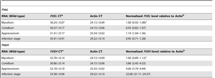
To gain insight into functions of PdeL and PdeH, we first examined the gene expression profiles at different stages of fungal development by qRT-PCR. There was no significant difference in the abundance of PDEL transcripts. However, higher levels of
PDEH expression were found in conidia, appressoria, and infection stages than in mycelia. In the infection stage, the expression ofPDEHincreased by almost 23-fold (Table 1). These observations suggested that PdeH might have an important role in
in plantainfection.
TargetedPDELandPDEHgene replacement
To examine the roles of PdeL and PdeH,PDELandPDEHgene replacement constructs (Figure S1A) were generated (see Materials and Methods) and transformed into the protoplasts of the wild-type strain Guy11. The resulting hygromycin-resistant transfor-mants were screened by PCR and confirmed by Southern blotting and RT-PCR analysis. No fragment was detected in theDpdeLor DpdeH mutants when hybridizing with gene-specific probes (probe1). RT-PCR analysis indicated that there was noPDELor
PDEH transcript in the respective mutants (Figure S1B). The DpdeLDpdeH mutant, as well as DpdeHDmagB and DpdeHDpka1
double mutants, was also confirmed by Southern blotting and RT-PCR analysis (Figure S1B).
PdeL but not PdeH is indispensable for conidiogenesis
To examine functions of PdeL and PdeH, conidiation of Guy11, DpdeL, DpdeH mutants, and complemented mutant strains were examined. Conidiation in 10-day-old cultures of theDpdeLmutant was markedly reduced to approximately 8% of the wild-type strain, while conidia in theDpdeHmutant were only reduced approximately 80% (Table 2). In contrast, the wild-type strain and complement strains showed normal sporulation under the same conditions. The data suggested that PdeL is required for conidiation inM. oryzae.
PdeL and PdeH are involved in conidial morphogenesis but dispensable for mycelial growth
other control strains. Surprisingly, both DpdeLand pdeH mutants produced elongated and thinner conidia, which were uniform and readily detected under the microscope (Figure 1A). The conidia of the mutants were, on average, longer by ,7mm and thinner by ,4mm than those of the controls (Figure 1B). Frequent branching and curly tips were also observed at the terminal hyphae of the DpdeHdeletion mutant. However, Calcofluor white (CFW) staining of mycelial cell walls showed that the septa were normal except for shorter intervals (Figure 1C). To determine whether PdeL and PdeH are involved in vegetative growth, the mutant and control strains were cultured on a variety of media including CM, OMA, SDC, or V8 juice agar media. No significant difference in colony morphology or growth rate was observed (Table 2). Combined, these findings suggested that PdeL and PdeH are indispensable for conidium morphology and but dispensable for mycelial growth.
PdeH is essential for maintenance of cell wall integrity
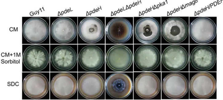
While the growth of theDpdeHmutant appeared normal on CM agar plates (Table 2), theDpdeHmutant did undergo progressive autolysis of mycelia after prolonged incubation for at least 14 days
(Figure 2, top panel). Autolysis began at the center of the colony and radiated outward. The autolysis of theDpdeHmutant can be suppressed by addition of 1 M sorbitol to the culture medium (Figure 2, middle panel). The autolysis tended to be more severe in the DpdeLDpdeH mutant than DpdeH, similar to the Dmps1 and Dmck1mutants that exhibited a defect in cell wall integrity [62,63]. Interestingly, theDpdeHmutant did not undergo autolysis on DSC medium under the same conditions (Figure 2, bottom panel), indicating that PdeH may also be involved in sensing nutrients in the maintenance of cell wall integrity.
PdeH is required for surface hydrophobicity
In previous studies, disruption of severalM. oryzaehydrophobin genes, includingMPG1andMHP1, resulted in a water- or detergent-soaked easily wettable phenotype [64,65,66,55,67,68,69]. To deter-mine whether PdeL and PdeH are involved in surface hydrophobic-ity, the mutant and wild type strains were tested with water and detergent solutions. UnlikeDmpg1, theDpdeHmutant did not show an easily wettable phenotype when incubated with water droplets (10ml) for 24 hours. However, aerial hyphae ofDpdeHmutants that were
Table 1.Real-time RT-PCR quantification of PdeL and PdeH expression inM. oryzae.
PdeL
RNA (Wild-type) PDELCTa Actin CT NormalizedPDELlevel relative to Actinb
Mycelium 30.2460.07 24.1360.09 1.00 (0.92–1.08)c
Conidium 30.3760.17 24.1560.06 0.93 (0.82–1.07)
Appressorium 31.4160.17 25.5460.02 1.19 (1.04–1.36)
Infection stage 35.4160.41 29.2260.14 0.95 (0.71–1.28)
PdeH
RNA (Wild-type) PDEHCTa Actin CT NormalizedPDEHlevel relative to Actinb
Mycelium 32.7860.14 24.1360.09 1.00 (0.89–1.12)c
Conidium 30.8660.14 24.1560.06 3.86 (3.45–4.32)
Appressorium 32.1860.10 25.5460.02 4.06 (3.70–4.44)
Infection stage 33.3860.06 29.2260.14 22.68 (21.11–24.37)
aCycle number at which the fluorescence crossed the threshold. Mean and standard deviation were calculated with data from three replicates. bRelative quantity of PDE at different developmental stages of the wild-type strain Guy11.
cThe mean and range of three replicates. doi:10.1371/journal.pone.0017241.t001
Table 2.Comparison of mycological characteristics among strains.
Mycelial growtha Biomassb Conidiationc Appressorium formationd%
Strain (cm) (mg) (x100/cm2) Hydrophobic Hydrophylic
Guy11 5.1060.15 0.072160.0018 103.8626.0 97.565.5 0
DpdeL 5.2060.21 0.102560.0025 8.866.5 98.864.2 0
DpdeH 5.0560.18 0.068460.0015 82.4629.7 99.263.8 88.763.8
DpdeLDpdeH 4.5060.05 0.040560.0010 - -
-DpdeHDpka1 5.1560.32 0.065560.0015 8.064.5 45.661.6 3.460.2
DpdeHDmagB 5.2060.25 0.077260.0023 7.065.3 70.362.7 10.262.7
DpdeL/PDEL 5.1560.20 0.076260.0020 100.3619.5 99.064.5 0
DpdeH/PDEH 5.1060.04 0.073860.0019 102.8621.0 99.262.7 3.260.7
aDiameter of hyphal radii at day 10 after incubation on complete medium agar plates at room temperature.
bDry weight of hyphal at day 2 after incubation in liquid complete medium at room temperature by shaken at 150 rpm. cNumber of conidia harvested from a 9 cm SDC plate at day 10 after incubation at room temperature.
grown on CM agar were more readily wettable by a solution containing both 0.02% SDS and 5 mM EDTA within 5 min (Figure 3A), as seen in theDmhp1mutant. The detergent-wettable phenotype shown byDpdeHmutant was stably maintained up to four successive generations, suggesting that this phenotype is mitotically stable (data not shown). Based on the results described above, we suggested that the surface hydrophobicity defect of DpdeH and DpdeLDpdeHmutants might be related to Mpg1 and Mhp1. To assess this, we examined the levels ofMPG1andMHP1expression in the mutant and wild-type strains. TheMPG1expression level showed a 50% decrease in the DpdeL mutant compared with the wild-type control, while the expression reduced more than 90% in theDpdeH
and DpdeLDpdeH mutants (Figure 3B). In contrast, there was no apparent difference in MHP1 expression in any of the mutants compared with the wild-type control (data not shown). These results indicated that the surface hydrophobicity defects of DpdeH and DpdeLDpdeHmutants were likely due to the lowedMPG1expression.
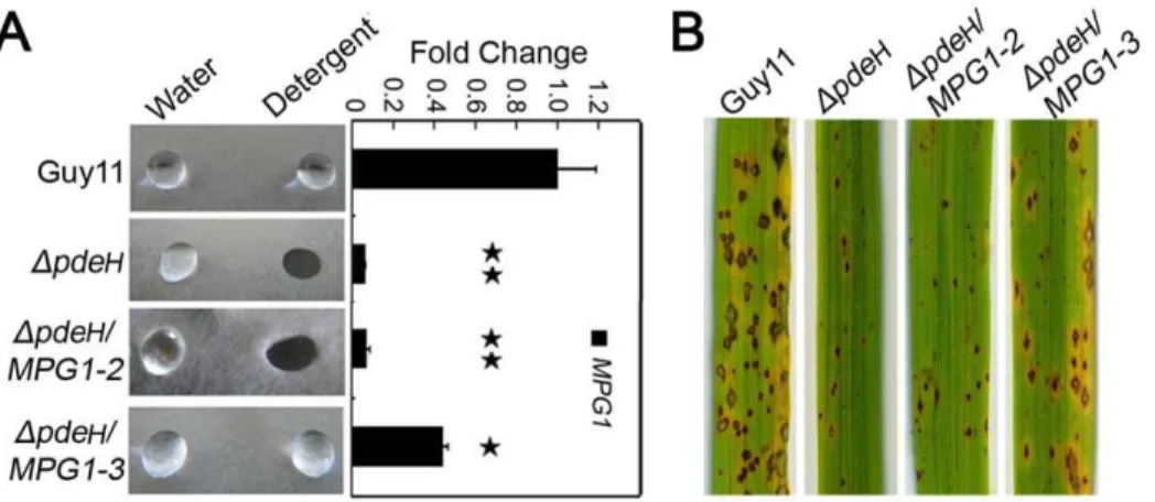
Over-expression ofMPG1partially restores the surface hydrophobicity and pathogenicity to theDpdeHmutant
As mentioned above, the expression level of MPG1 was significantly decreased in theDpdeHmutant. Additionally, Mpg1
is known as an important pathogenicity factor inM. oryzae[55]. We over-expressed MPG1 in the DpdeH mutant to determine whether Mpg1 could restore the defects in surface hydrophobicity and pathogenicity. We screened theDpdeH/MPG1transformants by qRT-PCR to check theMPG1expression level and obtained four MPG1-over-expressing transformant strains. One of these, DpdeH/MPG1-3, in which the MPG1 expression level was increased to 150% of that in the wild-type, was able to restore the defects of surface hydrophobicity and pathogenicity of the DpdeH mutant. However, the transformant DpdeH/MPG1-2, in whichMPG1expression level was almost the same as that of the DpdeHmutant, could not complement the defects (Figure 4A and 4B). We concluded that the hydrophobicity and pathogenicity defects in theDpdeHmutant were primarily due to the low level of
MPG1expression, and it is likely that the expression level ofMPG1
must remain at 150% of wild-type or more to maintain the surface hydrophobicity.
PdeL and PdeH regulate intracellular cAMP levels
Abundant studies have shown that PDEases can regulate intracellular cAMP levels in various organisms [3,4,5,6,7,8,9,10]. To determine whether PdeL and PdeH also regulate cAMP levels
Figure 1. TheDpdeLandDpdeHmutants have defects in conidial morphology and hyphal branching.(A) Conidia of the wild type and mutants were observed under an epi-fluorescence microscope. Conidia were stained with 1mg/ml Calcofluor white (CFW) for 5 min in dark. (B) Conidial size of the wild type and mutants. Values are the mean6SD from 100 conidia for each strain, which were measured using a microscope ruler. Length is the distance from the base to apex of conidia. And width is the size of the longest septum. (C) Branching patterns of mycelia on complete media slides at day 2 after incubation. Frequent branching occurs at the terminal mycelia ofDpdeHandDpdeHDpdeLdouble mutants. Calcofluor staining of mycelia is used to show the distance of septa.
in M. oryzae, we measured the intracellular cAMP levels in the hyphal stage. The results indicated that the DpdeL mutant accumulated only ,1.5-fold higher levels of cAMP than the wild-type strain, while the DpdeH mutant and the DpdeLDpdeH
double mutant accumulated,3-fold and,4.5-fold higher levels cAMP compared with the wild-type, respectively (Figure 5).
PdeH is required for appressorium differentiation, full virulence and elicitation of plant defense responses
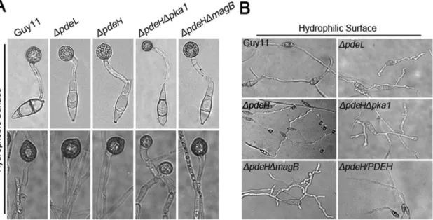
Physical cues of inductive surfaces, such as hardness and hydrophobicity, are necessary for appressorium formation [53]. The wild-type strain is unable to form appressoria on non-inductive surfaces, except in the presence of exogenous cAMP or inhibitors of PDEases [70]. To verify the effects of increased cAMP levels in the DpdeL and DpdeH mutants, we examined appressorium formation on inductive and non-inductive surfaces. The conidia and hyphal tips of the DpdeL and DpdeH mutants formed normal melanized appressoria on inductive surfaces, similar to the wild-type strain (Figure 6A, Table 2). However, theDpdeHmutant was able to form normal melanized appressoria on non-inductive surfaces (Figure 6B, Table 2), indicating that PdeH is an important negative regulator of appressorium formation.
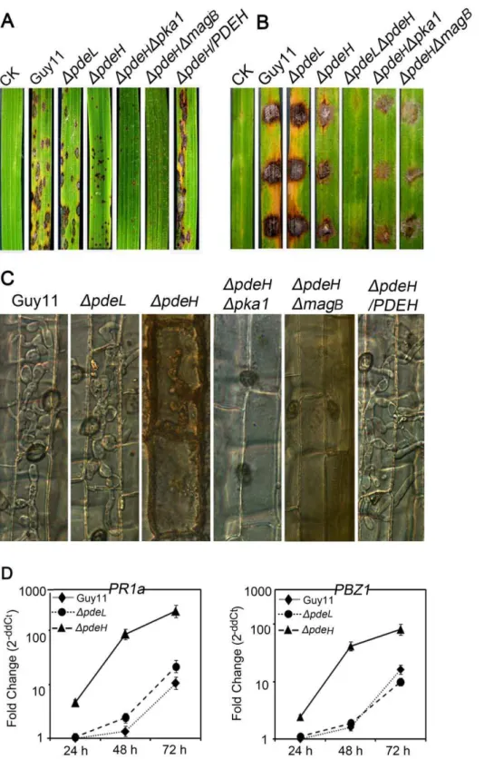
To determine whether PDEL and PDEH are involved in pathogenesis, conidial suspensions were sprayed onto susceptible rice seedlings and hyphal plugs were placed onto detached rice seedling leaves (CO-39 cv.oryzae) to develop rice blast lesions. In a spray-inoculation test, the DpdeH mutant produced tiny and restricted lesions, whereas the DpdeL mutant, wild-type, and complement strains showed susceptible-type spreading lesions (Figure 7A). In a detached-inoculation test, lesions caused by DpdeHmutant hyphal plugs spread very slowly and the lesion sizes were small, while those caused byDpdeLmutant, wild type, and complement strains spread rapidly and the neighboring lesions joined together by 5 days after inoculation (Figure 7B).
Because no defects in appressorium development by theDpdeH
mutant were observed on hydrophobic surface, the development of infectious hyphae within the host cells was examined using excised leaf sheaths. In the wild-type, DpdeL mutant, and
Figure 2.DpdeHmutants have a defect in cell-wall integrity.Growth of wild type and mutant strains on complete media (CM) without 1 M sorbitol (top); growth of strains on CM with 1 M sorbitol (middle); growth of strains on straw decoction and corn media (SDC) without sorbitol (bottom). TheDpdeHandDpdeHDpdeLmutants undergo progressive autolysis on CM in the absence of osmotic stabilization. Radial growth rates are identical to those of the wild-type strains.
doi:10.1371/journal.pone.0017241.g002
Figure 3. Detergent wettable phenotype of DpdeH and
DpdeHDpdeL mutants. (A) Ten microliters of water or detergent solution containing 0.02% SDS and 5 mM EDTA were placed on the colony surfaces of the wild type and mutants strains and photographed after 5 min. (B) Expression analysis ofMPG1 gene in wild type and mutant strains. The error bars indicate SD of three replicates. Asterisk indicates significant differences atP= 0.01.
complement strains, infectious hyphae grew actively and extended into 3–4 cells neighboring the primary infected cells by 48 hours after inoculation (Figure 7C). However, infectious hyphae of the DpdeHmutant was mostly restricted to the primary infected cells and almost no infectious hyphae extended into neighboring cells, and accumulation of dark brown granules was seen along the infectious hyphae (Figure 7C). These results were consistent with those of rice infection assays. Additionally, the expression levels of the rice defense-related genes PR1a and PBZ1 in response to DpdeH infection were much higher than those associated with infection by the wild-type,DpdeLmutant, and complement strains when analyzed by quantitative RT-PCR (Figure 7D). These observations indicated that the induction of plant defense responses in rice challenged by DpdeH mutant might contribute to the retardation of infectious hyphal development.
PdeL and PdeH regulate laccase activities
A previous study indicated that laccases are involved in the pathogenicity of certain fungi [71,97], and the laccase activity can
be detected readily using the specific substrate ABTS (2,2’-azino-di-3-ethylbenzthiazoline-6-sulfonate) [72,73]. To determine whether PDEases are involved in the regulation of laccase activity, we testedDpdeLandDpdeHmutant strains on CM agar plates and in liquid medium supplemented with 0.2 mM ABTS. In each case, decreases in laccase activity were observed inDpdeL,DpdeH, and DpdeLDpdeH mutants, with a less-oxidized dark purple stain around the colonies of the mutants and a lower level of the laccase activity in the culture filtrate compared with the wild-type strain (Figure 8A and 8B). Consistent with these observations, the expression of two extracellular peroxidase genes, MGG_08200.6 and MGG_01924.6, was significantly down-regulated in all the mutants (Figure 8C). Meanwhile, the results also indicated that the laccase activity was decreased to a greater extent in the DpdeL
mutant than in theDpdeLDpdeH and DpdeH mutants (Figure 8A and 8B), suggesting that PdeL may have a more prominent role in regulating the enzyme activity.
Genome-wide identification of genes regulated by PdeL and PdeH
To identify genes regulated by PdeL and PdeH, we compared gene expression profiles ofDpdeL,DpdeH, andDpdeLDpdeHmutants with the wild-type strain during the hyphal stage. In total, 1582 genes were up-regulated and 1724 genes were down-regulated in DpdeL,DpdeH, andDpdeLDpdeHmutants (Figure 9A). Of the 1582 up-regulated genes, we found 373 that were regulated by PdeL, 459 by PdeH, and 1248 by PdeL and PdeH together. Of the 1724 down-regulated genes, the corresponding numbers were 509, 652, and 1450, respectively. Those genes in which the expression ratio was greater than 2-fold were functionally grouped into GO categories as described in the Materials and Methods (Figure 9B, Tables S2, S3 and S4). Overall, we noted that genes associated with protein and amino acid degradation, lipid degradation, secondary metabolism including melanin biosynthesis, and cellular transportation exhibited marked decreases in expression. Among genes that were up-regulated by PdeL, PdeH, or PdeL and PdeH, we were able to positively assign two genes, MGG_07881.6 and MGG_03508.6, to PdeH, in contrast to many by PdeL or PdeL and PdeH (Table S5).
To confirm gene expression patterns derived from our microarray experiments, we performed real-time RT-PCR with
Figure 4. Over-expressionMPG1in theDpdeHmutant partially restores the surface hydrophobicity and pathogenicity defects.(A) Ten microliters of water or detergent solution containing 0.02% SDS and 5 mM EDTA were placed on the colony surfaces of wild type, mutants and Mpg1 over-expression strains and photographed after 5 min. Expression analysis ofMPG1gene in wild type and mutants andMPG1over-expression strains. The error bars indicate SD of three replicates. Asterisk indicates significant differences atP= 0.01. (B) Spraying assay. Five milliliters of conidial suspension (56104spores/ml) of each strain were sprayed on rice seedlings. Diseased leaves were photographed 7 days after inoculation. doi:10.1371/journal.pone.0017241.g004
four selected genes that were down-regulated in theDpdeHmutant. Two genes, MPG1 and PTH11, which are required for pathogenesis, were significantly down-regulated (,10-fold; Figure 4B and 10). Two laccase genes, MGG_11608.6 and MGG_13464.6, were down-regulated to a greater extent (,100-fold) in theDpdeHmutant (Figure 8C). Although the magnitudes of fold changes in laccase gene expression were much higher than the microarray results, the real-time RT-PCR data supported those of the microarray analyses.
Characterization of novel genes regulated by PdeH
To further explore the role of PdeH inM. oryzae, we deleted nine down-regulated genes identified from the DpdeH mutant microarray profile. MGG_00311.6 encodes an acid protease involved in protein and amino acid degradation, MGG_11608.6 encodes an oxidase enzyme laccase that is found in many plants, fungi, and microorganisms, MGG_10631.6 encodes a glycoside hydrolase involved in carbohydrate metabolism, MGG_07571.6 encodes a putative cell wall degrading protein with a LysM domain, MGG_07218.6 encodes a transcription factor containing a GAL4-like Zn2-Cys6-binuclear cluster DNA-binding domain at
the N-terminal, MGG_06326.6 encodes a vacuolar ATP synthase 16-kDa proteolipid subunit, and, finally, MGG_12214.6encodes a polyketide synthase. Two putative ion transporters, P-type ATPase MGG_04852.6 and sugar transporter MGG_10293.6, were also characterized. Overall, mutant conidia produced normal mela-nized appressoria on hydrophobic surfaces, and none of them formed appressoria on noninductive surfaces (Figure 11 and data not shown). However, the mutant strain for transcription factor MGG_07218.6 showed significantly reduced pathogenicity with tiny and slow-spread blast lesions, a phenotype similar to that of theDpdeHmutant (Figure 11).
Discussion
Cyclic AMP signaling plays an important role in regulating the growth and differentiation of eukaryotic organisms, including rice blast pathogen M. oryzae. A recent study by Ramanujam and
Naqvi [54] has demonstrated that phosphodiesterase PdeL and PdeH have both conserved and distinct functions in regulating cAMP levels and pathogenicity ofM. oryzae. Here, our presented our independent study that not only corroborates with that of recent publication but also further illustrates roles of PdeL and PdeH in this fungus. We found that deletion ofPDEHresulted in defects in conidial morphology, cell wall integrity, and surface hydrophobicity, as well as a significant reduction in pathogenicity. We also found thatPDEHdisruption partially rescued theDmagB
andDpka1mutant phenotypes. Moreover, we propose that PdeH might function through a feedback mechanism to regulate the expression of Mpg1, which is a demonstrated pathogenicity factor involved in surface hydrophobicity and pathogenic development of
M. oryzae.
The fungal cell wall plays an important role in maintaining cell shape and mediating exchanges between the cell and its environment [74]. Although it is rigid, its organization and structure must be remodeled constantly for growth and develop-ment [63]. Thus, in pathogenic fungi, the ability to maintain cell wall integrity is critical to the establishment of disease within the host. Several cell wall integrity-associated genes have been characterized in S. cerevisiae, including MEKK (Bck1), a pair of redundant MEKs (Mkk1/2), and a MAP kinase (Slt2) [75]. The Bck1 and Slt2 homologs Mck1 and Mps1 were described as also essential for cell wall integrity and pathogenicity of M. oryzae
[76,63]. In both S. cerevisiae and C. albicans, deletion of PDE2
affected cell wall integrity, stress response, hyphal development, and/or virulence [25,77,78]. As we failed to reveal any phenotypic changes between theDpdeHmutant and the wild-type control in the response to stress agents, such as ionic stress (Na+and Cu2+),
oxidative stress (H2O2), heavy metal (CoCl2) stress, osmotic stress
(sorbitol), or cell wall-disturbing agents (CFW, SDS, and lysing enzyme)M. oryzaemay have an alternative approach of responding to stress signals or may possess different stress sensors.
We found that DpdeLDpdeH double mutant undergo faster autolysis than the DpdeH mutant on CM agar plates, and the DpdeLDpdeHmutant also showed a higher level of cAMP level than theDpdeHmutant. This suggests that cAMP plays an important
Figure 6. Appressorium formation assays.(A) Conidia of each strain were incubated on hydrophobic surfaces for 24 hours (up panel); hyphal plugs of each strain were incubated on hydrophobic surfaces for 48 hours (bottom panel). (B) Conidia of each strain were incubated on hydrophilic surfaces for 24 hours.
Figure 7. The loss ofPDEHleads to reduced pathogenicity and induction of strong plant defense responses.(A) Spraying assay. Five milliliters of conidial suspension (56104spores/ml) of each strain were sprayed on rice seedlings. Diseased leaves were photographed 7 days after inoculation. (B) Detached leaf assay. The hyphal plugs of each strain were placed onto the upside of detached rice seedling leaves. Diseased leaves were photographed 5 days after inoculation. (C) Observation of infectious growth. Excised rice sheath from 4-week-old rice seedlings was inoculated with conidial suspension (56104spores/ml of each strain). Infectious growth was observed 48 hours after inoculation. (D) The expression of rice pathogenesis-related (PR) genes over time after inoculation. The transcriptional expression ofPR1aandPBZ1in the infected rice was analyzed using quantitative RT-PCR. The graph was generated with three replicates in a representative data set, and similar results were obtained in another independent biological repetition. The error bars indicate SD of three replicates.
role in the maintenance of cell wall integrity and, while both are involved in regulating intracellular cAMP levels and cell wall integrity, PdeH might have a more prominent role. Moreover, the DpdeH single and DpdeLDpdeH double mutants did not undergo autolysis or showed only very slight autolysis when cultured on SDC media (poor nutrition), suggesting that PdeH may act as a nutrition sensor in modulating intracellular cAMP levels, which may affect cell wall integrity. Regardless, further studies are required to determine whether and how PdeH senses nutritional signals.
Most hydrophobins confer surface hydrophobicity on fungi forming a spore rodlet layer. Disruption of several hydrophobin genes, including Mpg1, resulted in a water- or detergent-soaked easily wettable phenotype [64,65,66,55,67,68]. TheMpg1mutant is defective in conidiation and appressorium formation, and less pathogenic than wild-type strain, indicating that Mpg1 plays an important role in multiple infection-related processes. Here, deletion of PDEH resulted in a significantly reduced level of
MPG1 expression and defects in surface hydrophobicity and pathogenicity [55]. InDmpg1mutants, the defect in appressorium formation can be restored by adding exogenous cAMP, suggesting that Mpg1 functions upstream of cAMP signal for appressorium formation [55]. Here, over-expression of MPG1 in the DpdeH
mutant partially rescued the surface hydrophobicity and pathoge-nicity defects, suggesting that Mpg1 could function downstream of PdeH and cAMP signaling. Thus, we propose that PdeH may function through a feedback mechanism to activate Mpg1 and regulates its expression for surface hydrophobicity and pathogenic development.
Several induced genes in plants have been reported to affect fungal pathogenicity, directly or indirectly [79,55,80,81,69]. The high level ofPDEHexpression at late infection stages in infected rice leaves also suggests its roles in infectious growth and pathogenicity. Targeted disruption ofPDEHsignificantly reduced virulence in infection assays. TheDpdeHmutant produced smaller and less numerous lesions than the wild-type strain. The results presented above indicated that appressoria formed by DpdeH
mutants are probably defective in penetration. It is likely that PdeH regulates the processes involved in a late stage of appressorium development, such as turgor generation or appres-sorium pore formation. The qRT-PCR results, indicating that
PDEH was expressed at high levels in the appressoria, also supported this possibility. We assayed appressorium turgor using the cytorrhysis method described previously [44]. Preliminary data do not indicate any difference in cytorrhysis between appressoria formed byDpdeHmutant and wild-type strains (data not shown), suggesting that appressoria ofDpdeHmutants do not have defects in the generation of turgor pressure. Another possibility is that PdeH may regulate the early stages of appressorium penetration, such as development of the penetration peg or differentiation of infectious hyphae. The reduction in pathogenicity may be due to the reduction in development at the pre-penetration stages or a reduction in infectious growth ofDpdeHmutants in host cells. This may also explain the high level ofPDEHexpression during the late stages of infection.
Defense responses induced by the recognition of microbe-associated molecules (pathogen-microbe-associated molecular patterns, PAMPs) are often associated with cell wall strengthening, rapid
Figure 8. PdeL and PdeH are related to the activity of extracellular laccases.(A) Laccase activity was tested on CM agar medium containing 0.2 mM ABTS at final concentration. Discoloration was observed on day 2. (B) Laccase activity measured by ABTS oxidizing test (see Materials and Methods for details). (C) Quantitative RT-PCR analysis of two laccase genes in wild type and mutants. Expression data were normalized using the ACTINgene. Error bars represent standard deviation.
production of reactive oxygen species (ROS), and the transcrip-tional activation of PR genes [82]. In plants, accumulation of ROS at the site of infection is considered one of the earliest responses during plant PAMP-triggered immunity (PTI) [83,82]. Plant-derived ROS have various functions during plant–microbe interactions [84,85,86,87,88,89]. For pathogens to survive in harsh environments and successfully invade host cells, they must develop mechanisms to scavenge ROS and protect against ROS-induced damage [90,91,92,93]. In M. oryzae, several well-characterized genes have been reported to suppress basal host defenses and to be responsible for ROS detoxification at the site of infection [58,59,94]. As rice cells infected by the DpdeH mutant showed brown granule accumulation and cell death, it is likely that plant defense responses are involved in virulence attenuation of DpdeHmutant. Thus, the defense responses against the wild-type and mutant strains were compared with regard to two genes,PR1a
and PBZ1. These two genes are important components of the jasmonic acid (JA) and salicylic acid (SA) pathways, which are involved in JA- and SA-induced plant defense, respectively [95,96]. The stronger activation of both PR1a and PBZ1 in
mutant-inoculated plants compared with wild-type controls suggested thatDpdeHmutant can still elicit plant defenses, which may be involved in attenuation of the virulence of the DpdeH
mutant.
In M. oryzae, many evidences suggest that MagB may sense surface cues, stimulate cAMP synthesis, and activate the cAMP signal pathway [50,51,52]. Because PdeH can degrade intracel-lular cAMP, we hypothesize that the decrease in the PdeH activity in theDmagB mutant may maintain the balance of intracellular cAMP levels and restore its defects. The result of partially restoration in appressorium formation in theDpdeHDmagBdouble mutant supported this hypothesis (Table 2). Additionally, the appressorium formation rate on hydrophylic surfaces was reduced to 10% compared withDpdeHmutants, suggesting that MagB and PdeH can complement each other in some defects. However, no other defect was restored in theDpdeHDmagBdouble mutant, all of which were similar to those in theDpdeHmutant (Figure 1, 2, 3 and 8). These observations may have been due to the high cAMP level in theDpdeHDmagBdouble mutant, because the cAMP level was still much higher in DpdeH DmagB than in the wild-type
Figure 9. Functional categorization of the consensus genes.(A) Expression profiles were combined and showing PdeL- and PdeH-dependent. (B) Up-regulation and down-regulation of more than two-fold change genes were grouped according to their putative function (see Supplemental data for details).
(Figure 5). Since cAMP-dependent protein kinase PKA plays a pivotal role in cAMP-dependent pathways of M. oryzae, we compared the phenotype ofDpka1with that of theDpdeHDpka1
double mutant. TheDpdeHDpka1mutant formed small, misshapen appressoria on hydrophobic surfaces similar to theDpka1mutant, while very few appressoria were formed on hydropholic surfaces (Figure 6). However, other phenotypes, such as cell wall integrity and surface hydrophobicity, which may also be due to the high cAMP level, were similar to those of theDpdeHmutant (Figure 1, 2, 3 and 8). Based on these results, we conclude that, while MagB and Pka1 activate the cAMP signaling pathway to mediate appressorium formation and pathogenicity, PdeH plays a more critical role in regulating intracellular cAMP levels to affect cell wall integrity and surface hydrophobicity, bypassing the PKA signaling pathway.
Genome-wide analysis of gene expression changes during spore germination and appressorium formation on a hydrophobic surface compared with induction by cAMP revealed new insight into appressorium formation and function in M. oryzae [98]. During appressorium formation, the genes that respond to both stimuli are known to be involved in protein and amino acid
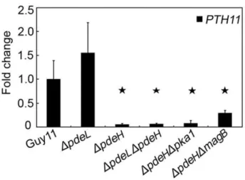
Figure 10.PTH11gene expression inDpdeHandDpdeLmutants.
RNA was extracted from mycelia cultured in liquid CM medium for 2 days.ACTINwas used for normalization, and the values were calculated by 2-ddCT methods with quantitative RT-PCR data. Values represent mean6SD from two independent experiments with three replicates each. Asterisk indicates significant differences atP= 0.01.
doi:10.1371/journal.pone.0017241.g010
degradation, lipid metabolism, secondary metabolism, and cellular transportation. In this study, the levels of expression of two pathogenicity-related genes were significantly decreased in the appressoria treated with cAMP. Targeted deletion of several other changed genes, such as polyketide synthase (MGG_07219.6), subtilisin-like protease (MGG_03670.6), and a transcription factor (MGG_07218.6), affected virulence and other characteristics related to pathogenicity. Similar to these results, our microarray data also provided some novel insight to identify pathogenicity factors. Three genes, MPG1 (MGG_10315.6), PTH11 (MGG_ 05871.6), andCOS1(MGG_03977.6), which have been reported to be involved in pathogenicity [55,99,100], were significantly down-regulated in the DpdeH single and DpdeLDpdeH double mutants, but not in theDpdeLmutant. Furthermore, deletion of a down-regulated transcription factor, from the DpdeH microarray data, also showed reduced pathogenicity, similar to that of the DpdeH mutant. These data may explain why DpdeH and DpdeLDpdeHmutants attenuated virulence.
In summary, we continued the characterization of the low- and high-affinity cAMP PDEases PdeL and PdeH in M. oryzae. We showed that, while PdeL and PdeH share certain functions, PdeH indeedplays a more prominent role than PdeL in regulating cell wall integrity, surface hydrophobicity and pathogenicity through modulation of intracellular cAMP levels. Our findings also reveal that PdeH may function through a feedback mechanism to regulate the expression of pathogenicity factor Mpg1, which is involved in surface hydrophobicity and pathogenic development in
M. oryzae.
Supporting Information
Figure S1 Generation of DpdeL and DpdeH deletion mutants.(A) Restriction map of thePDELandPDEHgenomic region and knockout construct. Thick arrows indicate orientations of the PDEL, PDEH and hygromycin phosphotransferase (hph)
genes. Thin lines below the arrows indicate the probe sequences of each gene. The restriction enzymes are EcoRV (EV), EcoRI (EI),
XbaI (XI) andKpnI (KI). (B) Southern blot and RT-PCR analyses ofDpdeL,DpdeH(top) and double-gene (bottom) knockout mutants. Genomic DNA of the wild-type strain and the knockout mutants
was digested with corresponding restriction enzymes. Total RNAs of the wild-type strain and the knockout mutants were isolated and the expression levels of target gene were detected usingACTINas control. WT: wild type; T: transformant.
(TIF)
Figure S2 Confirmation of target gene replacement by PCR.
(PDF)
Table S1 Primers used in this study. (DOC)
Table S2 Categorization ofPDELregulated genes with known function.
(DOC)
Talbe S3 Categorization of PDEHregulated genes with known function.
(DOC)
Table S4 Categorization of PDEL&PDEH regulated genes with known function.
(DOC)
Table S5 Categorization of genes only dependent on
PDEL,PDEHandPDEL & PDEH, respectively. (DOC)
Acknowledgments
We thank Dr. Amy Whittington, Louisiana State University Health Sciences Center, New Orleans, LA USA, for commenting on the manuscript.
Author Contributions
Conceived and designed the experiments: HZ X. Zheng ZZ. Performed the experiments: HZ KL X. Zhang WT JW MG QZ. Analyzed the data: HZ X. Zheng PW ZZ. Contributed reagents/materials/analysis tools: HZ KL X. Zhang WT JW MG QZ. Wrote the manuscript: HZ PW ZZ.
References
1. Daniel PB, Walker WH, Habener JF (1998) Cyclic AMP signaling and gene regulation. Annu Rev Nutr 18: 353–83.
2. Charbonneau H, Beier N, Walsh KA, Beavo JA (1986) Identification of a conserved domain among cyclic nucleotide phosphodiesterases from diverse species. Proc Natl Acad Sci USA 83: 9308–9312.
3. D’Angelo MA, Sanguineti S, Reece JM, Birnbaumer L, Torres HN, et al. (2004) Identification, characterization and subcellular localization of TcPDE1, a novel cAMP-specific phosphodiesterase fromTrypanosoma cruzi. Biochem J 378: 63–72. 4. De Voti JSG, Beach D, McLeod M (1991) Interaction between ran1+protein kinase and cAMP dependent protein kinase as negative regulators of fission yeast meiosis. EMBO J 10: 3759–3768.
5. Dunlap PV, Callahan SM (1993) Characterization of a periplasmic 3_:5_-cyclic nucleotide phosphodiesterase gene,cdpP, from the marine symbiotic bacterium
Vibrio fischeri. J Bacteriol 175: 4615–4624.
6. Hoyer LL, Cieslinski LB, McLaughlin MM, Torphy TJ, Shatzman AR, et al. (1994) A Candida albicans cyclic nucleotide phosphodiesterase: cloning and expression inSaccharomyces cerevisiae and biochemical characterization of the recombinant enzyme. Microbiology 140: 1533–1542.
7. Kunz S, Kloeckner T, Essen LO, Seebeck T, Boshart M (2004) TbPDE1, a novel class I phosphodiesterase ofTrypanosoma brucei. Eur J Biochem 271: 637–647. 8. Lacombe ML, Podgorski GJ, Franke J, Kessin RH (1986) Molecular cloning and
developmental expression of the cyclic nucleotide phosphodiesterase gene of
Dictyostelium discoideum. J Biol Chem 261: 16811–16817.
9. Nikawa J, Sass P, Wigler M (1987) Cloning and characterization of the low affinity cAMP phosphodiesterase gene of S. cerevisiae. Mol Cell Biol 7: 3629–3636.
10. Rascon A, Viloria ME, De-Chiara L, Dubra ME (2000) Characterization of cyclic AMP phosphodiesterases inLeishmania mexicanaand purification of soluble form. Mol Biochem Parasitol 106: 283–292.
11. Lemaire K, Van de Velde S, Van Dijck P, Thevelein JM (2004) Glucose and sucrose act as agonist and mannose as antagonist ligands of the G proteincoupled receptor Gpr1 in the yeastSaccharomyces cerevisiae. Mol Cell 16: 293–299.
12. Jones DL, Petty J, Hoyle DC, Hayes A, Oliver SG, et al. (2004) Genome-Wide Analysis of the Effects of Heat Shock on aSaccharomyces cerevisiaeMutant With a Constitutively Activated cAMP-Dependent Pathway. Comp Funct Genomics 5: 419–431.
13. Jones DL, Petty J, Hoyle DC, Hayes A, Ragni E, et al. (2003) Transcriptome profiling of aSaccharomyces cerevisiaemutant with a constitutively activated Ras/ cAMP pathway. Physiol Genomics 16: 107–118.
14. Pan X, Heitman J (1999) Cyclic AMP-dependent protein kinase regulates pseudohyphal differentiation inSaccharomyces cerevisiae. Mol Cell Biol 19: 4874–4887. 15. Park JI, Grant CM, Dawes IW (2005) The highaffinity cAMP phosphodiesterase ofSaccharomyces cerevisiaeis the major determinant of cAMP levels in stationary phase: involvement of different branches of the Ras-cyclic AMP pathway in stress responses. Biochem Biophys Res Commun 327: 311–319.
16. Schneper L, Krauss A, Miyamoto R, Fang S, Broach JR (2004) The Ras/protein kinase A pathway acts in parallel with the Mob2/Cbk1 pathway to effect cell cycle progression and proper bud site selection. Eukaryot Cell 3: 108–120. 17. Davey J (1998) Fusion of a fission Yeast. Yeast. pp 1529–1566.
19. Higuchi T, Watanabe Y, Yamamoto M (2002) Protein kinase A regulates sexual development and gluconeogenesis through phosphorylation of the Zn finger transcriptional activator Rst2p in fission yeast. Mol Cell Biol 22: 1–11. 20. Hoffman CS (2005) Glucose sensing via the protein kinase A pathway in
Schizosaccharomyces pombe. Biochem Soc Trans 33: 261–264.
21. Ma P, Wera S, Dijck PV, Thevelein JM (1999) ThePDE1-encoded low-affinity phosphodiesterase in the yeastSaccharomyces cerevisiaehas a specific function in controlling agonist-induced cAMP signaling. Mol Biol Cell 10: 91–104. 22. Nikawa JI, Cameron S, Toda T, Ferguson KM, Wigler M (1987) Rigorous
feedback control of cAMP levels inSaccharomyces cerevisiae. Genes Dev 1: 931–937. 23. Meima ME, Weening KE, Schaap P (2003) Characterization of a cAMP-stimulated cAMP phosphodiesterase inDictyostelium discoideum. J Biol Chem 278: 14356–14362.
24. Bahn YS, Staab J, Sundstrom P (2003) Increased high-affinity phosphodiesterase
PDE2gene expression in germ tubes counteractsCAP1-dependent synthesis of cyclic AMP, limits hypha production and promotes virulence ofCandida albicans. Mol Microbiol 50: 391–409.
25. Jung WH, Warn P, Ragni E, Popolo L, Nunn CD, et al. (2005) Deletion of
PDE2, the gene encoding the high-affinity cAMP phosphodiesterase, results in changes of the cell wall and membrane inCandida albicans. Yeast 22: 285–294. 26. Hicks JK, Bahn YS, Heitman J (2005) Pde1 phosphodiesterase modulates cyclic AMP levels through a protein kinase A-mediated negative feedback loop in
Cryptococcus neoformans. Eukaryot Cell 4: 1971–1981.
27. Larraya LM, Boyce KJ, So A, Steen BR, Jones S, et al. (2005) Serial analysis of gene expression reveals conserved links between protein kinase A, ribosome biogenesis, and phosphate metabolism in Ustilago maydis. Eukaryot Cell 4: 2029–2043.
28. Regenfelder E, Spellig T, Hartmann A, Lauenstein S, Bolker M, et al. (1997) G proteins in Ustilago maydis: transmission of multiple signals? EMBO J 16: 1934–1942.
29. Durrenberger F, Wong K, Kronstad JW (1998) Identification of a cAMP-dependent protein kinase catalytic subunit required for virulence and morphogenesis inUstilago maydis. Proc Natl Acad Sci USA 95: 5684–5689. 30. Gold SE, Brogdon SM, Mayorga ME, Kronstad JW (1997) The Ustilago maydis
regulatory subunit of a cAMP-dependent protein kinase is required for gall formation in maize. Plant Cell 9: 1585–1594.
31. Lee N, Kronstad JW (2002) ras2 Controls morphogenesis, pheromone response, and pathogenicity in the fungal pathogen Ustilago maydis. Eukaryot Cell 1: 954–966.
32. Banno S, Ochiai N, Noguchi R, Kimura M, Yamaguchi I, et al. (2005) A catalytic subunit of cyclic AMP-dependent protein kinase, PKAC-1, regulates asexual differentiation inNeurospora crassa. Genes Genet Syst 80: 25–34. 33. Bencina M, Panneman H, Ruijter GJ, Legisa M, Visser J (1997)
Characteriza-tion and overexpression of theAspergillus nigergene encoding the cAMPdepen-dent protein kinase catalytic subunit. Microbiology 143 (Pt 4): 1211–1220. 34. Brakhage AA, Liebmann B (2005)Aspergillus fumigatusconidial pigment and
cAMP signal transduction: significance for virulence. Med Mycol 43(Suppl 1): S75–82.
35. Grosse C, Heinekamp T, Kniemeyer O, Gehrke A, Brakhage AA (2008) Protein kinase A regulates growth, sporulation, and pigment formation inAspergillus fumigatus. Appl Environ Microbiol 74: 4923–4933.
36. Ivey FD, Kays AM, Borkovich KA (2002) Shared and independent roles for a Galpha(i) protein and adenylyl cyclase in regulating development and stress responses inNeurospora crassa. Eukaryot Cell 1: 634–642.
37. Kays AM, Rowley PS, Baasiri RA, Borkovich KA (2000) Regulation of conidiation and adenylyl cyclase levels by the Galpha protein GNA-3 in
Neurospora crassa. Mol Cell Biol 20: 7693–7705.
38. Kays AM, Borkovich KA (2004) Severe impairment of growth and differentiation in aNeurospora crassamutant lacking all heterotrimeric G alpha proteins. Genetics 166: 1229–1240.
39. Liebmann B, Gattung S, Jahn B, Brakhage AA (2003) cAMP signaling in
Aspergillus fumigatusis involved in the regulation of the virulence gene pksP and in defense against killing by macrophages. Mol Genet Genomics 269: 420–435. 40. Saudohar M, Bencina M, van de Vondervoort PJ, Panneman H, Legisa M, et al.
(2002) Cyclic AMP-dependent protein kinase is involved in morphogenesis of
Aspergillus niger. Microbiology 148: 2635–2645.
41. Liebmann B, Muller M, Braun A, Brakhage AA (2004) The cyclic AMP-dependent protein kinase a network regulates development and virulence in
Aspergillus fumigatus. Infect Immun 72: 5193–5203.
42. Lafon A, Han KH, Seo JA, Yu JH, d’Enfert C (2006) G-protein and cAMP mediated signaling inAspergilli: a genomic perspective. Fungal Genet Biol 43: 490–502.
43. Talbot NJ (2003) On the trail of a cereal killer: Exploring the biology of
Magnaporthe grisea. Annu Rev Microbiol 57: 177–202.
44. Howard RJ, Ferrari MA, Roach DH, Money NP (1991) Penetration of hard substrates by a fungus employing enormous turgor pressures. Proc Natl Acad Sci USA 88: 11281–11284.
45. Howard RJ, Valent B (1996) Breaking and entering: host penetration by the fungal rice blast pathogenMagnaporthe grisea. Annu Rev Microbiol 50: 491–512. 46. Choi WB, Dean RA (1997) The adenylate cyclase geneMAC1ofMagnaporthe grisea controls appressorium formation and other aspects of growth and development. Plant Cell 9: 1973–1983.
47. Adachi K, Hamer JE (1998) Divergent cAMP signaling pathways regulate growth and pathogenesis in the rice blast fungusMagnaporthe grisea. Plant Cell 10: 1361–1373.
48. Mitchell TK, Dean RA (1995) The cAMP-dependent protein kinase catalytic subunit is required for appressorium formation and pathogenesis by the rice blast pathogenMagnaporthe grisea. Plant Cell 7: 1869–1878.
49. Xu JR, Urban M, Sweigard JA, Hamer JE (1997) The CPKA gene of
Magnaporthe griseais essential for appressorial penetration. Mol Plant-Microbe Interact 10: 187–194.
50. Liu SH, Dean RA (1997) G protein alpha subunit genes control growth, development, and pathogenicity of Magnaporthe grisea. Mol Plant-Microbe Interact 10: 1075–1086.
51. Fang EG, Dean RA (2000) Site-directed mutagenesis of themagBgene affects growth and development inMagnaporthe grisea. Mol Plant-Microbe Interact 13: 1214–1227.
52. Ebbole DJ (2007) Magnaporthe as a model for understanding hostpathogen interactions. Annu Rev Phytopathol 45: 437–56.
53. Liu H, Suresh A, Willard FS, Siderovski DP, Lu S, et al. (2007) Rgs1 regulates multiple Galpha subunits in Magnaporthe pathogenesis, asexual growth and thigmotropism. EMBO J 26: 690–700.
54. Ramanujam R, Naqvi NI (2010) PdeH, a high-affinity cAMP phosphodiesterase, is a key regulator of asexual and pathogenic differentiation inMagnaporthe oryzae.
PLoS Pathogen 6: e1000897.
55. Talbot NJ, Ebbole DJ, Hamer JE (1993) Identification and characterization of
MPG1, a gene involved in pathogenicity from the rice blast fungusMagnaporthe grisea. Plant Cell 5: 1575–1590.
56. Stringer MA, Timberlake WE (1995) dewA encodes a fungal hydrophobin component of theAspergillusspore wall. Mol Microbiol 16: 33–44.
57. Zhang HF, Zhao Q, Liu KY, Zhang ZG, Wang YC, et al. (2009)MgCRZ1, a transcription factor ofMagnaporthe grisea, controls growth, development and is involved in full virulence. FEMS Microbiol Lett 2: 160–169.
58. Guo M, Guo W, Chen Y, Dong SM, Zhang X, et al. (2010) The bZIP transcription factor Moatf1 mediates oxidative stress responses and is necessary for full virulence of the rice blast fungusMagnaporthe oryzae. Mol Plant-Microbe Interactions 23: 1053–1068.
59. Chi MH, Park SY, Kim S, Lee YH (2009) A novel pathogenicity gene is required in the rice bast fungus to suppress the basal defenses of the host. PLoS Pathogen 5: e1000401.
60. Livak KJ, Schmittgen TD (2001) Analysis of relative gene expression data using real-time quantitative PCR and the 2-DDCtmethod. Methods 25: 402–408. 61. Ashburner M, Ball CA, Blake JA, Botstein D, Butler H, et al. (2000) Gene
Ontology: tool for the unification of biology. Nat Genet 25: 25–29. 62. Xu JR, Staiger CJ, Hamer JE (1998) Inactivation of the mitogen-activated
protein kinaseMPS1from the rice blast fungus prevents penetration of host cells but allows activation of plant defence responses. Proc Natl Acad Sci USA 95: 12713–12718.
63. Jeon J, Goh J, Yoo S, Chi MH, Choi J, et al. (2008) A putative MAP kinase kinase kinase, MCK1, is required for cell wall integrity and pathogenicity of the rice blast fungus,Magnaporthe oryzae. Mol Plant-Microbe Interact 21: 525–534. 64. Stringer MA, Dean RA, Sewell TC, Timberlake WE (1991)Rodletless, a new
Aspergillusdevelopmental mutant induced by direct gene inactivation. Gene Dev 5: 1161–1171.
65. Bell-Pedersen D, Dunlap JC, Loros JJ (1992) TheNeurosporacircadian clock-controlled gene, ccg-2, is allelic to eas and encodes a fungal hydrophobin required for formation of the conidial rodlet layer. Genes Dev 6: 2382–2394. 66. Lauter FR, Russo VE, Yanofsky C (1992) Developmental and light regulation of
eas, the structural gene for the rodlet protein of Neurospora. Genes Dev 6: 2373–2381.
67. van Wetter MA, Schuren FHJ, Schuurs TA, Wessels JGH (1996) Targeted mutation of theSC3hydrophobin gene ofSchizophyllum communeaffects formation of aerial hyphae. FEMS Microbiol Lett 140: 265–269.
68. Spanu PD (1998) Deletion ofHCf-1, a hydrophobin gene ofCladosporium fulvum, does not affect pathogenicity on tomato. Physiol Mol Plant–Pathol 52: 323–334. 69. Kim S, Ahn IlP, Rho HS, Lee YH (2005) MHP1, a Magnaporthe grisea
hydrophobin gene, is required for fungal development and plant colonization. Molecular Microbiology 57: 1224–1237.
70. Lee YH, Dean RA (1993) cAMP regulates infection structure formation in the plant pathogenic fungusMagnaporthe grisea. Plant Cell 5: 693–700.
71. Bar-Nunn N, Tal-Lev A, Harel E, Mayer AM (1988) Repression of laccase formation in Botrytis cinerea and its possible relation to phytopathogenicity. Phytochemistry 27: 2505–2509.
72. Song WW, Dou XY, Qi ZQ, Wang Q, Zhang X, et al. (2010) R-SNARE homolog MoSec22 is required for conidiogenesis, cell wall integrity, and pathogenesis ofMagnaporthe oryzae. PLoS ONE 5: e13193.
73. Dou XY, Wang Q, Qi ZQ, Song WW, Wang W, et al. (2011) MoVam7, a conserved SNARE component involved in vacuole assembly, is required for growth, endocytosis, chitin distribution, ROS accumulation, and pathogenesis of
Magnaporthe oryzae. PLoS ONE 6: e16439.
74. Cabib E, Roh DH, Schmidt M, Crotti LB, Varma A (2001) The yeast cell wall and septum as paradigms of cell growth and morphogenesis. J Biol Chem 276: 19679–19682.
75. Levin DE (2005) Cell wall integrity signaling inSaccharomyces cerevisiae. Microbiol Mol Biol Rev 69: 262–291.
77. Park JI, Grant CM, Dawes IW (2005) The high-affinity cAMP phosphodies-terase ofSaccharomyces cerevisiaeis the major determinant of cAMP levels in stationary phase: involvement of different branches of the Ras-cyclic AMP pathway in stress responses. Biochem Biophys Res Commun 327: 311–319. 78. Wilson D, Tutulan-Cunita A, Jung WH, Hauser, NC, Hernandez R, et al.
(2007) Deletion of the high-affinity cAMP phosphodiesterase encoded by PDE2 affects stress responses and virulence inCandida albicans. Molecular Microbiology 65: 841–856.
79. Pieterse CMJ, Riach MBR, Bleker T, van den Berg-Verthuis GCM, Govers F (1993) Isolation of putative pathogenicity genes of the potato late blight fungus
Phytophthora infestansby differential hybridization of a genomic library. Physiol Mol Plant Pathol 43: 69–79.
80. Lauge R, Joosten MHA, van den Ackerveken GFJM, van den Broek HWJ, De Wit PJGM (1997) Thein planta-produced extracellular proteins ECP1 and ECP2 ofCladosporium fulvumare virulence factors. Mol Plant–Microbe Interact 10: 725–734.
81. Stephenson SA, Hatfield J, Rusu AG, Maclean DJ, Manners JM (2000)CgDN3: an essential pathogen city gene ofColletotrichum gloeosporioidesnecessary to avert a hypersensitive-like response in the hostStylosanthes guianensis. Mol Plant-Microbe Interact 13: 929–941.
82. Nu¨rnberger T, Brunner F, Kemmerling B, Piater L (2004) Innate immunity in plants and animals: striking similarities and obvious differences. Immunol Rev 198: 249–266.
83. Apostol I, Heinstein PF, Low PS (1989) Rapid stimulation of an oxidative burst during elicitation of cultured plant cells: Role in defense and signal transduction. Plant Physiol 90: 109–116.
84. Abramovitch RB, Kim YJ, Chen S, Dickman MB, Martin GB (2003)
Pseudomonas type III effector AvrPtoB induces plant disease susceptibility by inhibition of host programmed cell death. EMBO J 22: 60–69.
85. Bradley DJ, Kjellbom P, Lamb CJ (1992) Elicitor- and wound-induced oxidative cross-linking of a proline-rich plant cell wall protein: A novel, rapid defense response. Cell 70: 21–30.
86. Levine A, Tenhaken R, Dixon R, Lamb C (1994) H2O2from the oxidative burst
orchestrates the plant hypersensitive disease resistance response. Cell 79: 583–593.
87. Lin CH, Yang SL, Chung KR (2009) The YAP1 homolog-mediated oxidative stress tolerance is crucial for pathogenicity of the necrotrophic fungusAlternaria alternatain citrus. Mol Plant-Microbe Interact 22: 942–952.
88. Tanaka A, Christensen MJ, Takemoto D, Park P, Scott B (2006) Reactive oxygen species play a role in regulating a fungus-perennial ryegrass mutualistic interaction. Plant Cell 18: 1052–1066.
89. Torres MA, Dangl JL (2005) Functions of the respiratory burst oxidase in biotic interactions, abiotic stress and development. Curr Opin Plant Biol 8: 397–403. 90. Apel K, Hirt H (2004) Reactive oxygen species: metabolism, oxidative stress, and
signal transduction. Annu Rev Plant Biol 55: 373–399.
91. Miller RA, Britigan BE (1997) Role of oxidants in microbial pathophysiology. Clin. Microbiol Rev 10: 1–18.
92. Moye-Rowley WS (2003) Regulation of the transcriptional response to oxidative stress in fungi: Similarities and differences. Eukaryotic Cell 2: 381–389. 93. Toone WM, Jones N (1999) AP-1 transcription factors in yeast. Curr Opin
Genet Dev 9: 55–61.
94. Yi M, Chi MH, Khang CH, Park SY, Kang S, et al. (2009) The ER chaperone LHS1 is involved in asexual development and rice infection by the blast fungus
Magnaporthe oryzae. Plant Cell 21: 681–695.
95. Mei C, Qi M, Sheng G, Yang Y (2006) Inducible overexpression of a rice allene oxide synthase gene increases the endogenous jasmonic acid level,PR gene expression, and host resistance to fungal infection. Mol Plant-Microbe Interact 19: 1127–1137.
96. Qiu D, Xiao J, Ding X, Xiong M, Cai M, et al. (2007) OsWRKY13 mediates rice disease resistance by regulating defense-related genes in salicylate- and jasmonate-dependent signaling. Mol Plant-Microbe Interact 20: 492–499. 97. Zhang HF, Zhang X, Liu KY, Song WW, Zhao Q, et al. (2010) A
two-component histidine kinase, MoSLN1, is required for cell wall integrity and pathogenicity of the rice blast fungus,Magnaporthe oryzae. Current genetics 56: 517–528.
98. Oh YY, Donofrio N, Pan HQ, Coughlan S, Brown DE, et al. (2008) Transcriptome analysis reveals new insight into appressorium formation and function in the rice blast fungusMagnaporthe oryzae.Genome Biology 9: R85. 99. DeZwaan TM, Carroll AM, Valent B, Sweigard JA (1999)Magnaporthe grisea