DOI: 10.5935/2359-4802.20170037
ORIGINAL ARTICLE
Mailing Address: Priscila Neri Lacerda
Rua Reitor Miguel Calmon, s/n. Postal Code: 40110-100. Vale do Canela, Salvador, BA – Brazil E-mail: [email protected]
Assessment of Right Ventricle Function and Myocardial Fibrosis by Cardiovascular
Magnetic Resonance in Patients with Inferior Wall Myocardial Infarction
Priscila Neri Lacerda,1 Rafael Fernandes Almeida,2 Fernanda Gabriella Figueiredo Pinto,2 Adilson Machado Gomes Júnior,1 Jéssica Mendes Santos,1 Cristiano Ricardo Bastos de Macêdo,2 André Maurício Souza Fernandes,2 Roque Aras Júnior2
Universidade Federal da Bahia (UFBA);1 Hospital Ana Neri,2 Salvador, BA – Brazil
Manuscript received October 20, 2016; revised manuscript February 22, 2017; accepted February 28, 2017.
Abstract
Background: Right ventricular dysfunction (RVD) can be found in 30-50% of patients with inferior wall myocardial infarction (I-MI) and predicts early mortality. Myocardial fibrosis is associated with progressive ventricular dysfunction and severe prognosis. In these patients, cardiovascular magnetic resonance (CMR) is an important risk stratification method.
Objectives: This study sought to evaluate the association between RVD and myocardial fibrosis in patients with I-MI, using CMR.
Methods: Cohort study conducted in a prominent center of cardiology. Forty individuals with I-MI were included in the study. CMR was performed during hospitalization to estimate parameters of right ventricle function and to quantify myocardial fibrosis through late gadolinium enhancement (LGE) technique. Patients were stratified by ventricular function, and clinical characteristics were compared between study groups.
Results: Forty patients were included in the study, 75% were male and 43% elderly (age ≥ 60 years). Hypertension (45%) and smoking (33%) were the most prevalent cardiovascular risk factors. RVD was found in 33% of patients. Mean fibrosis mass was 22 ± 12 g in patients with RVD compared with 15 ± 8 g in patients with preserved ventricular function (p = 0.051).
Conclusions: The findings of our study indicate a possible association between RVD and myocardial fibrosis in patients with I-MI. However, further studies with larger series are needed to confirm our findings. (Int J Cardiovasc Sci. 2017;30(2):109-116)
Keywords: Myocardial Infarction; Spectroscopy; Magnetic Resonance Imaging; Fibrosis.
Introduction
Right ventricular dysfunction (RVD) can be observed in 30% to 50% of patients with inferior wall myocardial infarction (I-MI) and it might be associated with atrioventricular block, hemodynamic instability and
in-hospital mortality.1,2 In these patients, early detection
of right ventricle involvement plays a key role in planning the most appropriate treatment strategy and
in determining favorable prognosis.1-3
The assessment of right ventricle (RV) by echocardiography is technically difficult due to the lack of an adequate acoustic window and its peculiar anatomical
conformation.3,4 The capability to precisely visualize the
RV makes cardiac magnetic resonance (CMR) the method of choice for estimating the extent of myocardial damage and the functional impairment by means of highly
accurate and reproducible measures of RV.3-6
CMR has been used extensively in a large number of studies as the technique of choice for detection and measurement of myocardial fibrosis.9-12
In patients with I-MI, therefore, CMR has been established as the gold standard imaging method for the
assessment of RV function and myocardial fibrosis.9-12
This study aimed to evaluate the association between myocardial fibrosis and RVD in patients with I-MI, using CMR.
Methods
Study population
A total of fifty-seven patients with acute ST segment elevation myocardial infarction with inferior wall involvement (ST segment elevation in D2, D3 and aVF derivations on the electrocardiography) were prospectively recruited at Ana Neri Hospital, Brazil, between January and December 2014. Patients were excluded if they had metallic implants incompatible with CMR, glomerular filtration rate (GFR) < 30 ml/min, severe claustrophobia or gadolinium hypersensitivity.
Clinical data including age, sex, family history, comorbidities and cardiovascular risk factors were retrospectively collected from patients’ medical records. CMR was performed during hospitalization to estimate parameters of RV function and to quantify myocardial fibrosis. Right ventricular ejection fraction (RVEF), end-systolic volume and end-diastolic volume were measured to estimate ventricular function. LGE-CMR technique was used to measure myocardial fibrosis in the inferior wall. Patients were stratified by ventricular function, considering RVD if RVEF < 40%.
The study was approved by the ethical, institutional review board (Ana Nery Hospital Ethics Committee) and the National Ethics Committee and all patients provided written informed consent.
CMR acquisition
Patients were scanned in the supine position and CMR studies were performed using a 1.5 T whole-body scanner (Avanto, Siemens Medical Solutions, Germany).
(SSFP) short-axis images of the ventricles were acquired during breath holds with 20 image frames per cardiac cycle. Acquisition parameters were: 8-mm slice thickness, FOV 300, matrix 128 x 128.
A stack of images, using a minimum of 8 and a maximum of 12 slices in short-axis plane (slice thickness 8-mm; gap 2-mm) was acquired, allowing coverage of the entire cardiac volume. Every effort was made to obtain adequate images with a satisfactory right ventricle depiction.
LGE-CMR enabled the assessment of myocardial fibrosis, as presented in Figure 1. After a bolus of 0.2 mmol/kg of contrast agent (Gadodiamide, Omniscan™, GE Healthcare), images were acquired using a T1-weighted segmented inversion-recovery turbo fast low-angle shot sequence (echo time 4.8 ms; voxel size 1.4×2.4×7 mm; flip angle, 20°). The inversion time was meticulously adjusted for optimal nulling of normal myocardium. A non-viable segment was one in which delayed enhancement comprised more than 50% of wall thickness.
CMR analysis
Ventricular mass, volume, and systolic function, including RVEF, were analyzed using the cine MR images and ARGUS 4D VF software. End-systolic and end-diastolic frames were identified by the smallest and largest cavity area, respectively. Ventricular contours were manually traced in both systolic and diastolic frames, for at least 8 slices from base to apex.
The regions of interest were manually traced along the areas of fibrosis (Figure 2). Fibrosis mass was obtained by multiplying this area by the slice thickness and by myocardium density (1.05 g/ml).
Statistical analysis
Continuous variables were expressed as mean ± SD if normally distributed and otherwise as median and
range. The Kolmogorov-Smirnov test was used to test
Figure 2 – Measurement of fibrosis by late gadolinium enhancement (1 represents the area of fibrosis).
Figure 1 – Late gadolinium enhancement-cardiac magnetic resonance images from patients with inferior wall myocardial infarction (white arrows show myocardial fibrosis in the inferior wall).
were made using Fisher’s exact test. The Pearson’s correlation test was applied to examine the association between RVD and fibrosis. Multivariate logistic regression was performed to determine predictors
Assessed for eligibility (n = 69)
Excluded (n = 12)
• Metallic implants • GFR: < 30 ml/min • Claustrophobia
• Gadolinium hypersensitivity
Patients recruited (n = 57)
Clinical data collected
CMR
YES NO
Analyzed (n = 40)
Excluded from analysis (n = 17)
• Non-attendance (n = 12) • Technical issues on CMR (n = 5)
Figure 3 – Flow chart of patients’ recruitment. GFR: glomerular filtration rate; CMR: cardiac magnetic resonance. CMR imaging exam was performed in forty individuals,
and seventeen participants were excluded due to impossibility of performing the exam or technical issues on their CMR (Figure 3).
Of the forty patients included in the study, 30 (75%) were male and 18 (45%) were elderly (age ≥ 60 years) (Table 1). Twenty-two patients (55%) had hypertension, 12 (30%) had
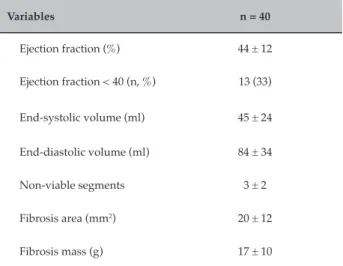
Table 2 describes RV function and the variables analyzed by LGE-CMR. Mean end-systolic volume was 45 ± 24 mL, mean end-diastolic volume was 84 ± 34 ml and mean ejection fraction was 44 ± 12%. Thirteen (33%) patients had RVD. Mean fibrosis area obtained by CRM
was 20 ± 12 mm2, mean fibrosis mass was 17 ± 10 g and
Hypertension
Diabetes
Dyslipidemia
Obesity
Smoking
Heart Failure
Graph 1 – Prevalence of cardiovascular risk factors.
Table 1 – Clinical characteristics of the study population
Variables n = 40
Demographic
Age (mean ± SD) 58 ± 8
Age ≥ 60 (n, %) 18 (45)
Male (n, %) 30 (75)
Clinical
Systemic arterial hypertension (n, %) 22 (55)
Diabetes mellitus (n, %) 10 (25)
Dyslipidemia (n, %) 8 (20)
Obesity (n, %) 10 (25)
Smoking (n, %) 16 (40)
Heart failure (n, %) 10 (25)
Stroke (n, %) 3 (8)
Coronary artery disease (n, %) 12 (30)
Table 2 – Ventricular function and variables analyzed by late gadolinium enhancement technique
Variables n = 40
Ejection fraction (%) 44 ± 12
Ejection fraction < 40 (n, %) 13 (33)
End-systolic volume (ml) 45 ± 24
End-diastolic volume (ml) 84 ± 34
Non-viable segments 3 ± 2
Fibrosis area (mm2) 20 ± 12
Fibrosis mass (g) 17 ± 10
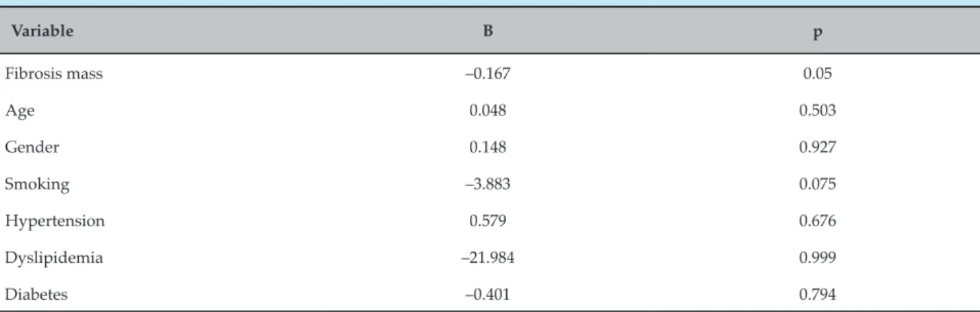
According to Pearson’s correlation, fibrosis mass and RVEF were indirectly correlated, although there was no statistical significance (r = -0.3; p = 0.08). Furthermore, multivariate logistical regression analysis showed that age, gender and hypertension were positively correlated, while smoking, dyslipidemia and diabetes
were negatively correlated with RVEF, without statistical significance (Table 3). Moreover, there was a negative correlation between RVEF and fibrosis mass (p = 0.05).
Fibrosis mass –0.167 0.05
Age 0.048 0.503
Gender 0.148 0.927
Smoking –3.883 0.075
Hypertension 0.579 0.676
Dyslipidemia –21.984 0.999
Diabetes –0.401 0.794
Table 4 – Comparison of clinical characteristics and cardiac magnetic resonance variables between study groups
Variable
Right ventricular dysfunction
p YES (n = 13) NO (n = 26)
Demographic, n (%)
Age ≥ 60 5 (38) 12 (46) 0.473
Gender (male) 9 (69) 20 (77) 0.613
Clinical, n (%)
Systemic arterial hypertension 5 (39) 16 (61) 0.358
Diabetes mellitus 2 (15) 7 (27) 0.475
Dyslipidemia 0 (0) 8 (30) 0.154
Obesity 2 (15) 5 (19) 0.953
Smoking 4 (31) 12 (46) 0.481
Heart failure 3 (23) 6 (23) 0.559
Stroke 1 (7) 2 (7) 0.537
Coronary artery disease 3 (23) 8 (31) 0.543
CMR variables (Mean ± SD)
Non-viable segments 4 ± 3 3 ± 2 0.464
Fibrosis area (mm2) 25 ± 14 18 ± 10 0.092
Fibrosis mass (g) 22 ± 12 15 ± 8 0.051
Discussion
Recent studies have been focused in the negative impact of RVD in patients with I-MI, as it is considered an important independent predictor of mortality in
these patients.13 The assessment of RV function and its
predictors enables early identification of individuals who tend to have worse outcomes and poor prognosis. The present study, in agreement with previous
reports,14-17 confirms the ability of CMR to precisely
evaluate RV function and quantify myocardial fibrosis.
In our study group, composed mostly of male and elderly patients, hypertension was the most prevalent cardiovascular risk factor (55%) followed by smoking (40%), diabetes mellitus (25%) and heart failure (25%).
Smarz et al.18 have reported a similar prevalence of these
cardiovascular risk factors in 90 patients with I-MI,except
for the prevalence of dyslipidemia of 70%, which was different from that found in our study (20%).
In the present study, RVD was evident in 33% of cases with I-MI, which was similar to the prevalence of 32%
reported in previous studies on 50 patients with I-MI.19,20
Considering that similar clinical features were observed between patients with RVD and patients with preserved ventricular function, our study could investigate, with relative precision, the association between RV function and myocardial fibrosis.
Our study revealed a negative correlation between RVEF and variables such as smoking, dyslipidemia, diabetes and fibrosis mass. The analysis showed a strong trend towards the association between RVEF and fibrosis mass (p = 0.05), indicating that grater mass of fibrosis is related to lower RVEF. This finding suggests that fibrosis is a possible predictor of RVD.
Furthermore, the current study showed an important trend towards higher mean fibrosis mass in patients with RVD compared to patients with preserved ventricular function (22 ± 12g vs 15 ± 8 g, p = 0.051). This result indicates a possible association between RVD and myocardial fibrosis within inferior wall, with clinical and prognostic significance in patients with I-MI. A similar association has been reported in a study
by Kaandorp et al.,21 which results showed higher values
of RV end-diastolic volume in the group of patients with
higher mean fibrosis mass.3
The small sample size of our study population is the main limitation to the present findings. Moreover, the amount of patients excluded (30%) due to the lack of CMR imaging data is an important limitation of the present study. Therefore, further studies with larger series are needed to confirm our findings.
Conclusions
The CMR seems to be an adequate method for risk stratification of patients with I-MI and RV dysfunction. The findings of our study indicate a possible association between myocardial fibrosis in the left ventricular inferior wall and RVD in patients with I-MI. Nevertheless, further studies with larger series are needed to confirm our findings.
Author contributions
Conception and design of the research and Critical revision of the manuscript for intellectual content: Lacerda PN, Macêdo CRB, Aras Júnior R, Fernandes AMS; Acquisition of data: Gomes Júnior AM, Lacerda PN, Pinto FGF, Almeida RF, Santos JM; Analysis and interpretation of the data: Lacerda PN, Pinto FGF, Almeida RF, Gomes Júnior AM, Santos JM, Fernandes AMS; Statistical analysis and Writing of the manuscript: Lacerda PN, Pinto FGF, Almeida RF, Gomes Júnior AM, Santos JM, Macêdo CRB, Aras Júnior R, Fernandes AMS.
Potential Conflict of Interest
No potential conflict of interest relevant to this article was reported.
Sources of Funding
There were no external funding sources for this study.
Study Association
2. Kakouros N, Cokkinos DV. Right ventricular myocardial infarction: pathophysiology, diagnosis, and management. Postgrad Med J. 2010;86(1022):719-28.
3. Galea N, Francone M, Carbone I, Cannata D, Vullo F, Galea R, et al. Utility of cardiac magnetic resonance in the evaluation of right ventricular involvement in patients with myocardial infarction. Radiol Med. 2013;119(5):309-17.
4. Rallidis LS, Makavos G, Nihoyannopoulos P. Right ventricular involvement in coronary artery disease: role of echocardiography for diagnosis and prognosis. J Am Soc Echocardiogr. 2014;27(3):223-9.
5. Rambihar S, Dokainish H. Right ventricular involvement in patients with coronary artery disease. Curr Opin Cardiol. 2010;25(5):456-63.
6. Todiere G, Aquaro GD, Piaggi P, Formisano F, Barison A, Masci PG, et al. Progression of myocardial fibrosis assessed with cardiac magnetic resonance in hypertrophic cardiomyopathy. J Am Coll Cardiol. 2012;60(10):922-9.
7. Ambale-Venkatesh B, Lima JA. Cardiac MRI: a central prognostic tool in myocardial fibrosis. Nat Rev Cardiol. 2015;12(1):18-29.
8. Allman KC, Shaw LJ, Hachamovitch R, Udelson JE. Myocardial viability
testing and impact of revascularization on prognosis in patients with coronary artery disease and left ventricular dysfunction: a meta-analysis. J Am Coll Cardiol. 2002;39(7):1151-8.
9. Mewton N, Liu Chia Y, Croisille P, Bluemke D, Lima JA. Assessment of myocardial fibrosis with cardiac magnetic resonance. J Am Coll Cardiol. 2012;57(8):891-903.
10. Barranhas AD, Santos AS, Coelho-Filho OR, Marchiori E, Rochitte CE, Nacif MS. Cardiac magnetic resonance imaging in clinical practice. Radiol Bras. 2014;47(1):1-8.
11. Simonetti OP, Kim RJ, Fieno DS, Hillenbrand HB, Wu E, Bundy JM, et al. An improved MR imaging technique for the visualization of Myocardial infarction. Radiology 2001;218(1):215-23.
1994;330(17):1211-7.
14. Masci PG, Francone M, Desmet W, Ganame J, Todiere G, Donato R, et al. Right ventricular ischemic injury in patients with acute ST-segment elevation myocardial infarction: characterization with cardiovascular magnetic resonance. Circulation. 2010;122(14):1405-12.
15. Kelle S, Roes SD, Klein C, Kokocinski T, de Roos A, Fleck E. Prognostic value of myocardial infarct size and contractile reserve using magnetic resonance imaging. J Am Coll Cardiol. 2009;54(19):1770-7.
16. Jensen CJ, Jochims M, Hunold P, Sabin GV, Schlosser T, Bruder O. Right ventricular involvement in acute left ventricular myocardial infarction: prognostic implications of MRI findings. AJR Am J Roentgenol. 2010;194(3):592-8.
17. Gerber BL, Rousseau MF, Ahn SA, le Polain de Waroux JB, Pouleur AC, Phlips T. Prognostic value of myocardial viability by delayed-enhanced magnetic resonance in patients with coronary artery disease and low ejection fraction: impact of revascularization therapy. J Am Coll Cardiol. 2012;59(9):825-35.
18. Smarz K, Zaborska B, Jaxa-Chamiec T, Maciejewski P, Budaj A. Right ventricular dysfunction and exercise capacity after inferior (posterior) wall acute myocardial infarction. Am J Cardiol. 2012;110(6):784-9.
19. Liaqat A, Asghar N, Rehman A. In hospital Outcome of acute inferior with right ventricular or posterior wall myocardial infarction. Ann Pak Inst Med Sci. 2013;9(4):219-24.
20. Iqbal A, Muddarangappa R, Shah SKD, Vidyasagar S. A study of right ventricular infarction in inferior wall myocardial infarction. J Clin Sci Res. 2013;2:66-71.