Review Article
Magnetic Resonance Imaging as Image Diagnosis in Heart
Valve Disease
Marcelo Nigri, Carlos Eduardo Rochitte, Flávio Tarasoutchi, Max Grinberg
Instituto do Coração do Hospital das Clínicas - FMUSP - São Paulo, SP, BrazilMailing address: Marcelo nigri •
Dr. Enéas de Carvalho Aguiar, 44 - O5403-000 • São Paulo, SP • Brazil E-mail: [email protected]
Received on 08/12/05 • Accepted on 10/17/05
In most recent years, magnetic resonance imaging (MRI) has been establishing itself as one of the major non-invasive complementary exams in Cardiology. Among the major benefits to be pointed out are the excellent anatomic definition between tissues, the possibility of three-dimensional acquisition and reconstruction, the absence of ionizing radiation, and the use of non-nephrotoxic contrast (gadolinium) in the doses clinically used.
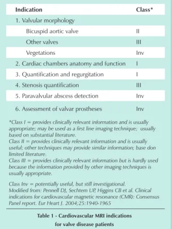
MRI has been used to assess valve disease whenever echocardiographic technique is hard to be used (improper acoustic window, for instance) and whenever there are conflicts with other exams, as heart catheterism. With the rise of new image acquisition techniques, MRI collects additional information related to accurate measure of heart chamber dimensions, ventricular function and mass, valvular regurgitation volumes and characterization of myocardial fibrosis associated to valve diseases (Tab. 1).
MRI major techniques to assess heart valves are static anatomy sequences (fast spin-echo), cine magnetic resonance,
indication class*
1. Valvular morphology
Bicuspid aortic valve II
Other valves III
Vegetations Inv
2. Cardiac chambers anatomy and function I
3. Quantification and regurgitation I
4. Stenosis quantification III
5. Paravalvular abscess detection Inv
6. Assessment of valvar prostheses Inv
*Class I = provides clinically relevant information and is usually
appropriate; may be used as a first line imaging technique; usually based on substantial literature.
Class II = provides clinically relevant information and is usually useful; other techniques may provide similar information; base don limited literature.
Class III = provides clinically relevant information but is hardly used because the information provided by other imaging techniques is usually appropriate.
Class Inv = potentially useful, but still investigational.
Modified from: Pennell DJ, Sechtem UP, Higgins CB et al. Clinical indications for cardiovascular magnetic resonance (CMR): Consensus Panel report. Eur Heart J. 2004;25:1940-1965
table 1 - cardiovascular Mri indications for valve disease patients
flow mapping through phase contrast or flow map, and more recently, myocardial delayed enhancement MRI. Cine magnetic resonance is the most commonly used technique for the assessment of heart valves. The sequence is based on the segmented acquisition of dynamic images along some heart cycles, therefore allowing the assessment of structures movement in any anatomic plane. Cine magnetic resonance allows accurate measuring of cardiac chambers diameters and volumes, as well as ventricular mass and function. Ventricular ejection fraction is calculated based on systolic and diastolic volumes of different views transversally to heart main axis, thus covering all ventricular extension, following Simpson’s1
method. As compared to bidimensional echocardiography, one advantage is that no ventricular geometric assumptions are made for this calculation. Additionally, the technique is not limited by patient’s thoracic format; it presents low intra and interobserver’s variability, and also allows right ventricle analysis, a chamber usually hard to access through echocardiogram. All those qualities make MRI an excellent exam for the follow-up of valve disease patients’ ventricular function and mass (with aortic stenosis, for instance) as well as for functional comparison after a surgical intervention2.
The technique – also known as velocity coding – is an extension to the cine magnetic resonance, and allows the determination of blood flow velocity from a vase or a cardiac valve (Fig. 1). It is an equivalent to Doppler in echocardiography, but it has the advantage of allowing flow access in any direction with no acoustic window limitation3.
Through the Bernoulli modified equation, it is also possible to determine transvalvular pressure gradient. This technique is very useful to quantify valvular disorders (stenosis or failure). However, despite technique accuracy, wide availability, and fast acquisition, it demands experience on the part of the operator in selecting most appropriate technical parameters and anatomic view scan for each clinical scenario.
Based on a detailed description of gadolinium kinetics in the myocardium that is infarcted, normal or with microvascular obstruction4 (Fig. 2) an MRI technique was developed to
clearly point out gadolinium concentration of myocardial tissue different injury levels. Described approximately 4 years ago, the delayed enhancement technique consists in the acquisition of images about 10 to 20 minutes after intravenous contrast injection (gadolinium), preceded by an inversion-recovery pulse5.
Thus, it allows the visualization of minimal myocardial necrosis/fibrosis areas, and provides excellent correlation with pathologic anatomy6 in different cardiopathies7-9.
The delayed enhancement technique has recently been
Review Article
nigri et alMAgnetic resonAnce iMAging As iMAge DiAgnosis in heArt vAlve DiseAse
Arq Bras Cardiol 2006; 87 : 485-488
Fig. 1 - Cine magnetic resonance technique showing an aortic valve with its three leaflets for transvalvular flow calculation.
Fig. 2 - Late enhancement technique in detecting infarcted myocardium shown by arrows.
used in patients with marked aortic valve disease (stenosis or failure) and normal coronary arteries (Fig. 3). Patients have been assessed through MRI before and after valvular surgery, with myocardial fibrosis having been observed in 60% of cases.
Fig. 3 - Late Enhancement Technique in aortic stenosis – myocardial fibrosis shown by arrows; A- Short axis; B- Long axis; RV(Right Ventricle).
The presence of myocardial fibrosis at MRI had correlation with the level of left ventricular dysfunction and good accuracy when compared to the myocardial biopsy carried out during the surgery10-14.
For valvular assessment a view plane perpendicular to valvular plane is used to allow for the observation of thickened, fusioned and/or lower mobility valves. Cine magnetic resonance allows the visualization of the systolic turbulence squirt generated by valvular stenosis and the measuring of cardiac chambers dimensions, thickness, and function. In aortic stenosis ventricular hypertrophy due to pressure overload, post-stenotic arterial dilation, and systolic turbulence squirt in ventricular exit can be observed. In atrioventricular valves stenosis, in its turn, major findings are atrial enlargement and diastolic turbulence squirt in ventricular entry site. There are different ways to quantify valvular stenosis using MRI. Systolic turbulence squirt observed through cine magnetic resonance may provide a clue of stenosis intensity (semi-quantitative method), but it should not be considered as an isolated parameter for valvular disorder severity. Cine magnetic resonance also helps determine valvular area through direct planimetry. Calcification – which generates MRI signal loss – may result in overestimating valvular opening area through planimetry, although generally speaking the method provides excellent correlation with echocardiography15. Flow velocity, valvular area, and transvalvular pressure gradient measures through MRI with phase contrast technique show close correlation with Doppler and catheterism in patients with mitral and aortic stenosis16,17.
One of MRI strengths in valve disease assessment is its ability in accurately measuring regurgitation volume and fraction in valvular failure. For such purpose, MRI has been considered first line imaging method in the recently published cardiovascular MRI consensus of the Magnetic Resonance Imaging Society and of the European Society of Cardiology18.
One of cardiologists’ major concern is the safety of MRI in patients with mechanic valvular prosthesis. Today, it has been made clear that it is safe to expose those using that kind of prosthesis to the magnetic fields used for MRI (1.5 Tesla)19. Magnetic power over the material is very small if compared to that of prosthesis surgical fixation. On the other hand, those prostheses result in a signal loss artifact (dark area in image) due to magnetic field distortion from its metallic content. That artifact usually extends to adjacent structures, which varies according to the pulse sequence used (usually smaller extension in spin-echo sequence). Consequently, the assessment of valvular flow turbulence squirts – especially those of low magnitude – is jeopardized. With biological prostheses, that effect is usually restricted to the valvular ring and does not significantly interfere in exam interpretation.
However, as in every exam, MRI faces limitations that must be taken into account when indicated. All cardiac pulse sequences are obtained through electrocardiographic synchronization. In patients with arrhythmia – a common condition among valve disease patients – that synchronization is jeopardized, which may compromise exam quality standard. Additionally, image acquisition is almost always carried out in some seconds’ apnea. Dyspneic or decompensated heart
A
b
Review Article
nigri et al MAgnetic resonAnce iMAging As iMAge DiAgnosis in heArt vAlve DiseAsefailure patients are usually not tolerant, which also reduces image quality. Approximately 2% of patients cannot tolerate the exam due to claustrophobia. Brain metallic implantations are a formal contra-indication to MRI, unless there is proof of no ferromagnetic properties. Finally, a pacemaker or an
implantable cardioversor-defibrillator is also considered contra-indication to the exam, although some studies have shown it to be safe in 1.5 Tesla equipment20.
Therefore, MRI has shown to be a safe method to be incorporated to the propedeutic arsenal for valve diseases.
References
1. Rehr RB, Malloy CR, Filipchuk NG, et al. Left ventricular volumes measured by MR imaging. Radiology. 1985;156:717-9.
2. Mohiaddin RH, Kilner PJ. Valvular Heart Disease. In: Manning WJ, Pennell DJ, editors. Cardiovascular Magnetic Resonance. Philadelphia, PA: Churchill Livingstone, 2002: 387-404.
3. Mohiaddin RH, Longmore DB. Functional aspects of cardiovascular nuclear magnetic resonance imaging. Techniques and application. Circulation. 1993;88:264-81.
4. Rochitte CE, Lima JA, Bluemke DA, et al. Magnitude and time course of microvascular obstruction and tissue injury after acute myocardial infarction. Circulation. 1998;98:1006-14.
5. Simonetti OP, Kim RJ, Fieno DS, et al. An improved MR imaging technique for the visualization of myocardial infarction. Radiology. 2001;218:215-23.
6. Wagner A, Mahrholdt H, Holly TA, et al. Contrast-enhanced MRI and routine single photon emission computed tomography (SPECT) perfusion imaging for detection of subendocardial myocardial infarcts: an imaging study. Lancet. 2003;361:374-9.
7. Wilson JM, Villareal RP, Hariharan R, et al. Magnetic resonance imaging of myocardial fibrosis in hypertrophic cardiomyopathy. Tex Heart Inst J. 2002;29:176-80.
8. Mahrholdt H, Goedecke C, Wagner A, et al. Cardiovascular magnetic resonance assessment of human myocarditis: a comparison to histology and molecular pathology. Circulation. 2004;109:1250-58.
9. Serra JJ, Monte GU, Mello ES, et al. Images in cardiovascular medicine. Cardiac sarcoidosis evaluated by delayed-enhanced magnetic resonance imaging. Circulation. 2003;107:e188-e189.
10. Nigri M. Fibrose miocárdica em valvopatia aórtica: estudo comparativo entre a ressonância magnética e biópsia intra-operatória miocárdica – Tese apresentada ao Departamento de Cardiopneumologia da FMUSP para obtenção do titulo de Doutor em 16/12/2004. 2004.
11. Nigri M, Rochitte C, Tarasoutchi F, et al. Myocardial fibrosis detected by MRI and biopsy is associated with worse left ventricular function in patients with severe chronic aortic valve disease. Circulation. 2003;108 [17 (S-IV)]:567 (abstract).
12. Nigri M, Rochitte C, Tarasoutchi F, et al. Delayed-enhanced magnetic resonance imaging detects myocardial fibrosis in patients with chronic aortic valve disease. J Am Coll Cardiol. 2004;41 [6 (SA)]:450 (abstract).
13. Nigri M, Grimberg M, Tarasoutchi F, et al. Myocardial delayed enhancement MRI detects myocardial fibrosis and is associated with worse left ventricular function and biopsy increased collagen content
Review Article
nigri et alMAgnetic resonAnce iMAging As iMAge DiAgnosis in heArt vAlve DiseAse
Arq Bras Cardiol 2006; 87 : 485-488
in patients with severe chronic aortic valve disease. J Cardiovasc Magn Reson. 2004;6:86 (abstract).
14. Nigri M, Rochitte C, Tarasoutchi F, et al. Relationship of myocardial fibrosis by delayed enhancement MRI with biopsy-quantified collagen content in patients with severe chronic aortic valve disease. Circulation. 2004;110:572 (abstract).
15. John AS, Dill T, Brandt RR, et al. Magnetic resonance to assess the aortic valve area in aortic stenosis: how does it compare to current diagnostic standards? J Am Coll Cardiol. 2003;42:519-26.
16. Kilner PJ, Manzara CC, Mohiaddin RH, et al. Magnetic resonance jet velocity mapping in mitral and aortic valve stenosis. Circulation. 1993;87:1239-48.
17. Caruthers SD, Lin SJ, Brown P et al. Practical value of cardiac magnetic resonance imaging for clinical quantification of aortic valve stenosis: comparison with echocardiography. Circulation. 2003;108:2236-43.
18. Pennell DJ, Sechtem UP, Higgins CB, et al. Clinical indications for cardiovascular magnetic resonance (CMR): Consensus Panel report. Eur Heart J. 2004;25:1940-65.
19. Edwards MB, Taylor KM, Shellock FG. Prosthetic heart valves: evaluation of magnetic field interactions, heating, and artifacts at 1.5 T. J Magn Reson Imaging. 2000;12:363-9.
20. Martin ET, Coman JA, Shellock FG, et al. Magnetic resonance imaging and cardiac pacemaker safety at 1.5-Tesla. J Am Coll Cardiol. 2004;43:1315-24.