Use of an Osteoplastic Flap for the Prevention of
Mastoidectomy Retroauricular Defects
Ricardo Ferreira Bento
1Robinson Koji Tsuji
1Anna Carolina de Oliveira Fonseca
1Ricardo Dourado Alves
11Department of Otolaryngology, Universidade de São Paulo, São Paulo, São Paulo, Brazil
Int Arch Otorhinolaryngol 2017;21:151–155.
Address for correspondence Ricardo Dourado Alves, MD, Department of Otolaryngology, Universidade de São Paulo, Av Dr Enéas de Carvalho Aguiar, 255, sala 6167, São Paulo, SP, 05403-0000, Brazil
(e-mail: [email protected]).
Introduction
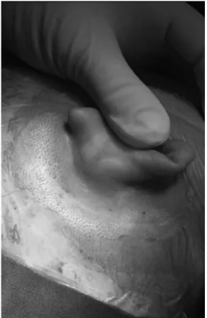
A most common complaint of patients after mastoidectomy is depressions in the mastoid cortical bone in the retroauricular region, which is the surgical site (►Fig. 1). This depression
occurs mainly in procedures that require wide openings on the mastoid, such as: open or closed mastoidectomy for chronic ear and cholesteatoma; translabyrinthine approach for tumors of the acoustic nerve; congenital cholesteatomas;
decompression of the facial nerve; approaches to endolym-phatic sac; and cochlear implants, among others.
For closing the cavity resulting from cell removal, most surgeons simply overlay the cavity with connective tissue and skin, and then suture it in two layers, while some overlay it with a muscle periostealflap.1
Invariable retractions arise after surgery, with more or less intensity in all patients and remain even after years of surgery, even when using materials such as muscle or fat
Keywords
►
mastoid
►
ear deformities
►
acquired
►
otologic surgical
procedures
►
vascularized bone
fl
ap
►
wound closure
techniques
Abstract
Introduction
After mastoidectomy, patients usually complain of bone depressions in
the retroauricular region in the surgical site, especially in procedures that require
extensive cortical resections. This causes inconveniences such as dif
fi
culty wearing
glasses, cleaning, and aesthetics complaints.
Objective
This study aims to describe a vascularized
fl
ap surgical technique that uses
the mastoid cortical bone adhered to the periosteum, which is pedicled on the anterior
portion and repositioned at the end of the surgery. This ensures the coverage of the
mastoid cavity generated by surgery and prevents ear retraction into the cavity. This
preliminary report describes the technique and intraoperative and immediate
postop-erative complications.
Methods
After retroauricular incision, periosteal exposure is performed. A U-shaped
incision is required for the procedure and delimits a periosteum area appropriate to the
size of the mastoidectomy. The cortical bone is opened using a 2.5 mm drill around the
perimeter of the
“
U,
”
at a 3 mm depth. A chisel is introduced through the surface cells of
the mastoid, and a hammer evolves into the anterior direction. The
fl
ap is lifted, leaving
the periosteum adhered to it and forming a cap. The
fl
ap is anteriorly
fi
xed to not hinder
the surgery, and repositioned at the end. The periosteum is then sutured to the adjacent
periosteum.
Results
The
fi
rst 14 cases had no intraoperative complications and were
fi
rm and
stable when digital pressure was applied during the intraoperative and immediate
postoperative periods.
Conclusion
The osteoplastic
fl
ap pedicle is a safe and simple procedure, with good
results in the immediate postoperative period.
received January 5, 2016 accepted April 12, 2016 published online May 30, 2016
DOI http://dx.doi.org/ 10.1055/s-0036-1584266. ISSN 1809-9777.
Copyright © 2017 by Thieme-Revinter Publicações Ltda, Rio de Janeiro, Brazil
underneath the muscle periostealflap to obliterate the cavity and close dura mater defects or prevent cerebrospinalfluid leaks.
The most common complaints of patients include pain and discomfort to the touch, uncomfortable feeling while wearing glasses because of the contact of the temple tips with the depression caused by the surgery, accumulation of skin peeling, difficulty of dirt removal, and aesthetic problems. It is common tofind dirt accumulation in bone depressions when examining patients who underwent mastoidectomy surgery (►Fig. 2).
In addition to these complaints and aesthetic problems, we know that the mastoid cavity maintains the middle ear
pressure, acting as a reservoir of air, and aids in the effective conduction of sound as a sound box.1Reconstruction of the mastoid defect is desirable to maintain the mastoid cavity closest to its physiological function, preventing the growth of
fibrous tissue into the surgical cavity.
Several types offlaps have been described to minimize depression, as reported by Yanagihara et al2who found good results with mastoid cortical bone reconstructions using bone pate. Muscular or periostealflaps were also used, but favor the formation and invasion of the cavity byfibrosis and do not prevent depression.1
Yuen and Chen3synthesized the main reconstruction options available for the correction of bone defects in schwannoma resection surgeries via the presigmoid approach to prevent further depression of the region. These options are divided between endogenous and allogeneic materials. For the alloge-neic materials, Jung and Park4describe the correction of these depressions in revision surgeries after complaints from patients on whom they used a titanium mesh to close the resulting bone cavity. Another option is allogeneic hydroxyapatite cements, which can be prepared and molded intraoperatively. These favor osseointegration and do not cause foreign body reaction, but entail a high cost and hamper revision surgeries.3The following are endogenous options: local muscleflaps, which also exhibit volumetric shrinkage over time,5and vascularized and non-vascularized bone grafts.3
Couldwell and Fukushima6and Rica et al7described local non-vascularized bone graft techniques. In their cases, the whole periosteum was peeled off, exposing the cortical bone to be removed. The cortical bone was removed with the aid of craniotomy blades, kept in saline solution outside the body during surgery, and repositioned and fixed with titanium plates and screws at the end of the surgery. Yuen et al8 described a cranioplasty technique in schwannoma surgeries via the presigmoid approach by using an inferior pedicle osteomuscularflap, with the periosteum and sternocleido-mastoid muscle preserved, and the flap attached to bone cortical mastoid. Theflap was folded inferiorly to the tip of mastoid process and repositioned at the end of the surgery without fixation by plates and screws, just suturing the periostealflap to the periosteum around it.
The aim of this study was to describe the surgical tech-nique of using a pedicle osteoperiostealflap in the anterior region of the mastoid approach to prevent retroauricular depression and evaluate the intraoperative and immediate postoperative outcomes.
Methods
This technique was performed to date in 14 patients, of whom 5 underwent primary surgery for schwannoma vestibular resection via the pre-sigmoid approach and 9 underwent cochlear implant surgery (►Table 1). All the patients
under-went computed tomography (CT) of the temporal bones for surgical planning and preoperative evaluation of the position of the sigmoid sinus. We excluded patients with sigmoid sinuses that were lateralized and prominent due to the risk of injury duringflap creation.
Fig. 1 Late retroauricular depression in a patient who underwent schwannoma resection via the presigmoid approach.
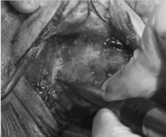
After retroauricular incision, the periosteum of the retro-auricular region was exposed. We performed a U-shaped incision, delimiting the periosteum area appropriate to the size of the mastoidectomy to be achieved (►Fig. 3). With a
small elevator, the periosteum was deviated from the entire incision to expose the mastoid cortical bone to achieve drilling (►Fig. 4).
We opened the cortical bone with a 2.5-mm drill around the perimeter of the“U,”at a depth of3mm (►Fig. 5). Then,
with the drill acting at an angle of20 degrees to the cortical bone, a boneflap3 mm thick was created around, deepen-ing to1 cm parallel to the cortical bone (►Fig. 6). We gently
introduced a chisel through the surface cells of the mastoid
Table 1 Patients already subjected to the osteoplastic flap technique
Patient Surgery Type Age (years) Sex
1 Schwannoma excision 31 Female
2 Schwannoma excision 35 Male
3 Schwannoma excision 54 Female
4 Cochlear Implant 29 Male
5 Cochlear Implant 4 Male
6 Cochlear Implant 46 Male
7 Cochlear Implant 52 Male
8 Schwannoma excision 66 Male
9 Schwannoma excision 62 Male
10 Cochlear Implant 38 Male
11 Cochlear Implant 26 Female
12 Cochlear Implant 56 Female
13 Cochlear Implant 5 Male
14 Cochlear Implant 72 Female
Fig. 3 U-shaped incision of the periosteum.
Fig. 4 Exposure of the cortical bone with the aid of a curette, removing the periosteum.
Fig. 5 Boring of the mastoid cortical bone in the“U”region.
and hammered to an anterior direction until it got close to the ear canal (►Fig. 7).
When the entire length of the osteoperiosteal flap detached from the cortical bone, it was lifted, leaving the periosteum adhered to it and forming a“cap”of bone, with the periosteum pedicled on the anterior portion (►Fig. 8).
The flap was fixed anteriorly to not hinder the surgical procedure (►Fig. 9), and the mastoid drilling was performed
normally.
After the procedure, theflap was folded over the cavity and the periosteum edges were sutured to the corresponding periosteum (►Fig. 10). The subcutaneous and skin sutures
were performed normally, in two layers.
Results
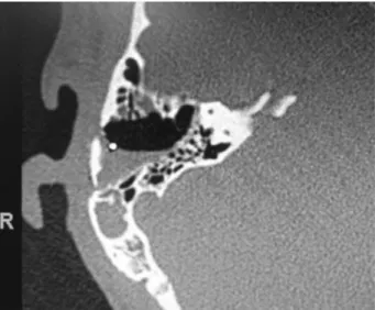
Thefirst 14 cases had no intraoperative complications, and their appearances in the intraoperative and immediate post-operative periods were adequate, staying firm and stable upon digital pressure. CT performed in the immediate post-operative period (the day after the procedure) revealed the boneflap in position and the aerated aspect of the mastoid
(►Fig. 11). The average time added to the surgical duration for
obtaining thisflap was10 minutes.
Discussion
Post-mastoidectomy retroauricular depressions are extreme-ly common and cause inconvenience to many patients. It is desirable to keep the aeration of the mastoid as close to normal so that the cavity is notfilled with fibrosis, which alters its natural function of volume balance and the gas pressure of the middle ear, as well as their compositions.9
The use of synthetic materials such as titanium meshes to cover the cavity has been successful. However, these materi-als can cause foreign body reaction, entail high costs, and require availability of different sizes during surgery.
Fig. 7 Separation of the mastoid osteoperiostealflap with the aid of a chisel and hammer.
Fig. 8 Lifting of the osteoperiostealflap.
Fig. 9 Flap anteriorfixation with suture to protect it and not hinder surgery.
Moreover, their management in revision surgery can pose a great difficulty and their reuse is not always possible. More-over, these materials are not recommended for use in surger-ies for infections such as chronic otitis media, as they may evolve with biofilm and need future removal for complete control of the infection.
The use of free grafts, bone pate, or even bone plates from bone stock has the disadvantage of not being pedicled and, therefore, non-vascularized. This makes their nutrition diffi -cult and facilitates resorption with time, becoming necrotic and easily infected.
The flap described by Yuen et al8 is also vascularized. However, unlike our proposedflap, it is inferiorly pedicled in the sternocleidomastoid muscle region. Because of this fea-ture, this technique cannot be used with minorflaps, which always require aflap that goes near the dura mater of the middle fossa to the tip of the mastoid, as this is unnecessary for most non-tumor ear surgeries. Another drawback of this technique is the handling of the sternocleidomastoid muscle, which can increase postoperative pain.
Theflap we proposed has the advantage of being obtain-able from the patient. It can be made to the size appropriate
for the intended procedure. Being pedicled, it allows for irrigation and thus minimizes the risk of infection and necrosis.
The risk of complications is minimal. The major one is the possible exposure of the sigmoid sinus when it is elevated and lateralized. Thorough evaluation of preoperative CT scans can prevent this exposure and aid inflap planning and selection of patients for whom it can be performed safely. Another advantage is that if found during surgery that it cannot be used, theflap can be converted to a periostealflap, as it has always been used.
Conclusion
The osteoplasticflap pedicle is a safe, fast, and simple proce-dure, presenting good results in the immediate postoperative period. Several cases are being followed up so that results from at least 1 year of follow-up can be reported in future publications.
References
1 Bento RF, Pinna MH, Martins G. Tratado de Otologia. 2nd edition. São Paulo, Brazil: Editora Atheneu; 2013
2 Yanagihara N, Hinohira Y, Sato H. Mastoid cortex plasty using bone pate. Otol Neurotol 2002;23(4):422–424
3 Yuen H-W, Chen JM. Reconstructive options for skull defects following translabyrinthine surgery for vestibular schwanno-mas. Curr Opin Otolaryngol Head Neck Surg 2008;16(4): 318–324
4 Jung TT, Park SK. Reconstruction of mastoidectomy defect with titanium mesh. Acta Otolaryngol 2004;124(4):440–442 5 Mehta RP, Harris JP. Mastoid obliteration. Otolaryngol Clin North
Am 2006;39(6):1129–1142
6 Couldwell WT, Fukushima T. Cosmetic mastoidectomy for the combined supra/infratentorial transtemporal approach. Technical note. J Neurosurg 1993;79(3):460–461
7 Rica CM, D’Osvaldo DH, Buchfelder M, Oviedo JD. En-bloc craniot-omy for the pre-sigmoid infra- and supratentorial approach: technical note. Acta Neurochir (Wien) 2011;153(12):2473–2478, discussion 2478
8 Yuen H-W, Thompson AL, Symons SP, Pirouzmand F, Chen JM. Vascularized mastoid boneflap cranioplasty after translabyrin-thine vestibular schwannoma surgery. Skull Base 2009;19(3): 193–201
9 Austin DF. On the function of the mastoid. Otolaryngol Clin North Am 1977;10(3):541–547