Plasma hydroxy-metronidazole/
metronidazole ratio in hepatitis
C virus-induced liver disease
1Unidade Integrada de Farmacologia e Gastroenterologia, and 2Departamento de Patologia, Faculdade de Medicina,
Universidade São Francisco, Bragança Paulista, SP, Brasil M.A.M. Marchioretto1,
C. Ecclissato1, C.M.F. da Silva1,
N.M. Cassiano1, S.A. Calafatti1,
S. Mendonça1, M.L. Ribeiro1,
G.C.R. Bernasconi1,
M.F. Degger1, H. Piovesan2
and J. Pedrazzoli Jr.1
Abstract
It has been suggested that the measurement of metronidazole clear-ance is a sensitive method for evaluating liver function. The aim of this study was to evaluate the usefulness of plasma hydroxy-metronida-zole/metronidazole ratios as indicators of dynamic liver function to detect changes resulting from the various forms of chronic hepatitis C virus (HCV) infection. A total of 139 individuals were studied: 14 healthy volunteers, 22 healthy, asymptomatic, consecutive anti-HCV-positive HCV-RNA negative subjects, 81 patients with chronic hepa-titis C(49 with moderate/severe chronic hepatitis and 34 with mild hepatitis), and 20 patients with cirrhosis of the liver. HCV status was determined by the polymerase chain reaction. Plasma concentrations of metronidazole and its hydroxy-metabolite were measured by re-verse-phase high-performance liquid chromatography with ultraviolet detection in a blood sample collected 10 min after the end of a metronidazole infusion. Anti-HCV-positive HCV-RNA-negative in-dividuals demonstrated a significantly reduced capacity to metabolize intravenously infused metronidazole compared to healthy individuals (0.0478 ± 0.0044 vs 0.0742 ± 0.0232). Liver cirrhosis patients also had a reduced plasma hydroxy-metronidazole/metronidazole ratio when compared to the other groups of anti-HCV-positive individuals (0.0300 ± 0.0032 vs 0.0438 ± 0.0027 (moderate/severe chronic hepatitis) vs 0.0455 ± 0.0026 (mild chronic hepatitis) and vs 0.0478 ± 0.0044 (anti-HCV-positive, HCV-RNA-negative individuals)). These results sug-gest an impairment of the metronidazole metabolizing system induced by HCV infection that lasts after viral clearance. In those patients with chronic hepatitis C, this impairment is paralleled by progression of the disease to liver cirrhosis.
Correspondence
J. Pedrazzoli Jr.
Unidade Integrada de Farmacologia e Gastroenterologia
Faculdade de Medicina Universidade São Francisco Av. São Francisco de Assis, 218 12916-900 Bragança Paulista, SP Brasil
Fax: +55-11-4034-1825 E-mail:
pedrazzoli@saofrancisco.edu.br
Research supported by FAPESP (to J. Pedrazzoli Jr.).
Received June 2, 2003 Accepted November 11, 2004
Key words
Introduction
Hepatitis C virus (HCV) is a blood-borne pathogen apparently endemic in most parts of the world (1), and, therefore a major public health problem (2). HVC infection is a frequent cause of chronic liver disease, progressing in the majority of patients to persistent viremia and chronic hepatitis (3-5). Mortality associated with chronic hepati-tis C results mainly from liver cirrhosis and complications such as liver carcinoma (6). The common course of chronic hepatitis C involves slow progression. However, the spectrum of liver disease is broad and pro-gression rates are extremely variable (5,7). The long-term outcome is difficult to predict since patients with chronic hepatitis are sel-dom symptomatic and end-stage liver dis-ease, when it occurs, can take more than three decades to develop (8).
Physical evaluation, the measurement of biochemical parameters, imaging procedures (such as ultrasound or computed tomogra-phy) and liver biopsy are used routinely to evaluate chronic HCV carriers and to deter-mine the degree and extension of liver injury and the prognosis of the disease. However, the extent to which these variables estimate hepatic reserve is controversial. Parameters such as albumin and coagulation factors may also be used, but they lack sensitivity and occur late in the course of chronic liver disease (9,10). Thus, a test that could dis-criminate more subtle impairments of he-patic function would be extremely useful.
Dynamic liver function tests such as in-docyanine green clearance (11), caffeine elimination (12) and lidocaine metabolism (13) have been used to complement standard liver assessment in patients with liver dis-ease. Metronidazole clearance has been re-cently proposed as a highly sensitive method for evaluating liver function (14). This method consists of measuring plasma hy-droxy-metronidazole/metronidazole ratio by high-performance liquid chromatography
(HPLC) following the intravenous adminis-tration of a single dose of metronidazole. The test has been used to show that anti-HCV-positive blood donors and chronic hepatitis C patients have a decreased capac-ity to metabolize metronidazole compared to healthy individuals, thus demonstrating its usefulness for detecting impaired liver func-tion in HCV-infected individuals, even in the absence of liver cirrhosis (14). These findings suggest that the evaluation of met-ronidazole metabolism may provide an easy-to-perform, dynamic liver function test. The present study was conducted in order to determine whether the hydroxy-metronida-zole/metronidazole test could discriminate between different stages of hepatic func-tional impairment during the various stages of chronic HCV infection as well as the impact of viral load and genotype on metro-nidazole metabolism.
Material, Subjects and Methods
Material
Metronidazole and tinidazole were pur-chased from Sigma (St. Louis, MO, USA). Hydroxy-metronidazole was synthesized at Unidade Integrada de Farmacologia e Gas-troenterologia, Universidade São Francisco, Bragança Paulista, SP, Brazil. Analytical grade potassium, dihydrogen phosphate, so-dium hydroxide, phosphoric acid, and HPLC-grade methanol were obtained from Merck S.A. Indústrias Químicas (Rio de Janeiro, RJ, Brazil). Analytical grade zinc sulfate and tetrahydrofuran were purchased from Reagen (Rio de Janeiro, RJ, Brazil) and Riedel de Haen AG (Seelze, Germany), respectively.
Subjects
pa-tients (N = 34), papa-tients with HCV-associ-ated chronic hepatitis (N = 49), and patients with HCV-associated liver cirrhosis and pre-served liver function (N = 20). All patients with circulating HCV-RNA were submitted to a percutaneous liver biopsy, unless con-traindicated.
Patients with abnormal bilirubin, albu-min, prothrombin time, ascites, and encepha-lopathy and/or those diagnosed with liver tumors were excluded from the study. Indi-viduals using drugs which could influence cytochrome P450 function were also ex-cluded.
The clinical protocol was approved by the São Francisco University Ethics Com-mittee, and the study was conducted in ac-cordance with the Declaration of Helsinki. All individuals gave written informed con-sent prior to entering the study.
Volunteer selection
Healthy volunteers were enrolled follow-ing assessment of their medical history, nor-mal physical examination and laboratory tests, including negative serology for hepati-tis B and C and the absence of alcoholism. A total of 139 individuals were included in the study: 14 healthy volunteers (males = 11, median age = 32 years, range 19-48 years), 22 healthy, asymptomatic, consecutive anti-HCV-positive HCV-RNA-negative individu-als (males = 14, median age = 37 years, range 20-63 years), 81 anti-positive HCV-RNA-positive individuals (males = 61, me-dian age = 36 years, range 22-62 years), and 20 liver cirrhosis patients (males = 13, me-dian age 44, range 29-44 years). Genotype 1 was the most prevalent. Forty-nine patients with circulating HCV-RNA had moderate/ severe chronic hepatitis (males = 37, median age 38 years, range 23-57 years), and 34 subjects had mild hepatitis (males = 24, median age 35 years, range 22-62 years) according to the criteria of Desmet et al. (15).
Anti-HCV-positive HCV-RNA-negative individuals
Asymptomatic, healthy consecutive anti-HCV-positive HCV-RNA-negative individu-als, without previous treatment for HCV in-fection, were tested for anti-HCV antibodies using an enzyme immunoassay (Abbott HCV EIA, 2nd generation, Abbott Laboratories Diagnostics Division, Chicago, IL, USA). All positive results were confirmed using a gelatin particle agglutination test with re-combinant antigens C22-3 and C200 (Serodia-HCV, Fujirebio Inc., Tokyo, Ja-pan). Anti-HCV-positive individuals were considered to have cleared HCV if serum levels of enzymes synthesized by the liver such as alanine aminotransferase (ALT), as-partate aminotransferase (AST), alkaline phosphatase, and gamma-glutamyl transpep-tidase were within the normal reference range, and if no circulating HCV-RNA could be detected by the polymerase chain reaction (PCR). Each patient was tested on three different occasions with at least one month between tests.
Non-cirrhotic hepatopathy
Anti-HCV-positive HCV-RNA individu-als had anti-HCV-positive serum (as meas-ured by EIA, circulating HCV-RNA and per-sistently abnormal liver enzymes, but no other forms of viral hepatitis (negative serol-ogy for HBV). The absence of liver cirrhosis was assessed by clinical evaluation, ultra-sonography and/or a CT scan, and was con-firmed by a liver biopsy.
HCV-associated liver cirrhosis
con-firmed by a liver biopsy. The patients in-cluded were classified as A according to the Child-Pugh scale (11). Patients with liver tumors, an ongoing bacterial infection, or renal failure (as determined by serial serum creatinine determinations) were excluded from the study. The presence of an ongoing bacterial infection was assessed by clinical evaluation, chestradiology and urine analy-sis.
Clinical laboratory analysis
Serum chemical analysis, urinalysis, a complete blood cell and platelet count, pro-thrombin, albumin, serum AST, ALT, alka-line phosphatase, γ-glutamyl transpeptidase, total bilirubin, prothrombin time, hemato-crit, and total/differential white cell counts were determined in all subjects.
Histology
Biopsy specimens were fixed in 10% formalin. Sections, 3 to 4 µm thick, were cut and stained with hematoxylin-eosin and Masson trichrome or silver for reticulin fi-bers. For each biopsy specimen, necro-in-flammation was graded and the stage of fibrosis was classified according to Desmet et al. (15). The sum of the necro-inflamma-tory and fibrosis score was considered to be the final score and used to define mild and moderate/severe chronic hepatitis. Chronic hepatitis is defined as mild if the final score was 5 or less and moderate or severe if >5.
Plasma hydroxy-metronidazole/metronidazole ratio
The study consisted of a single intrave-nous administration of metronidazole (Flagyl®, 5 mg/ml, Rhodia, São Paulo, SP, Brazil) for 20 min. At the end of the infusion, the can-nula was washed with 20 ml sterile saline. A blood sample (5 ml) was collected from an antecubital vein into EDTA-containing tubes
before and 10 min after the infusion. Blood samples were centrifuged at 2,000 g for 5 min and plasma was stored at -20ºC until assayed. The participants remained in the Clinical Pharmacology Unit during metroni-dazole administration and blood sampling.
Plasma metronidazole concentration
The plasma concentrations of metronida-zole and its hydroxy-metabolite were meas-ured by a validated reversed-phase HPLC method (14). The separation was performed on a reversed-phase Luna C18 column (250 x 4.6 mm ID, 10 µm; Phenomenex, Tor-rance, CA, USA) protected by a Security-guard C18 Security-guard column (4.0 x 3.0 mm; Phenomenex). The mobile phase consisted of 85% methanol and 15% 0.01 M potas-sium dihydrogen phosphate buffer adjusted to pH 4.5 and was eluted at a flow rate of 1.8 ml/min. The detector wavelength was set at 324 nm and peak heights were measured. For the extraction, 50 µl of the internal stan-dard (tinidazole, 100 µl in methanol) was added to 200 µl of plasma. After mixing, deproteinization was carried out by adding 20 µl 0.6 M ZnSO4 and 20 µl 0.4 M NaOH. The samples were vortex mixed and the tubes were then centrifuged at 2000 g for 5 min. Twenty microliters of each supernatant was injected into the HPLC system. The plasma hydroxy-metabolite/metronidazole ratios were determined in all participants. This method has a sensitivity of 100 ng/ml and the mean intra-assay coefficient of varia-tion (up to 25 µg/ml of both compounds) is 4%.
HCV-RNA detection, quantification and genotyping
and 211 as previously described (16). Using the enzymes RsaI-HaeIII and BstNI-HinfI, followed by digestion with ScrFI or BstUI, we were able to identify and distinguish the HCV genotypes 1a, 1b, 2a, 2b, 3a, 3b, 4, 5, and 6.
Viral load was evaluated by means of real-time quantitative RT-PCR, as previously described (17-19). The use ofa sequence detector (ABI Prism 7700; Applied Biosys-tems, Foster City, CA, USA) allows meas-urement of theamplified product in direct proportion to the increase in fluorescence emission continuously during PCR amplifi-cation. A value above or below 800,000 IU/ ml was used to define a high or low viral load (20).
Statistical analysis
Data are reported as means ± SEM unless otherwise stated. All variables were ana-lyzed by one-way ANOVA followed by the Student t-test for multiple comparisons among groups, with the level of significance set at P < 0.001.
Results and Discussion
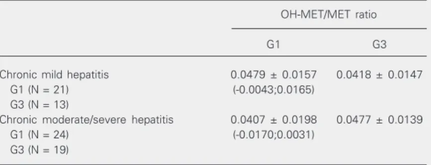
The data in Table 1 show that the plasma hydroxy-metronidazole/metronidazole ratio revealed a reduced capacity of HCV-infected individuals to metabolize intravenously in-fused metronidazole compared to healthy individuals, regardless of their HCV-RNA status, as indicated by the lower ratio com-pared to volunteers. Liver cirrhosis patients also had a reduced plasma hydroxy-metroni-dazole/metronidazole ratio when compared to the other groups of anti HCV-positive individuals (Table 1). The HCV genotype was not related to differences in the plasma hydroxy-metronidazole/metronidazole ratios among patients with chronic hepatitis (Table 2). Similar data for viral load were found (0.0443 ± 0.017 vs 0.0449 ± 0.0163 for low and high viral load, respectively; 95% CI:
Table 1. Plasma hydroxy-metronidazole/metronidazole ratios 10 min after intravenous administration of 500 mg metronidazole in patients with hepatitis C virus-induced liver disease.
Group OH-MET/MET ratio
Healthy volunteers (N = 14) 0.0742 ± 0.0232
Anti-HCV-positive HCV-RNA-negative 0.0478 ± 0.0207*+
individuals (N = 22) (0.0115;0.0414)a
(0.0072;0.0029)b
Mild chronic hepatitis (N = 34) 0.0455 ± 0.0152*+
(0.0154;0.0419)a (0.0075;0.0237)b
Moderate/severe chronic hepatitis (N = 43) 0.0438 ± 0.0187*+
(0.0183;0.0445)a (0.0050;0.0207)b
Liver cirrhosis (N = 20) 0.0300 ± 0.0143*
(0.0306;0.0579)a
(0.0072;0.0285)c
Data are reported as means ± SEM. The numbers in parentheses indicate the upper and lower limits of the 95% confidence intervals of the significant differences com-pared to ahealthy volunteers, bpatients with liver cirrhosis and canti-HCV + RNA individuals. OH-MET/MET ratio = hydroxy-metronidazole/metronidazole ratio. *P < 0.001 vs healthy volunteers, +P < 0.001 vs patients with liver cirrhosis (one-way ANOVA followed by the Student t-test).
-0.0080; 0.0070). Therefore, only two groups of patients were formed: individuals with mild hepatic histological alterations and in-dividuals with moderate/severe hepatitis.
Chronic viral hepatitis is a silently pro-gressive disease with a heterogeneous course,
Table 2. Plasma hydroxy-metronidazole/metronidazole ratios according to genotype and histological score.
OH-MET/MET ratio
G1 G3
Chronic mild hepatitis 0.0479 ± 0.0157 0.0418 ± 0.0147
G1 (N = 21) (-0.0043;0.0165)
G3 (N = 13)
Chronic moderate/severe hepatitis 0.0407 ± 0.0198 0.0477 ± 0.0139
G1 (N = 24) (-0.0170;0.0031)
G3 (N = 19)
making the clinical outcome difficult to pre-dict (7). Since standard liver tests are not useful for predicting the progress of fibrosis in chronic hepatitis patients, a truly quantita-tive test for liver function could allow a prognostic assessment of various liver dis-eases (21).
A reliable quantitative liver test would be useful for clinicians and researchers when deciding on therapeutic strategies. The search for such a test has been going on for many years and is complicated by the lack of a universally accepted standard. The forma-tion of monoethylglycinexylidide (MEGX), the main lidocaine metabolite, has been sug-gested as a simple and valuable liver func-tion test (22-24). However, the administra-tion of lidocaine may produce side effects in a high percentage of individuals tested (22), the formation of MEGX may be influenced by age and gender (24,25), and the lidocaine dose to be administered and the time point for measuring the MEGX concentration are still controversial (26-28). The MEGX test has also failed to discriminate between healthy volunteers and patients with chronic hepatitis (10,29).
The present results demonstrate the ability of the hydroxy-metronidazole/metronidazole ratio to discriminate between healthy indi-viduals and patients with Child-Pugh class A liver cirrhosis, chronic C hepatitis, or even those with a past HCV infection (Table 2).
Regarding the potential usefulness of the hydroxy-metronidazole/metronidazole ratio in early HCV chronic liver disease, we have shown that this test can discriminate be-tween Child-Pugh class A liver cirrhosis and chronic hepatitis C or anti-HCV-positive and RNA-negative individuals, but not between
the latter patient group and those with chronic hepatitis C, regardless of the severity of the disease (Table 2). These results suggest that HCV infection, regardless of viral load or genotype, causes abnormalities in liver func-tion, even though viral clearance may have already occurred, and that these abnormali-ties progress with further progression of liver disease.
Several viral and bacterial infections or interferon-inducing agents have been asso-ciated with impaired cytochrome P450-me-diated drug metabolism. This effect may be partly mediated by the release of cytokines or related to autoimmune-mediated phenom-ena which might be triggered by HCV (30-37). This immunological imbalance could lead to interference with drug metabolizing systems during the acute phase of hepatitis C and may persist, even when the infection is self-limited.
References
1. Wasley A & Alter MJ (2000). Epidemiology of hepatitis C: geo-graphic differences and temporal trends. Seminars in Liver Disease, 20: 1-16.
2. Wiese M, Berr F, Lafrenz M, Porst H & Oesen U (2000). Low frequency of cirrhosis in a hepatitis C (genotype 1b) single-source outbreak in Germany: a 20-year multicenter study. Hepatology, 32: 91-96.
3. Alter MJ, Margolis HS, Krawczinski K et al. (1992). The natural history of community-acquired hepatitis C in the United States. The sentinel counties chronic non-A, non-B hepatitis study team. New England Journal of Medicine, 327: 1899-1905.
4. National Institutes of Health Consensus Development Conference Panel Statement (1997). Management of hepatitis C. Hepatology, 26 (Suppl 1): 2S-10S.
5. EASL International Consensus Conference on Hepatitis C (1999). Consensus Conference. Journal of Hepatology, 30: 956-961. 6. Poynard T, Bedossa P & Opolon P (1997). Natural history of liver
fibrosis progression in patients with chronic hepatitis C. The Obsvirc, Metavir, Clinivir, and Dosvirc groups. Lancet, 349: 825-832. 7. Alberti A, Chemello L & Benvegnu L (1999). Natural history of
hepatitis C. Journal of Hepatology, 31 (Suppl 1): 17-24.
8. Tong MJ, el-Farra NS, Reikes AR & Co RL (1995). Clinical outcome after transfusion-associated hepatitis C. New England Journal of Medicine, 332: 1463-1466.
9. Herold C, Heinz R, Niedobitek G, Schneider T, Hahn EG & Schuppan D (2001). Quantitative testing of liver function in relation to fibrosis in patients with chronic hepatitis B and C. Liver, 21: 260-265. 10. Elin RJ, Fried MW, Sampson M, Ruddel M, Kleiner DE & DiBisceglie
AM (1997). Assessment of monoethylglycinexylidide as measure of liver function for patients with chronic viral hepatitis. Clinical Chem-istry, 43: 1952-1957.
11. Albers I, Hartmann H, Bircher J & Creutzfeldt W (1989). Superiority of the Child-Pugh classification to quantitative liver function tests for assessing prognosis of liver cirrhosis. Scandinavian Journal of Gastroenterology, 24: 269-276.
12. Wang T, Kleber G, Stellaard F & Paumgartner G (1985). Caffeine elimination: a test of liver function. Klinische Wochenschrift, 63: 1124-1128.
13. Shiffman ML, Fisher RA, Sanyal AJ, Edinboro LE, Luketic VA, Purdum 3rd PP, Raymond P & Posner MP (1993). Hepatic lidocaine metabolism and complications of cirrhosis. Implications for assess-ing patient priority for hepatic transplantation. Transplantation, 55: 830-835.
14. da Silva CMF, David FL, Muscara MN, Sousa SS, Ferraz JG, de Nucci G, Polimeno NC & Pedrazzoli Jr J (1998). Plasma hydroxy metronidazole/metronidazole ratio in anti-HCV carriers with and with-out apparent liver disease. British Journal of Clinical Pharmacology, 46: 176-180.
15. Desmet VJ, Gerber M, Hoofnagle JH, Manns M & Scheuer PJ (1994). Classification of chronic hepatitis: diagnosis, grading and staging. Hepatology, 19: 1513-1520.
16. Chan SW, McOmish F, Holmes EC, Dow B, Peutherer JF, Follett E, Yap PL & Simmonds P (1992). Analysis of a new hepatitis C virus type and its phylogenetic relationship to existing variants. Journal of General Virology, 73 (Part 5): 1131-1141.
17. Holland PM, Abramson RD, Watson R & Gelfand DH (1991). Detec-tion of specific polymerase chain reacDetec-tion product by utilizing the 5'3'-exonuclease activity of thermus aquaticus DNA polymerase. Proceedings of the National Academy of Sciences, USA, 88:
7276-7280.
18. Bukh J, Purcell RH & Miller RH (1994). Sequence analysis of the core gene of 14 hepatitis C virus genotypes. Proceedings of the National Academy of Sciences, USA, 91: 8239-8243.
19. Smith DB, Mellor J, Jarvis LM, Davidson F, Kolberg J, Urdea M, Yap PL & Simmonds P (1995). Variation of the hepatitis C virus 5’non-coding region: implications for secondary structure, virus detection and typing. The International HCV Collaborative Study Group. Jour-nal of General Virology, 76 (Part 7): 1749-1761.
20. Pawlotsky JM, Bouvier-Alias M, Hezode C, Darthuy F, Remire J & Dhumeaux D (2000). Standardization of hepatitis C virus RNA quan-tification. Hepatology, 32: 654-659.
21. Mion F, Rosseau M, Scoazec JY, Berger F & Minaire Y (1999). [C13 ]-Galactose breath test: correlation with liver fibrosis in chronic hepa-titis C. European Journal of Clinical Investigation, 29: 624-629. 22. Shiffman ML, Luketic VA, Sanyal AJ, Duckworth PF, Purdum 3rd
PP, Contos MJ, Mills AS, Edinboro LE & Poklis A (1994). Hepatic lidocaine metabolism and liver histology in patients with chronic hepatitis and cirrhosis. Hepatology, 19: 933-940.
23. Huang YS, Lee SD, Deng JF, Wu JC, Lu RH, Lin YF, Wang YJ & Lo KJ (1993). Measuring lidocaine metabolite-monoethylglycinexylidide as a quantitative index of hepatic function in adults with chronic hepatitis and cirrhosis. Journal of Hepatology, 19: 140-147. 24. Testa R, Caglieris S, Risso D, Arzani L, Campo N, Alvarez S, Giannini
E, Lantieri PB & Celle G (1997). Monoethylglycinexylidide formation measurement as a hepatic function test to assess severity of chronic liver disease. American Journal of Gastroenterology, 92: 2268-2273.
25. Orlando R & Palatini P (1997). The effect of age on plasma MEGX concentrations. British Journal of Clinical Pharmacology, 44: 206-208.
26. Fabris L, Jemmolo RM, Toffolo G et al. (1999). The monoethylgly-cinexylidide test for grading of liver cirrhosis. Alimentary Pharmacol-ogy and Therapeutics, 13: 67-75.
27. Reichel C, Wienkoop G, Nacke A, Sudhop T, Spengler U & Sauerbruch T (1995). Which lidocaine dose should be used for the MEGX liver function test? Journal of Hepatology, 22: 600. 28. Reichel C, Nacke A, Sudhop T, Wienkoop G, Luers C, Hahn C, Pohl
C, Spengler U & Sauerbruch T (1997). The low-dose monoethylgly-cinexylidide test: assessment of liver function with fewer side effects. Hepatology, 25: 1323-1327.
29. Testa R, Campo N, Caglieris S, Risso D, Alvarez S, Arzani L, Giannini E, Lantieri PB & Celle G (1998). Lidocaine elimination and monoeth-ylglycinexylidide formation in patients with chronic hepatitis or cir-rhosis. HepatoGastroenterology, 45: 154-159.
30. Renton KW & Knickle LC (1990). Regulation of hepatic cytochrome P-450 during infectious disease. Canadian Journal of Physiology and Pharmacology, 68: 777-781.
31. Thal C, el Kahwaji J, Loeper J, Tinel M, Doostzadeh J, Labbe G, Leclaire J, Beaune P & Pessayre D (1994). Administration of high doses of human recombinant interleukin-2 decreases the expres-sion of several cytochromes P-450 in the rat. Journal of Pharmacol-ogy and Experimental Therapeutics, 268: 515-521.
32. Abdel-Razzak Z, Loyer P, Fautrel A, Gautier JC, Corcos L, Turlin B, Beaune P & Guillouzo A (1993). Cytokines down-regulate expres-sion of major cytochrome P-450 enzymes in adult human hepato-cytes in primary culture. Molecular Pharmacology, 44: 707-715. 33. Tapner M, Liddle C, Goodwin B, George J & Farrell GC (1996).
primary cultures of well-differentiated rat hepatocytes. Hepatology, 24: 367-373.
34. Lunel F, Abuaf N, Frangeul L et al. (1992). Liver/kidney microsome antibody type 1 and hepatitis C virus infection. Hepatology, 16: 630-636.
35. Tran A, Quaranta JF, Benzaken S et al. (1993). High prevalence of thyroid autoantibodies in a prospective series of patients with chronic hepatitis C before interferon therapy. Hepatology, 18: 253-257.
36. Michel G, Ritter A, Gerken G, Meyerzum Buschenfelde KH, Decker R & Manns MP (1992). Anti-GOR and hepatitis C virus in autoim-mune liver diseases. Lancet, 339: 267-268.