ISSN 0100-879X
BIOMEDICAL SCIENCES
AND
CLINICAL INVESTIGATION
www.bjournal.com.br
www.bjournal.com.br
Volume 44 (8) 729-813 August 2011
Braz J Med Biol Res, August 2011, Volume 44(8) 729-737
doi: 10.1590/S0100-879X2011007500091
Liposomal photosensitizers: potential platforms for anticancer
photodynamic therapy
L.A. Muehlmann, G.A. Joanitti, J.R. Silva, J.P.F. Longo and R.B. Azevedo
Institutional Sponsors
The Brazilian Journal of Medical and Biological Research is partially financed by
Faculdade de Medicina de Ribeirão Preto Campus
Ribeirão Preto
Ex plor e H igh - Pe r for m a n ce M S Or bit r a p Te ch n ology I n Pr ot e om ics & M e t a bolom ics
Liposomal photosensitizers:
potential platforms for anticancer
photodynamic therapy
L.A. Muehlmann*, G.A. Joanitti*, J.R. Silva, J.P.F. Longo and R.B. Azevedo
Departamento de Genética e Morfologia, Instituto de Biologia, Universidade de Brasília, Brasília, DF, Brasil
Abstract
Photodynamic therapy is a well-established and clinically approved treatment for several types of cancer. Antineoplastic photo-dynamic therapy is based on photosensitizers, i.e., drugs that absorb photons translating light energy into a chemical potential that damages tumor tissues. Despite the encouraging clinical results with the approved photosensitizers available today, the prolonged skin phototoxicity, poor selectivity for diseased tissues, hydrophobic nature, and extended retention in the host organism shown by these drugs have stimulated researchers to develop new formulations for photodynamic therapy. In this context, due to their amphiphilic characteristic (compatibility with both hydrophobic and hydrophilic substances), liposomes have proven to be suitable carriers for photosensitizers, improving the photophysical properties of the photosensitizers. Moreover, as nanostructured drug delivery systems, liposomes improve the efficiency and safety of antineoplastic photodynamic therapy, mainly by the classical phenomenon of extended permeation and retention. Therefore, the association of photosensitizers with liposomes has been extensively studied. In this review, both current knowledge and future perspectives on liposomal carriers for antineoplastic photodynamic therapy are critically discussed.
Key words: Photodynamic therapy; Liposome; Antineoplastic therapy; Cancer; Photosensitizer
Introduction
Correspondence: L.A. Muehlmann, Departamento de Genética e Morfologia, Instituto de Ciências Biológicas, Campus Darcy Ribeiro, 70910-000 Brasília, DF, Brasil. E-mail: [email protected]
*These authors contributed equally to this study.
Received March 24, 2011. Accepted June 28, 2011. Available online July 22, 2011. Published August 19, 2011.
The cytotoxic effects of photodynamic therapy (PDT)
on tumor tissues were first described at the beginning of
the 20th century, but this therapy would be widely used in medical services only in the 1990s. Nowadays, PDT is a well-established antineoplastic therapy with several com-mercially available protocols approved for different types of cancer. It has proven to be a cost-effective treatment compared to other traditional therapies (1), and this may be due to the low costs of PDT equipment and hospital facilities in comparison to the requirements for other therapies such as surgery, chemotherapy and radiotherapy. All of these advantages have stimulated the emergence of several centers for PDT treatment worldwide, including emerging countries such as Brazil and China (1,2).
PDT is based on the delivery of photosensitizer drugs (PSs) to biological targets, which are then irradiated with
light at specific wavelengths (3). There are many different kinds of PS, each one with its specific excitation wavelength.
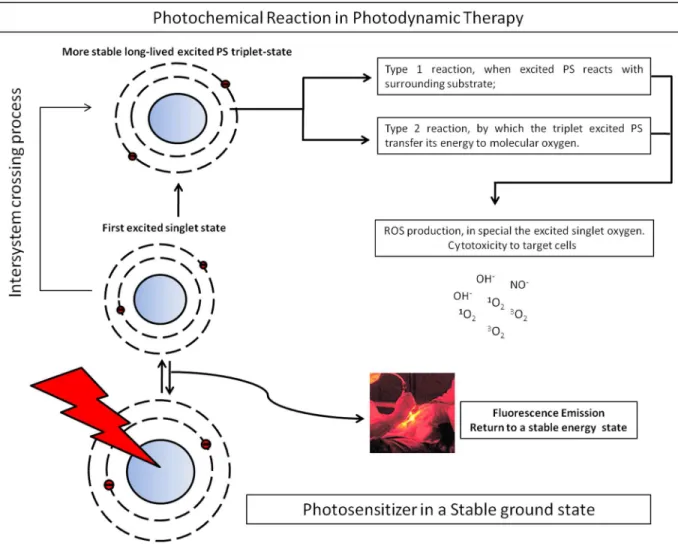
However, the useful spectral window for PDT application in
biological tissues is limited by the absorption of light by both water - at wavelengths above 1000 nm - and hemoglobin/ myoglobin - at wavelengths below 600 nm (4). Therefore, the excitation wavelength must be in the 600-1000-nm range so that the light can cross the cells without being ab-sorbed before reaching the PS. During PS photoactivation, an excitable electron is promoted to a higher energy level
(antibonding orbital) and the PS reaches the first excited
singlet state. This excited state decays to a lower energy
level by emitting fluorescence or, alternatively, it decays
730 L.A. Muehlmann et al.
nucleic acids and proteins - after PS irradiation. Didactically, the direct reaction between triplet PS and a biomolecule is called the Type 1 reaction and this process leads to PS blanching; the production of singlet oxygen by the triplet PS is called the Type 2 reaction and it regenerates the ground-state PS (4) (Figure 1).
PSs are classified as 1st, 2nd, and 3rd generation, or
according to their respective chemical class (e.g., phenoti-azine, porphyrins, chlorins, phthalocyanines) (3,5). Despite their effectiveness on tumor cells, the majority of these PSs have shown limitations to their use in clinical applications, which include extended retention in the host organism, substantial skin phototoxicity, low solubility in physiologi-cal solutions, low selective accumulation in the diseased tissue, and inappropriate half-lives within tissue, impairing
the optimization of illumination schedules (6).
Several approaches were tested in order to avoid the undesired effects described above and the employment of drug delivery systems has then emerged as a new
strat-egy to improve the efficacy and safety of PDT. The drug
delivery system is expected to incorporate high amounts of the PS without loss or alteration of its activity, to selec-tively accumulate the PS in the diseased tissue, to deliver therapeutic amounts of PS to the target site, to protect the PS from degradation and from premature clearance, to minimize drug leakage during transit to target, and to facilitate PS interaction with cells; moreover, the delivery system must be biodegradable and should induce little or no immunogenicity (7).
In this context, it is not by chance that liposomes are
Figure 1. When excited by light at a specific wavelength, the photosensitizer (PS) reaches its first excited singlet state. This state
one of the most extensively studied drug delivery systems
and represent a significant proportion of nanotechnology
research in biomedicine today (8). These lipid nanopar-ticles possess several features that are desired in a drug delivery system (9). Moreover, a large number of investi-gations on the subject of liposomes have provided some crucial improvements in liposomal formulations, such as: prolonged circulation time in the bloodstream (PEGylated liposomes), improved dose and target selectivity
(immuno-liposomes, tumor-specific ligands, pH-sensitive and mag
-netoliposomes) and enhanced cell internalization (cationic liposomes) (7). However, despite their advantages, some kinds of liposomes are not in current use inasmuch as none or few studies showing their application for PDT have
been reported since their first
description. Therefore, only those liposome approaches showing encouraging in vivo
results for anticancer PDT are described in the pres-ent review. In addition, new perspectives on liposome-based antineoplastic PDT are discussed.
General aspects of
liposomes
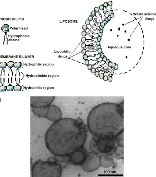
Liposomes are artificial
vesicles generally ranging in size from 20 to 1000 nm (8), composed mainly of a phos-pholipid bilayer surrounding an aqueous core (see Figure 2). The phospholipids em-ployed to produce liposomes can be synthetic or derived from natural sources. Their structure is characterized by 2 regions - polar head and hydrophobic long hydro-carbon acyl chains - which
can be modified to suit the
desired liposome size and shape (9).
The structures of both the hydrophobic and hydro-philic regions of a phospho-lipid determine its shape. For example, phospholipids such as phosphatidylcho-line, phosphatidylserine and sphingomyelin are known to be cylindrical-shaped lipids because the diameter of their
polar group is similar to that of their hydrophobic part, caus-ing them to spontaneously form bilayers when dispersed
in an aqueous medium. On the other hand, phosphati
-dylethanolamine and cardiolipin are cone-shaped lipids
because the diameter of their polar head is significantly
different from that of their hydrophobic region - these lipids are also called non-bilayer lipids as they preferentially form a hexagonal structure (depending on the conditions of the medium) (9).
Since there is a wide variety of phospholipids, it is possible to change the liposome size, charge, and surface properties by adding new ingredients to the lipid mixture. Therefore, it is important to select phospholipid combina-tions and to standardize appropriate PS concentracombina-tions
B
Figure 2.A, Liposomes are basically composed of a phospholipid bilayer surrounding an aque-ous core; they function as containers in which several chemically different compounds can be entrapped or to which they can be attached. Hydrophobic drugs can be associated with the lipid membrane while the hydrophilic ones can be dissolved in the aqueous core. B, Transmission electron microscopy images of liposomes. The small black particles are artifacts produced during sample preparation for microscopy.
732 L.A. Muehlmann et al.
and entrapment methods in order to improve PS-loaded liposomes. In general, the methods frequently employed to
encapsulate PSs are rehydration of a thin lipid film (classic
Bangham technique), ethanol injection and freeze/thawing (10-12). It should also be taken into account that the chosen method - sometimes involving mechanical energy, chemical reactions or temperature variation steps - should neither affect PS properties nor reduce its activity.
The stability of the liposomes should also be evaluated since the association of PS with liposomes has been shown to alter the thermal phase behavior of the latter (10,13). This parameter is intrinsically dependent on the
phospholipid-specific phase transition temperature at which the molecules are reorganized from a rigid bilayer to a fluid bilayer. It has been reported that the association of the PS temoporfin
resulted in a concentration-dependent decrease in the phase transition temperature of dipalmitoylphosphatidylcholine-based liposomes (13). When the lipid composition was
modified with phospholipids endowed with longer hydro
-phobic acyl chains (distearoylphosphatidylcholine), phase transitions were well above body temperature even at high PS load (13). Moreover, the lipid bilayer phase transition
can also be modified with the addition of cholesterol, where
increasing amounts of cholesterol increase liposome rigidity
and stability in biologic fluids (13).
Photosensitizer liposome formulations
One of the first PSs used for cancer therapy consisted
of a complex mixture of several partially unidentified por
-phyrins. Besides their effectiveness in PDT treatments, poor selectivity for diseased tissues, high dose, extended retention in the host organism, and low wavelength acti-vation (630 nm) were the main drawbacks (7,14). These drawbacks stimulated the development of new PSs show-ing fast elimination from the body, fewer side effects, and a higher absorption peak (650-800 nm) (6,7,15).
Despite the improvements achieved, these molecules present low solubility in physiological solutions (6,7,15). For instance, both phthalocyanines and chlorins are hydropho-bic PSs that form aggregates in physiological solutions (16).
When aggregated, the PSs are far less efficient in translating
light energy into a chemical potential. This happens because
the effect of quenching is amplified in these aggregates,
i.e., the aggregated molecule is not able to absorb light or, even though it absorbs light, the photoexcited PS decays to the ground-state before producing singlet oxygen (16). Moreover, PS aggregates also hamper parenteral admin-istration (such as intravenous injection) and the delivery of
the PS drugs to targeted tissues (6,7). One may think that
the use of a hydrophilic PS should minimize the drawbacks shown by hydrophobic PSs, but the hydrophilicity raises problems related to the interaction with biological tissues. In fact, it has been reported that the hydrophilic photosensitizer aminolevulinic acid (ALA) is not well internalized by cells
and has no specificity for diseased tissues (6). Therefore,
the use of PSs associated with nanoparticles could be an interesting approach in order to avoid PS aggregation and to deliver the PS into the target site.
In this context, liposomes are suitable delivery systems for carrying hydrophilic or hydrophobic PSs, improving their clinical application (17). The liposome structure enables the entrapment of hydrophilic PSs in the aqueous core and the attachment of the hydrophobic ones to the phospholipid bilayer; consequently, the interaction of hydrophilic PSs with cells is enhanced and the hydrophobic PSs are kept
in their monomeric configuration even in physiological
aqueous media. In general, hydrophobic PSs have been attached to small unilamellar vesicles (20 to 100 nm) - whereas hydrophilic PSs have been entrapped in large unilamellar vesicles (100-500 nm) because of their large aqueous core (9,18).
Dermal delivery
Superficial cutaneous tumor lesions are directly acces
-sible for topic application of PS molecules onto the disease site (12), minimizing eventual concerns involved in
intrave-nous administration. Nevertheless, the efficient penetration
of the PS into skin layers is essential to achieve successful PDT results (12). Some studies have shown that liposomal formulations, associated with organic solvents, terpenes, edge activators (sodium cholate, polysorbate 80 or polysor-bate 20) and ethanol, enhance PS penetration through the skin, even to an extent comparable to that of subcutaneous administration (12,19). This enhanced penetration feature
is attributed to the high elasticity, small size and high flex
-ibility shown by these vesicles, which are therefore able to squeeze out through narrow constrictions of the skin layers and/or to interact with skin stratum corneum lipids, to solubilize these molecules and to create some channels enabling their permeation through the skin (12,19).
Fang et al. (20), using an animal skin permeation protocol, evaluated ethosomes - liposomes containing a relatively high concentration of ethanol - as PS dermal delivery systems. The PS used was ALA, a precursor of the PS protoporphyrin IX (PpIX), formed in vivo after exogenous ALA application. The results showed that PpIX concentration in the inner skin layers was enhanced when ALA was carried by ethosomes compared to conventional liposomes. The same group investigated the permeation ability of topically applied ALA-containing ethosomes in a hyperproliferative mouse skin model. Ethosome vehicle formulation showed a 3.64-fold increase in PpIX detection in mouse skin when compared to ALA aqueous formulation vehicle (20). Dragicevic-Curic et al. (21) showed that the PS meta-tetrahydroxyphenylchlorin (mTHPC) associated with conventional liposomes showed lower skin penetration than ethosomes formulated with 20% ethanol. The same
growth of the very invasive human colorectal carcinoma HT29 in mice after topical application of mTHPC associated with liposomes obtained by a combination of phospholipids, ethanol and terpenes (12).
In order to improve dermal delivery, the association of
liposome with ionophoresis (to enhance the flux of ionic
compounds across the skin) has been suggested (22). Taken together, all the experiments described here show that it is possible to improve the topical delivery of PSs simply by incorporating new components into the liposome formulation, once more proving the versatility of this drug
delivery system. Other liposomal formulations for topi
-cal drug delivery and their related patents are described elsewhere (23).
Intravenous delivery
The interaction of conventional liposomes with a diseased tissue after intravenous administration is not
immediate. Once administered by the intravenous route,
the liposomes are exposed to several proteins, cells and tissues that can reduce or even totally prevent them from reaching their target site. For example, it is known that liposomes are rapidly eliminated from the bloodstream due to surface opsonization and later phagocytosis by the reticuloendothelial system (RES), a physiological system mainly consisting of macrophages resident in the liver, spleen and lymphatic system, responsible for the elimina-tion of several macromolecules and particles from the body (22,24). Moreover, the interaction of liposomes with plasma
lipoproteins - which destabilize the vesicles - significantly
reduces the circulation time of liposomes in the bloodstream and promotes the leakage of the PS before it can reach the target site (17).
The knowledge about the dynamics of particles adminis-tered to the human body has led many researchers to
sug-gest interesting modifications in the structure/composition
of liposomes in order to improve not only their stability but also their tissue selectivity and internalization. Particularly, there are some well-known questions regarding both the vasculature and the lymphatic clearance of particles at the tumor site that have created an entire line of improvements in antineoplastic liposome formulations.
It is known that tumors are supplied by leaky vessels pre-senting gaps between their endothelial cells (25). Because of this leaky vasculature, compounds and particles tend to migrate easily from the bloodstream to the diseased tissue. Furthermore, the presence of poor lymphatic drainage in these lesions also contributes to the enhanced retention time of the particles. Both the increased permeation and reduced clearance of particles in the tumor account for the passive accumulation of particles at the disease site, known as the ‘enhanced permeability and retention’ (EPR) effect (26). When the PSs are associated with liposomes ranging in size from 100 to 400 nm, the passive PS accumulation
in the disease tissue is optimized (27).
Nevertheless, the short circulation time of conventional liposomes (tens of minutes) in the bloodstream impairs their
efficient accumulation at the disease site, which depends
on the frequency at which the liposomes pass through the
tumoral vessels. Some surface modifications have been
suggested in order to reduce the interaction of liposomes with the immune system. Studies have demonstrated that the addition of a polymeric hydrophilic coat to the liposome
surface significantly increases its half-life in the bloodstream
by reducing the recognition and interaction with plasma proteins and RES macrophages (28,29). These long-circulation liposomes are known as PEGylated liposomes, due to the use of polyethylene glycol as a coating agent. In fact, PEGylated liposomes have an extended half-life in the bloodstream in comparison to conventional liposomes and subsequently enhanced PS accumulation in diseased tissues by the EPR effect, as observed by Sadzuka et al. (30). These investigators reported a higher accumulation rate of the PS coproporphyrin I incorporated into PEGylated liposomes and an improved response to PDT treatment in mice when compared to conventional liposomes and/or to
free PS. Similarly, Oku et al. (31) showed that 80% of mice
bearing a subcutaneous sarcoma were cured after PDT treatment with intravenous injection of the benzoporphyrin derivative monoacid ring A (BPD-MA) incorporated into
glucuronide-modified liposomes, while only a 20% cure rate
was observed for injected PS-conventional liposomes.
Selectivity and dose improvement
Besides prolonged bloodstream circulation time and notable tumor accumulation, some reports have claimed that the cell interaction/uptake of PS-PEGylated liposomes
is low and consequently may lead to reduced PDT efficiency. For instance, Oku et al. (31) reported that the PEGylation of
BPD-MA-liposomes enhanced the passive accumulation of the liposomal drug in tumor tissues at 3 h after
administra-tion, but did not enhance the PDT efficacy, suggesting that
the liposomes were not effectively taken up by the tumor cells before laser irradiation. Moreover, the low interaction of PEGylated liposomes with cells also reduces the amount
of reactive oxygen species (ROS) that reach the targeted cells, since ROS are generated at the PS activation site
and then diffuse through short distances (about 0.02 µm) due to their short half-life in biological tissues (3).
Besides the passive accumulation obtained by the EPR effect with liposomes within a certain size range, the conjugation of targeting molecules to the liposomal surface
is an interesting alternative for obtaining a more specific
localization of the PS, not only improving tissue selectivity but also reducing the effective PS concentration necessary to achieve successful PDT results (32). In contrast to the passive accumulation process, this approach is known as
734 L.A. Muehlmann et al.
tumor cells and vessels. Both the passive and active target-ing can work together in accumulattarget-ing the PS-containtarget-ing
liposome at the tumor site and specifically directing the PS
to the tumor cells, respectively.
The active targeting to tumor cells can be based on the fact that tumor cells aberrantly overexpress certain tumor-associated antigens that are found in low amounts or are even absent in non-tumor cells. Tumor cells overexpress, for example, receptors for vitamins (folate), growth factors, glycoproteins (transferrin) and glycolipids to support their high metabolic activity (7). Liposomes conjugated with these
tumor-specific ligands have been developed for PS delivery
in antineoplastic PDT. The treatment of cervical cancer cells overexpressing transferrin receptors with PEGylated
liposomes conjugated with transferrin significantly increased
the PS AlPcS4 cell uptake and was ten times more effective than the free AlPcS4 (33).
Several tumor-specific ligands are widely available,
relatively inexpensive and minimally immunogenic (17). Nevertheless, their use demands a careful evaluation of the overexpressed receptors present in the tumor microenvi-ronment to avoid reaching normal cells that often express receptors for the same ligands (17).
Internalization improvement
After the initial interaction of the liposome with the surface of the target cell, it is important that PS reaches the subcellular location that causes the desired damage to the target cell, be it cell death by apoptosis or by necrosis, or even just cellular stress. Internalization, instead of just adsorption, of the PS-containing liposomes by the cell is generally desired in order to induce more intense cell dam-age. Most of the internalization strategies are focused on endocytotic processes, such as phagocytosis, or on the in-vading mechanisms used by some intracellular parasites. Viruses, for example, are endowed with fusogenic pro-teins that are able to bypass the plasma membrane allowing the internalization of their genetic material (34). Fusogenic viral envelope proteins and cell penetrating peptides, such as TAT (from the HIV virus), are positively charged molecules able to adhere to the target cell by electrostatic attraction since the cell membrane possesses an overall negative charge (35). This viral strategy has inspired researchers to design liposomes containing cationic lipids or cationic peptides/polymers coupled to their surface, which have been shown to be interesting models for the delivery of PS due to the enhanced effective tissue penetration (7).
Molinari et al. (36) reported that the internalization of
the PS m-THPC in malignant glioma cells was significantly
enhanced when cationic liposomes were used as carriers. Takeuchi et al. (37) designed a cationic
polyethylenimine-modified liposome encapsulating the PS BPD-MA for anti
-angiogenic PDT and reported that the cationic liposomes maintained their phototoxicity to endothelial cells while the
effect of non-modified liposomes was suppressed (37,38).
Another study by the same research group also reported the destruction of angiogenic vessels and the subsequent apoptosis of tumor cells after iv injection of a low dose of cationic BPD-MA-liposomes on sarcoma-bearing mice (39).
Although the internalization mechanism for liposomes is not fully known, the process of endocytosis has been described as one of the most common pathways by which these particles are internalized by cells (35,40). A remark-able characteristic of this process, which must be taken into account when designing PS-containing liposomes, is that endosome vesicles entrapping liposomes fuse with the lysosome, which contains enzymes in an acid aqueous
en-vironment, before the PS is released in significant amounts
to the cytosol (40). Thus, the endosomal/lysosomal pathway can potentially damage the PS and, consequently, reduce their effectiveness. In this context, a potential advantage of cationic liposomes, which is also explored by some viruses, is that they can fuse with the endosome membrane and then release their content directly into the cytoplasm, preventing it from being degraded by lysosomal enzymes (35).
The use of cationic liposomes increases the efficiency of
S internalization for PDT treatment, thus reducing the total therapeutic dose needed. Nevertheless, cationic liposomes
do not present any selectivity for specific cells - unless that
mediated by surface charge - and they may in fact fuse with almost all types of mammalian cells (35). In order to obtain tumor selectivity and assure low side effects, the combina-tion of cacombina-tionic molecules and active-targeting moieties is necessary (41). In addition, the exact proportion of positively charged polymers and/or phospholipids to be added to a liposome formulation must be correctly calculated, since the fraction of positively charged molecules in a liposome is likely to be critical for its effective delivery, cell uptake and toxicity (35).
Liposomal formulations currently in clinical
use
Several pharmaceutical companies are investigating new PSs, PS delivery vehicles, and PDT protocols for the
treatment of different tumors (3,7). Only one liposomal for
caused by the ROS generated after photoactivation of the
liposomal PS (42).
New perspectives
PDT and hyperthermia
Tumor cells are more sensitive to temperatures in the range of 38-45°C than normal cells. This characteristic has led to the development of hyperthermia therapy, which is based on the heating of tumor tissue in order to kill neo-plastic cells. The association of PDT and hyperthermia in a single therapy has been proposed and encouraging results have already been published (43). This is an interesting approach because one treatment complements the other as oxygenated tumor cells are preferentially killed by PDT while hypoxic cells are preferentially killed by hyperthermia (44,45). Therefore, besides the advantages of liposomes in PDT itself, this type of drug delivery system could also be used to carry hyperthermia agents such as superpara-magnetic nanoparticles, associated with PSs, an approach that has a great potential to improve tumor cell death rates. Nevertheless, to our knowledge, such studies employing liposomes still remain to be done.
Magnetoliposomes
The entrapment of magnetic nanoparticles into PS-containing liposome carriers is another promising approach
to tissue-specific PS targeting, given that a magnetic field
can be used to selectively accumulate the PS-magnetic
lipo-some in a certain tissue (22). Oliveira et al. (46) described
the characterization of a magnetic liposome entrapping the PS zinc phthalocyanine and reported that the photophysical and photochemical properties of the PS were very close to the characteristics of an ideal PS compound. Aside from encouraging results, the effects of PS-magnetic liposomes in in vivo tumor models have not been reported, indicat-ing that further improvements are needed for this kind of liposomal formulations.
Immunization
Most of the antineoplastic treatments induce a large destruction of the tumoral mass, but generally they are not capable of totally eliminating it, often due to the tumor idiosyncrasy or to the limited range of treatment. PDT itself can lethally damage over 90% of the neoplastic cells in tumors (47), but the remaining viable neoplastic cells are still capable of maintaining the tumor growth after PDT, representing a potential possibility for tumor recurrence. Taking into account only this acute effect, PDT could be viewed only as a co-therapy but not as a main antineoplastic approach. However, the effects of PDT go far beyond the more discussed light-induced oxidative damage in the cells; they do affect the structure and function of the tissue in which the cell is located and, often, the whole organism.
The cellular debris along with the inflammatory environ
-ment elicited at the site of PDT application are responsible for a potential long-lasting effect, which can be more im-portant than the direct cell killing itself, i.e., the activation of the adaptive immune system against tumoral antigens (47). This event can be responsible for the destruction not only of all neoplastic cells in the treated tumor, but also of those metastatic neoplastic cells sharing antigens with the PDT-damaged ones. Experimental evidence supports this rationale (48). The classical study by Korbelik et al. (49) showed that, although the acute antineoplastic PDT effect was the same in both immunocompetent and
immunode-ficient individuals, the long-term protection against tumor
recurrence was obtained only in immunocompetent mice, demonstrating the crucial role played by the immune system after PDT application.
The mechanisms behind the PDT-induced immunization are not fully understood but several studies relate cellular stress and damage to the subsequent immunization. The cellular debris at the site of PDT application are strong induc-ers of the immune system and their immune adjuvanticity seems to be dependent on the danger signals, which are discussed in detail elsewhere (50,51). The danger signals are often related to cell stress/damage and are capable
of eliciting strong inflammatory responses (52). The cells
subjected to PDT undergo a characteristic stress response, increasing the expression of stress molecules, such as heat-shock proteins (53). The lysis of these stressed cells occurs within minutes to hours after PDT application, re-leasing several stress proteins along with other intracellular components such as ATP, which function as danger signals. These danger signals promote the activation of the dendritic cells (DCs) present at this site, which then become capable of migrating to the draining lymph nodes and activating
ef-fector functions on T cells specific for the tumoral antigens
(47,50). In addition to DCs, other antigen-presenting cells at the PDT application site can be responsible for the local reinforced activation of the adaptive immune system.
Another significant event following PDT is the direct
ROS-mediated activation of inflammatory signaling path
-ways, such as those leading to the formation of inflam
-masome complexes (47,52), which play a critical role in
immunization. The production of ROS can be directly or
indirectly promoted by PDT. The direct stimulation is re-lated to the classical generation of singlet oxygen after PS photoactivation, previously cited as Type II reaction. By the indirect pathway, the cellular debris, along with the danger signals released by damaged cells, elicit the oxidative burst
by inflammatory cells (47). Therefore, both the PDT-induced
oxidative burst and the danger signals can be viewed as important adjuvants for the process of immunization.
However, despite the important immune adjuvanticity exerted by the cellular debris, some studies have related the local application of exogenous immune adjuvants to a
significant improvement of PDT-induced tumor immuniza
736 L.A. Muehlmann et al.
References
1. Bagnato VS, Kurachi C, Ferreira J, Marcassa LG, Sibata CH, Allison RR. PDT experience in Brazil: a regional profile.
Photodiagnosis Photodyn Ther 2005; 2: 107-118.
2. Huang Z. Photodynamic therapy in China: Over 25 years of unique clinical experience. Part 1 - History and domestic photosensitizers. Photodiagnosis Photodyn Ther 2006; 3: 3-10.
3. Castano AP, Demidova TN, Hamblin MR. Mechanisms in photodynamic therapy: Part 1 - photosensitizers, photo-chemistry and cellular localization. Photodiagnosis
Photo-dyn Ther 2004; 1: 279-293.
4. Richards-Kortum R, Sevick-Muraca E. Quantitative optical spectroscopy for tissue diagnosis. Annu Rev Phys Chem
1996; 47: 555-606.
5. Allison RR, Downie GH, Cuenca R, Hu XH, Childs CJH, Sibata CH. Photosensitizers in clinical PDT. Photodiagnosis
Photodyn Ther 2004; 1: 27-42.
6. Chen B, Pogue BW, Hasan T. Liposomal delivery of pho-tosensitising agents. Expert Opin Drug Deliv 2005; 2: 477-487.
7. Konan YN, Gurny R, Allemann E. State of the art in the delivery of photosensitizers for photodynamic therapy. J
Photochem Photobiol B 2002; 66: 89-106.
8. Kim KY. Nanotechnology platforms and physiological chal-lenges for cancer therapeutics. Nanomedicine 2007; 3: 103-110.
9. Gregoriadis G, Florence AT. Liposomes in drug delivery. Clinical, diagnostic and ophthalmic potential. Drugs 1993; 45: 15-28.
10. Di Venosa G, Hermida L, Batlle A, Fukuda H, Defain MV, Mamone L, et al. Characterisation of liposomes containing aminolevulinic acid and derived esters. J Photochem
Pho-tobiol B 2008; 92: 1-9.
11. Cuomo V, Jori G, Rihter B, Kenney ME, Rodgers MA. Lipo-some-delivered Si(IV)-naphthalocyanine as a photodynamic sensitiser for experimental tumours: pharmacokinetic and phototherapeutic studies. Br J Cancer 1990; 62: 966-970. 12. Dragicevic-Curic N, Grafe S, Albrecht V, Fahr A. Topical
ap-plication of temoporfin-loaded invasomes for photodynamic therapy of subcutaneously implanted tumours in mice: a pilot study. J Photochem Photobiol B 2008; 91: 41-50.
13. Kuntsche J, Freisleben I, Steiniger F, Fahr A. Temoporfin-loaded liposomes: physicochemical characterization. Eur J
Pharm Sci 2010; 40: 305-315.
14. Dougherty TJ. Photosensitizers: therapy and detection of malignant tumors. Photochem Photobiol 1987; 45: 879-889.
15. Ris HB, Altermatt HJ, Inderbitzi R, Hess R, Nachbur B, Stew-art JC, et al. Photodynamic therapy with chlorins for diffuse malignant mesothelioma: initial clinical results. Br J Cancer
1991; 64: 1116-1120.
16. Damoiseau X, Schuitmaker HJ, Lagerberg JW, Hoebeke M. Increase of the photosensitizing efficiency of the Bacte -riochlorin a by liposome-incorporation. J Photochem
Photo-biol B 2001; 60: 50-60.
17. Derycke AS, de Witte PA. Liposomes for photodynamic therapy. Adv Drug Deliv Rev 2004; 56: 17-30.
18. Sharma A, Sharma US. Liposomes in drug delivery: prog-ress and limitations. Int J Pharm 1997; 154: 123-140. 19. El Maghraby GM, Barry BW, Williams AC. Liposomes and
skin: from drug delivery to model membranes. Eur J Pharm Sci 2008; 34: 203-222.
20. Fang YP, Huang YB, Wu PC, Tsai YH. Topical delivery of 5-aminolevulinic acid-encapsulated ethosomes in a hyper-proliferative skin animal model using the CLSM technique to evaluate the penetration behavior. Eur J Pharm Biopharm
2009; 73: 391-398.
21. Dragicevic-Curic N, Scheglmann D, Albrecht V, Fahr A. De-velopment of liposomes containing ethanol for skin delivery of temoporfin: characterization and in vitro penetration stud-ies. Colloids Surf B Biointerfaces 2009; 74: 114-122. 22. Torchilin VP. Recent advances with liposomes as
pharma-ceutical carriers. Nat Rev Drug Discov 2005; 4: 145-160. 23. Nounou MI, El-Khordagui LK, Khalafallah NA, Khalil SA.
Liposomal formulation for dermal and transdermal drug delivery: past, present and future. Recent Pat Drug Deliv
Formul 2008; 2: 9-18.
24. Ishida T, Harashima H, Kiwada H. Liposome clearance.
Biosci Rep 2002; 22: 197-224.
25. Tozer GM, Kanthou C, Baguley BC. Disrupting tumour blood vessels. Nat Rev Cancer 2005; 5: 423-435.
26. Maeda H, Sawa T, Konno T. Mechanism of tumor-targeted not only photosensitizer but also strong immune adjuvants
could improve the long-term effectiveness of PDT and also relating the use of immunoadjuvants with an improvement in the antineoplastic PDT (47,54).
Liposomes have proven to be good adjuvants and antigen vehicles for vaccines. Therefore, the use of lipo-somes in formulations designed for antineoplastic PDT applications can make it possible to concentrate both the PS and immunoadjuvants at the tumor site, eliciting the im-munization against tumoral antigens. This is an alternative for the development of a rational antineoplastic PDT-based immunization protocol in order to increase the long-term effectiveness of antineoplastic PDT.
As discussed throughout this review, the use of liposome
nano-carriers may be a suitable strategy to improve PS ef-fectiveness in PDT application. Due to its lipophilic nature, PS in association with liposomes can be useful to prevent PS aggregation in aqueous solutions and to increase PS delivery to tumor tissues by both passive (according to the di-ameter of vascular pores) and active (association with target molecules) accumulation. Furthermore, new perspectives point out the association of PS with different antineoplastic agents, such as hyperthermia agents or immunoadjuvants, in the same liposomal formulation. Taken together, all the facts discussed here indicate that PS-containing liposomes are a viable pharmaceutical option to improve both the
delivery of macromolecular drugs, including the EPR effect in solid tumor and clinical overview of the prototype poly-meric drug SMANCS. J Control Release 2001; 74: 47-61. 27. Nagayasu A, Uchiyama K, Kiwada H. The size of liposomes:
a factor which affects their targeting efficiency to tumors and therapeutic activity of liposomal antitumor drugs. Adv Drug
Deliv Rev 1999; 40: 75-87.
28. Chatterjee DK, Fong LS, Zhang Y. Nanoparticles in photody-namic therapy: an emerging paradigm. Adv Drug Deliv Rev
2008; 60: 1627-1637.
29. Lasic DD, Martin FJ, Gabizon A, Huang SK, Papahadjopou-los D. Sterically stabilized liposomes: a hypothesis on the molecular origin of the extended circulation times. Biochim
Biophys Acta 1991; 1070: 187-192.
30. Sadzuka Y, Iwasaki F, Sugiyama I, Horiuchi K, Hirano T, Ozawa H, et al. Phototoxicity of coproporphyrin as a novel photodynamic therapy was enhanced by liposomalization.
Toxicol Lett 2008; 182: 110-114.
31. Oku N, Saito N, Namba Y, Tsukada H, Dolphin D, Okada S. Application of long-circulating liposomes to cancer photody-namic therapy. Biol Pharm Bull 1997; 20: 670-673. 32. Torchilin VP. Drug targeting. Eur J Pharm Sci 2000; 11 (Suppl
2): S81-S91.
33. Gijsens A, Derycke A, Missiaen L, De Vos D, Huwyler J, Eberle A, et al. Targeting of the photocytotoxic compound AlPcS4 to Hela cells by transferrin conjugated PEG-lipo-somes. Int J Cancer 2002; 101: 78-85.
34. Mercer J, Schelhaas M, Helenius A. Virus entry by endocy-tosis. Annu Rev Biochem 2010; 79: 803-833.
35. Heitz F, Morris MC, Divita G. Twenty years of cell-penetrating peptides: from molecular mechanisms to therapeutics. Br J
Pharmacol 2009; 157: 195-206.
36. Molinari A, Colone M, Calcabrini A, Stringaro A, Toccacieli L, Arancia G, et al. Cationic liposomes, loaded with m-THPC, in photodynamic therapy for malignant glioma. Toxicol In Vitro
2007; 21: 230-234.
37. Takeuchi Y, Kurohane K, Ichikawa K, Yonezawa S, Ori H, Koishi T, et al. Polycation liposome enhances the endocytic uptake of photosensitizer into cells in the presence of serum.
Bioconjug Chem 2003; 14: 790-796.
38. Takeuchi Y, Ichikawa K, Yonezawa S, Kurohane K, Koishi T, Nango M, et al. Intracellular target for photosensitization in cancer antiangiogenic photodynamic therapy mediated by polycation liposome. J Control Release 2004; 97: 231-240. 39. Takeuchi Y, Kurohane K, Ichikawa K, Yonezawa S, Nango
M, Oku N. Induction of intensive tumor suppression by anti -angiogenic photodynamic therapy using polycation-modified
liposomal photosensitizer. Cancer 2003; 97: 2027-2034. 40. Wrobel I, Collins D. Fusion of cationic liposomes with
mam-malian cells occurs after endocytosis. Biochim Biophys Acta
1995; 1235: 296-304.
41. Allison RR, Sibata CH. Oncologic photodynamic therapy photosensitizers: a clinical review. Photodiagnosis Photodyn Ther 2010; 7: 61-75.
42. FDA Drug Approval Package - NDA 021119. http://www. accessdata.fda.gov/drugsatfda_docs/nda/2000/21-119_ Visudyne.cfm. Accessed March 15, 2011.
43. Chen Q, Chen H, Shapiro H, Hetzel FW. Sequencing of combined hyperthermia and photodynamic therapy. Radiat Res 1996; 146: 293-297.
44. Kong G, Anyarambhatla G, Petros WP, Braun RD, Colvin OM, Needham D, et al. Efficacy of liposomes and hyper -thermia in a human tumor xenograft model: importance of triggered drug release. Cancer Res 2000; 60: 6950-6957. 45. Chen WR, Liu H, Ritchey JW, Bartels KE, Lucroy MD,
Nordquist RE. Effect of different components of laser immu-notherapy in treatment of metastatic tumors in rats. Cancer Res 2002; 62: 4295-4299.
46. Oliveira DM, Lacava ZG, Lima EC, Morais PC, Tedesco AC. Zinc phthalocyanine/magnetic fluid complex: a promising dual nanostructured system for cancer treatment. J Nanosci
Nanotechnol 2006; 6: 2432-2437.
47. Nowis D, Stoklosa T, Legat M, Issat T, Jakóbisiak M, Golab J. The influence of photodynamic therapy on the immune re -sponse. Photodiagnosis Photodyn Ther 2005; 2: 283-298. 48. Korbelik M. Photodynamic therapy-generated cancer
vac-cines. Methods Mol Biol 2010; 635: 147-153.
49. Korbelik M, Krosl G, Krosl J, Dougherty GJ. The role of host lymphoid populations in the response of mouse EMT6 tumor to photodynamic therapy. Cancer Res 1996; 56: 5647-5652.
50. Matzinger P. Tolerance, danger, and the extended family.
Annu Rev Immunol 1994; 12: 991-1045.
51. Gallucci S, Matzinger P. Danger signals: SOS to the immune system. Curr Opin Immunol 2001; 13: 114-119.
52. Martinon F, Mayor A, Tschopp J. The inflammasomes: guard -ians of the body. Annu Rev Immunol 2009; 27: 229-265. 53. Uehara M, Inokuchi T, Tobita T, Ohba S, Asahina I. Expres
-sion of heat shock protein 47 in the fibrous tissue adjacent to mouse tumour subjected to photodynamic therapy. Oral
Oncol 2007; 43: 804-810.