ISSN 0100-879X
BIOMEDICAL SCIENCES
AND
CLINICAL INVESTIGATION
www.bjournal.com.br
www.bjournal.com.br
Volume 43 (01) 1-123 January 2010
Braz J Med Biol Res, January 2010, Volume 43(1) 8-12
Characterization of the interdependency between residues that
bind the substrate in a β-glycosidase
M.H. Tomassi, J.H.K. Rozenfeld, L.M. Gonçalves and S.R. Marana
Faculdade de Medicina de Ribeirão Preto Campus
Ribeirão Preto
Institutional Sponsors
Characterization of the interdependency
between residues that bind the
substrate in a β-glycosidase
M.H. Tomassi*, J.H.K. Rozenfeld*, L.M. Gonçalves and S.R. Marana
Departamento de Bioquímica, Instituto de Química, Universidade de São Paulo, São Paulo, SP, Brasil
Abstract
The manner by which effects of simultaneous mutations combine to change enzymatic activity is not easily predictable because these effects are not always additive in a linear manner. Hence, the characterization of the effects of simultaneous mutations of amino acid residues that bind the substrate can make a significant contribution to the understanding of the substrate specificity of enzymes. In the β-glycosidase from Spodoptera frugiperda (Sfβgly), both residues Q39 and E451 interact with the substrate and
this is essential for defining substrate specificity. Double mutants of Sfβgly (A451E39, S451E39 and S451N39) were prepared by site-directed mutagenesis, expressed in bacteria and purified using affinity chromatography. These enzymes were charac -terized using p-nitrophenyl β-galactoside and p-nitrophenyl β-fucoside as substrates. The kcat/Km ratio for single and double
mutants of Sfβgly containing site-directed mutations at positions Q39 and E451 was used to demonstrate that the effect on the free energy of ES‡ (enzyme-transition state complex) of the double mutations (∆∆G‡
xy) is not the sum of the effects resulting
from the single mutations (∆∆G‡
x and ∆∆G‡y). This difference in ∆∆G‡ indicates that the effects of the single mutations partially
overlap. Hence, this common effect counts only once in ∆∆G‡
xy. Crystallographic data on β-glycosidases reveal the presence
of a bidentate hydrogen bond involving residues Q39 and E451 and the same hydroxyl group of the substrate. Therefore, both thermodynamic and crystallographic data suggest that residues Q39 and E451 exert a mutual influence on their respective interactions with the substrate.
Key words: β-glycosidase; Substrate specificity; Site-directed mutagenesis; Spodoptera frugiperda
Introduction
Correspondence: S.R. Marana, Departamento de Bioquímica, Instituto de Química, USP, Caixa Postal 26077, 05513-970 São Paulo, SP, Brasil. Fax: +55-11-3818 2186. E-mail: [email protected]
*These authors contributed equally to this study.
Received August 18, 2009. Accepted November 27, 2009. Available online December 18, 2009. Published January 11, 2010.
The rational design of enzymes usually requires informa-tion from two or more simultaneous mutainforma-tions. However, the manner in which effects of simultaneous mutations com-bine to change enzymatic activity is not easily predictable because these effects on enzyme activity are not always additive in a linear manner (1-4).
The interaction between the residues that bind the substrate can be detected by comparing the effect of
double mutations (∆∆G‡xy) on the free energy of the ES‡ (enzyme-transition state complex) with that of the
cor-responding single mutations (∆∆G‡
x and ∆∆G‡y). The
difference between ∆∆G‡
xy and the sum of ∆∆G‡x and
∆∆G‡
y, known as “coupling energy” (∆G‡I), corresponds to the extent to which the interaction between two residues
(x and y) affects the stabilization of ES‡ and consequently
the substrate specificity of enzymes (1).
β-glycosidases (EC 3.2.1.21) from family 1 of the
glycoside hydrolases (GH1) catalyze the hydrolysis of
β-glycosidic bonds, releasing monosaccharides from the non-reducing end of glycosides (5). Their active site is di
-vided into several subsites, which are identified by positive
and negative numbers relative to the scissile bond of the substrate (6). Subsite -1 binds the monosaccharide from the non-reducing end of the substrate, also called glycone.
Spatial structures of complexes between β-glycosidases
and substrates or inhibitors show that a network of hydrogen bonds is formed between the hydroxyls of the glycone and amino acid residues from subsite -1 (7-11). The energetic contribution of these interactions to the substrate binding
has been studied for β-glucosidase from Agrobacterium
sp (10), lactase-phlorizin hydrolase from lamb (11) and
fru-Additivity of mutational effects on aβ-glycosidase 9
www.bjournal.com.br Braz J Med Biol Res 43(1) 2010
giperda (Sfβgly) (12).
The energy of interactions of glycone hydroxyls 4 and
6 with residues Q39 (glutamine 39) and E451 (glutamate 451) in the ES‡ was evaluated in Sfβgly (12) and it was shown that these residues are essential elements in
deter-mining subsite -1 specificity for fucosides, glucosides, and
galactosides. Single mutations of these residues showed
that replacement of Q39 with E and N (glutamate and
asparagines) causes a drastic decrease (about 70%) in the energy of the interactions with the glycone hydroxyls
4 and 6. In addition, substitution of E451 with Q did not
affect the interactions with glycone hydroxyl 4, whereas substitutions with D and S (aspartate and serine) produced large reductions in the energy of these interactions (13). However, these interactions were analyzed on a single site mutant so that the effect of mutation of one residue on the interactions formed by a second was not studied. Indeed, the manner in which multiple mutations in the active site
are combined and affect the substrate specificity has not been characterized for any β-glycosidase.
In the present study, in order to identify the influence of
interactions formed with the substrate glycone by residues
39 and 451, three double mutants of Sfβgly (A451E39, S451E39 and S451N39) were produced and characterized
using enzyme kinetic parameters (kcat/Km) and ∆∆G‡ was
compared to single mutant (A451, S451, E39, and N39) and wild-type Sfβgly.
Material and Methods
All reagents, unless otherwise specified, were pur
-chased from Sigma (USA) and Merck (Germany).
Site-directed mutagenesis
A pT7-7 vector coding the wild-type Sfβgly was used
as a template in site-directed mutagenesis experiments employing the kit “QuikChange site directed mutagenesis”
(Stratagene, USA). The primer used for mutation at
posi-tion E451 was 5’ GGAGTCTAATGGACAACTTTnnnTGG
ATGGAGGGTTATATTGAGCG 3’ with tcA and gcc as mutagenic codons (nnn) for S451 and A451, respectively.
The primer used for mutation at position Q39 was 5’ CGCT
ACAGCCTCCTACnnnATCGAAGGTGCTTGG 3’ with gAg and Acc as mutagenic codons for E39 and N39, respectively. Thus, to produce a double mutant two different pairs of primers were used in two sequential experiments.
The DNA segment coding the double mutant Sfβgly was then amplified by the polymerase chain reaction and cloned in the pET46 vector (Novagen, USA) using the “Ek/LIC
cloning” kit (Stratagene). The incorporation of the mutations
was confirmed by DNA sequencing.
Production and purification of the Sfβgly recombinant
BL21 DE3 cells transformed with pET46 vectors coding
the double mutant Sfβgly were grown in Luria-Bertani broth containing 50 µg/mL carbenicillin at 37°C and 150 rpm until A600 = 0.6-1.0 was obtained. The expression of the Sfβgly
recombinant was then induced using 1 mM isopropyl thio-β-D-galactoside for 3 h at 25°C and 150 rpm. The cells were
harvested at 7000 g for 20 min at 4°C and the identity of
the recombinant protein was checked by SDS-PAGE (14).
The induced bacteria were resuspended in 30 mL 20 mM
sodium phosphate, pH 7.4, containing 0.5 M NaCl, 20 mM
imidazole, 0.1% Triton X-100 (v/v) and 0.2% chicken hen
egg white lysozyme (w/v). After incubation for 30 min with
slow shaking (10 rpm), cells were disrupted by sonication
(5 pulses of 30 s at output 4 in a Branson sonifier adapted
with a microtip) and the suspension was centrifuged at 7000
g for 20 min at 4°C. The supernatant was collected and the
Sfβgly recombinant was purified by affinity chromatography on a HisTrap FF column (GE Healthcare, UK). About 15 mL of the supernatant was passed through 0.22-µm filters (Millex, Millipore, USA) and injected into the column. It was
eluted with 20 mM sodium phosphate, pH 7.4, containing
0.5 M NaCl and 20 mM imidazole (flow rate: 1 mL/min). The non-retained proteins were washed out with 15 column
volumes of the same buffer. The retained proteins were then
eluted with 15 column volumes of 20 mM sodium phosphate, pH 7.4, containing 0.5 M NaCl and 0.5 M imidazole. Sfβgly activity was detected using 7.2 mM NPβfuc (p-nitrophenyl β-D-fucopyranoside; Sigma) prepared in 100 mM citrate phosphate, pH 6.0. The purification of the Sfβgly mutant was confirmed by SDS-PAGE (14).
Protein was determined on the basis of absorbance
at 280 nm. The ε280 of each Sfβgly double mutant was
calculated according to Gill and von Hippel (15).
Enzyme kinetic and thermodynamic parameters
Steady-state kinetic parameters, kcat and Km, were
determined at 30°C using at least 10 different substrate concentrations [0.2 to 20 mM for NPβfuc and 0.02 to 8 mM for NPβgal (p-nitrophenyl β-D-galactopyranoside)]. Two independent experiments were performed. NPβgal and NPβfuc were prepared in 100 mM citrate-phosphate,
pH 6.0. Substrate hydrolysis was detected by the release of p-nitrophenolate by measuring absorbance at 420 nm.
Initial rate data were fitted to the Michaelis-Menten equation using the Enzfitter software (R.J. Leatherbarrow;
Elsevier-Biosoft, UK).
Changes in the free energy (∆∆G‡) of the ES‡ caused
by single or double mutations were calculated using Equa -tion 1 (16):
∆∆G‡ = -RT ln (k
cat/Kmmutant) / (kcat/Kmwild-type) [1]
where R is the gas constant, T is the absolute temperature (303 K), and kcat/Km is the apparent second-order rate
constant of hydrolysis of a substrate. ∆∆G‡ represents the
free enzyme and substrate (E + S).
The ∆∆G‡ caused by a double mutation (∆∆G‡ xy) can
be related to the ∆∆G‡ caused by the corresponding single
mutations (∆∆G‡
x and ∆∆G‡y) using Equation 2 (1):
∆∆G‡xy = ∆∆G‡x + ∆∆G‡y + ∆G‡I [2]
∆G‡
I, also known as coupling energy, corresponds to the extent to which the interaction between residues X and Y
affects the stabilization of ES‡ and consequently the rate of substrate hydrolysis.
thermal inactivation
Enzyme samples (wild-type Sfβgly, A451E39 and S451E39) were incubated at 50°C for different times (0 to
4 min). Next, the remaining activity was determined using
8 mM NPβfuc prepared in 100 mM citrate-phosphate, pH
6.0. Inactivation rates were compared by plotting the log of the relative remaining activity versus incubation time.
Results and Discussion
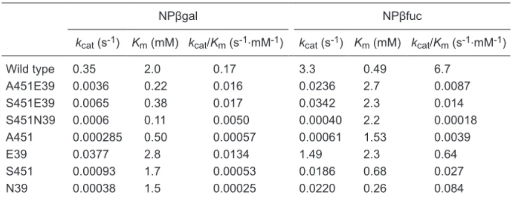
Mutants E39, N39, A451 and S451 (replacement of Sfβgly residues Q39 or E451 by E, N, A, and S, respec
-tively) were previously produced and purified. These single
mutations caused a large reduction in the kcat/Km ratio for
the hydrolysis of NPβglycosides due to a reduction of about
70% in the energy of the interactions formed by Q39 and
E451 with the substrate in the ES‡ complex (13).
Double mutants of Sfβgly (A451E39, S451E39 and S451N39) were prepared by site-directed mutagenesis,
expressed in BL21 DE3 cells in order to determine interac -tions between sites of mutation.
Steady-state kinetic parameters (kcat and Km) were
determined for the hydrolysis of NPβgal and NPβfuc us
-ing a purified Sfβgly double mutant (Table 1). The double
mutations modify kcat 102- to 103-fold, whereas Km presents smaller changes. This pattern was also observed for the
single mutations of residues Q39 and E451 (12,13). Thermal
inactivation experiments showed that the structures of the
A451E39 and S451E39 double mutants are similar to the wild-type Sfβgly (t½ ~1.2 min at 50°C), suggesting that the mutational effects on kcat did not result from large structural
modifications of the protein.
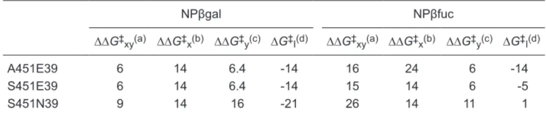
On the basis of kcat/Km data of the wild-type and double
mutant Sfβgly, changes in the transition state stabilization energy caused by a double mutation (∆∆G‡xy) and by
single mutations that compose a double mutant (∆∆G‡ x
and ∆∆G‡
y) were calculated as described in Material and
Methods (1,16) (Table 2). These ∆∆G‡
xy were compared
to the corresponding ∆∆G‡
x and ∆∆G‡y resulting in the
coupling energy (∆G‡I) between residues at sites 451 and
39 of Sfβgly (Table 2). ∆G‡
I is different from zero for all double mutants, except
for S451N39 when NPβfuc is used as substrate. Thus, the replacement of E451 and Q39 with S and N, respectively, has a simple additive effect on Sfβgly activity when NPβfuc
is the substrate, whereas in all remaining double mutants a complex additive effect is observed. Complex additivity
of mutational effects resulting in ∆G‡
I different from zero has been observed for other enzymes (1-4), but it has not
been described for GH1 β-glycosidases.
table 1. Steady-state kinetic parameters for the hydrolysis of NPβglycosides by the wild-type and
double mutant Sfβgly.
NPβgal NPβfuc
kcat (s-1) Km (mM) kcat/Km (s-1.mM-1) kcat (s-1) Km (mM) kcat/Km (s-1.mM-1)
Wild type 0.35 2.0 0.17 3.3 0.49 6.7
A451E39 0.0036 0.22 0.016 0.0236 2.7 0.0087 S451E39 0.0065 0.38 0.017 0.0342 2.3 0.014 S451N39 0.0006 0.11 0.0050 0.00040 2.2 0.00018 A451 0.000285 0.50 0.00057 0.00061 1.53 0.0039
E39 0.0377 2.8 0.0134 1.49 2.3 0.64
S451 0.00093 1.7 0.00053 0.0186 0.68 0.027 N39 0.00038 1.5 0.00025 0.0220 0.26 0.084
Additivity of mutational effects on aβ-glycosidase 11
www.bjournal.com.br Braz J Med Biol Res 43(1) 2010
Negative ∆G‡
I indicates that double mutations are less damaging to the enzyme activity (measured by kcat/Km) than the sum of the single mutations that compose them
(∆∆G‡
xy < ∆∆G‡x + ∆∆G‡y). This difference also indicates that the effects of the single mutations partially overlap.
Hence, this common effect counts just once in the ∆∆G‡ xy. Moreover, this common effect could result from the
pres-ence of a mutual influpres-ence between residues at positions 451 and 39, which affects their bonds with the substrate
and favors substrate hydrolysis.
In the specific case of NPβgal hydrolysis, ∆G‡
I presents similar values for all double mutants. In addition, the effects
of the double mutations (∆∆G‡xy) are less than those of the
most damaging single mutations (∆∆G‡
x), which involve
residue 451 (Table 2). Thus, mutations at residue 39 are
partially suppressing the damaging effect of the mutations
at residue 451, which is compatible with a mutual influence between residues at positions 451 and 39.
On the other hand, in the case of NPβfuc hydrolysis, ∆G‡
I changes depending on the double mutations (Table 2).
Variation of ∆G‡
I depending on the type of residue introduced
in the mutant enzyme was also observed for tyrosyl-tRNA synthetase and subtilisin BPN’ mutants (1). In the A451E39 mutant the observation of a negative ∆G‡
I and a ∆∆G‡xy
lower than ∆∆G‡
x suggests that the introduction of E39 is
reducing the damage caused by mutation A451, an effect similar to that described for the hydrolysis of NPβgal by this
same double mutant. The suppressing effect completely
disappears in the mutants S451E39 and S451N39 when NPβfuc is the substrate. In spite of that, the ∆∆G‡
xy is
still lower than ∆∆G‡
x + ∆∆G‡y for mutants S451E39 and
S451N39, confirming the presence of a mutual influence between residues at positions 451 and 39.
Therefore, the present data indicate a mutual effect of
table 2. Changes in the ES‡ complex energy resulting from double and single mutations in Sfβgly.
NPβgal NPβfuc
∆∆G‡
xy(a) ∆∆G‡x(b) ∆∆G‡y(c) ∆G‡I(d) ∆∆G‡xy(a) ∆∆G‡x(b) ∆∆G‡y(c) ∆G‡I(d)
A451E39 6 14 6.4 -14 16 24 6 -14
S451E39 6 14 6.4 -14 15 14 6 -5
S451N39 9 14 16 -21 26 14 11 1
Data are reported as kJ/mol. a∆∆G‡
xy was calculated by comparing the double mutant and wild-type
Sfβgly. b∆∆G‡
x was calculated by comparing a single mutant at position 451 and the wild-type Sfβgly. Data from Ref. 13. c∆∆G‡
y was calculated by comparing a single mutant at position 39 and the
wild-type Sfβgly. Data from Ref. 13. d∆G‡I was calculated using the equation ∆∆G‡xy = ∆∆G‡x + ∆∆G‡y
+ ∆G‡
I (Ref. 1) and corresponds to the coupling energy between residues at positions 451 and 39. NPβgal = p-nitrophenyl β-galactoside; NPβfuc = p-nitrophenyl β-fucoside;A = alanine; E = glutamate; N = asparagine; S = serine.
residues occupying positions 39 and 451 on their interac
-tions with the substrate. According to previous observa-tions, negative ∆G‡I occurs when the mutated residues form direct or indirect interactions (1). Crystallographic data for
β-glycosidases show that residues corresponding to Q39 and E451 do not interact directly because they are more
than 3 Å apart. Nevertheless these residues form bidentate hydrogen bonds with the hydroxyl 4 of the glycone
sub-strate (17-20). As the hydroxyl 4 is simultaneously bound to Q39 and E451, these interactions are not isolated. Thus,
interacting with the same group within the substrate rather than with different groups may result in different interac-tion energies. Hence, the sharing of the same group within
the substrate may be the source of the mutual influence between residues Q39 and E451 for Sfβgly.
Comparison of sequence data shows that residues forming the subsite -1 are highly conserved among the
GH1 β-glycosidases (17) and several of them share a
common group (glycone hydroxyl) in their interactions with
the substrate (9,17-20), suggesting that a mutual influence
between residues involved in substrate binding may be a common characteristic within this group of enzymes. Therefore, this property should be considered in the
char-acterization and/or modification of the substrate specificity of GH1 β-glycosidases.
Acknowledgments
References
1. Wells JA. Additivity of mutational effects in proteins. Bio-chemistry 1990; 29: 8509-8517.
2. Kuliopulos A, Talalay P, Mildvan AS. Combined effects of two mutations of catalytic residues on the ketosteroid isomerase reaction. Biochemistry 1990; 29: 10271-10280.
3. Mildvan AS, Weber DJ, Kuliopulos A. Quantitative inter -pretations of double mutations of enzymes. Arch Biochem Biophys 1992; 294: 327-340.
4. Mildvan AS. Inverse thinking about double mutants of en -zymes. Biochemistry 2004; 43: 14517-14520.
5. Coutinho P, Henrissat B. Carbohydrate-active enzymes: an integrated database approach. In: Gilbert HJ, Davies G, Henrissat B, Svensson B (Editors), Recent advances in car-bohydrate bioengineering. Cambridge: The Royal Society of Chemistry; 1999. p 3-12.
6. Davies GJ, Wilson KS, Henrissat B. Nomenclature for sugar-binding subsites in glycosyl hydrolases. Biochem J 1997;
321 (Part 2): 557-559.
7. Burmeister WP, Cottaz S, Driguez H, Iori R, Palmieri S, Henrissat B. The crystal structures of Sinapis alba myrosi-nase and a covalent glycosyl-enzyme intermediate provide insights into the substrate recognition and active-site ma-chinery of an S-glycosidase. Structure 1997; 5: 663-675.
8. Cicek M, Blanchard D, Bevan DR, Esen A. The aglycone specificity-determining sites are different in 2, 4-dihydroxy-7-methoxy-1,4-benzoxazin-3-one (DIMBOA)-glucosidase (Maize glucosidase) and dhurrinase (Sorghum beta-glucosidase). J Biol Chem 2000; 275: 20002-20011.
9. Gloster TM, Roberts S, Ducros VM, Perugino G, Rossi M, Hoos R, et al. Structural studies of the beta-glycosidase from Sulfolobus solfataricus in complex with covalently and noncovalently bound inhibitors. Biochemistry 2004; 43:
6101-6109.
10. Fernandez P, Canada FJ, Jimenez-Barbero J, Martin-Lomas M. Substrate specificity of small-intestinal lactase: study of the steric effects and hydrogen bonds involved in enzyme-substrate interaction. Carbohydr Res 1995; 271: 31-42.
11. Namchuk MN, Withers SG. Mechanism of Agrobacterium
beta-glucosidase: kinetic analysis of the role of
noncova-lent enzyme/substrate interactions. Biochemistry 1995; 34:
16194-16202.
12. Marana SR, Terra WR, Ferreira C. The role of amino-acid residues Q39 and E451 in the determination of substrate specificity of the Spodoptera frugiperda beta-glycosidase.
Eur J Biochem 2002; 269: 3705-3714.
13. Marana SR, Andrade EH, Ferreira C, Terra WR. Investiga -tion of the substrate specificity of a beta-glycosidase from
Spodoptera frugiperda using site-directed mutagenesis and bioenergetics analysis. Eur J Biochem 2004; 271:
4169-4177.
14. Laemmli UK. Cleavage of structural proteins during the as-sembly of the head of bacteriophage T4. Nature 1970; 227:
680-685.
15. Gill SC, von Hippel PH. Calculation of protein extinction coefficients from amino acid sequence data. Anal Biochem
1989; 182: 319-326.
16. Wilkinson AJ, Fersht AR, Blow DM, Winter G. Site-directed mutagenesis as a probe of enzyme structure and catalysis: tyrosyl-tRNA synthetase cysteine-35 to glycine-35 mutation.
Biochemistry 1983; 22: 3581-3586.
17. Marana SR. Molecular basis of substrate specificity in family 1 glycoside hydrolases. IUBMB Life 2006; 58: 63-73.
18. Sanz-Aparicio J, Hermoso JA, Martinez-Ripoll M, Lequerica JL, Polaina J. Crystal structure of beta-glucosidase A from
Bacillus polymyxa: insights into the catalytic activity in family 1 glycosyl hydrolases. J Mol Biol 1998; 275: 491-502.
19. Czjzek M, Cicek M, Zamboni V, Burmeister WP, Bevan DR, Henrissat B, et al. Crystal structure of a monocotyledon (maize ZMGlu1) beta-glucosidase and a model of its com-plex with p-nitrophenyl beta-D-thioglucoside. Biochem J
2001; 354: 37-46.