Received on 6 December 2007; revised 8 March 2008.
Address for correspondence: Dr Dahir Ramos de Andrade Júnior, PhD, Av. Paes de Barros 701, ap. 101, Mooca, São Paulo, São Paulo, Brazil. CEP=03115020 (ZIP Code). Phone=0115511 - 30617029. Fax=0115511 - 30617029. [email protected]. Financial support: Supported by FAPESP (Fundação de Amparo à Pesquisa no Estado de São Paulo). Informed consent was obtained from each patient or a relative according to U.S. Department of Health and Human Services.
The Brazilian Journal of Infectious Diseases 2008;12(3):226-233. © 2008 by The Brazilian Journal of Infectious Diseases and Contexto Publishing. All rights reserved.
Correlation Between Serum Tumor Necrosis Factor Alpha Levels and Clinical Severity of Tuberculosis
Dahir Ramos de Andrade Júnior1, Sânia Alves dos Santos1, Isac de Castro2 and Dahir Ramos de Andrade1
1Laboratoty of Medical Research - LIM 54, School of Medicine, University of São Paulo; 2Laboratoty of Medical Research - LIM 16, School of
Medicine, University of São Paulo; São Paulo, SP, Brazil
This study verified the correlation between the serum levels of TNF alpha and different clinical forms of tuberculosis. We described a group of 24 patients presenting several clinical forms of tuberculosis and a control group of 13 healthy individuals. The levels of TNF alpha were measured by bioassay method. The levels of TNF-alpha had significant differences between the tuberculosis and control groups. The patients with abnormal chest X-Ray findings had higher TNF alpha levels (15328.48 ± 4602.19 pg/mL) when compared to patients with normal X-Rays (3353.18 ± 1495.29 pg/mL) (p<0.05). Patients that lost weight had higher TNF alpha levels (15468.54 ± 4580.54 pg/ mL) than those that didn’t loose weight (2904.98 ± 1367.89) (p<0.05). The levels of TNF alpha were higher in patients with a positive PPD skin test than in those with a negative PPD test (p<0.05). There was a positive correlation between patients’ clinical severity and the serum levels of TNF alpha. In patients with successive measurements of TNF alpha, we observed that there was a drop in cytokine levels, and also a clinical improvement concomitantly. We concluded that there was a correlation between serum TNF alpha levels and chest X-Ray alterations, loss of weight, positive PPD skin test and clinical severity in patients with tuberculosis. There was evidence of a worse clinical outcome in patients with tuberculosis that presented higher TNF alpha serum levels.
Key-Words: TNF alpha, tuberculosis, cytokines, clinical severity.
Abbreviations: TNF=tumor necrosis factor, TB=tuberculosis, M. tuberculosis=Mycobacterium tuberculosis, IL=interleukin, CTLs=cytotoxic T lymphocytes, MHC=major histocompatibility complex, IFN=interferon, BAL=bronchoalveolar lavage, CT=computed tomographic, ADA=adenosine deaminase.
The pathogenesis of the inflammatory response in tuberculosis (TB), involving mainly cellular and molecular phenomena, remains unclear. Macrophage activation, phagocytosis mechanisms, and the survival of Mycobacterium
tuberculosis in phagocytic vesicles must be further studied.
When tuberculosis infection occurs, a variety of chemokines and cytokines are secreted from infected cells and tissue macrophages. TNF alpha increases early in the disease and takes part in the pathogenesis and prevention of mycobacterial infection. TNF alpha also appears crucial for the formation of M. tuberculosis-constraining granulomas, infection control and elimination of mycobacteria [1]. The production of TNF alpha in HIV-TB co-infected patients is down-regulated in T lymphocytes, and this may explain the cause of defective granuloma formation in this condition [2]. Serum levels of TNF alpha and IL-1 are proportional to the degree of tissue infection [3] and are also associated with induction of fever and other consumptive manifestations of the disease [4].
The cellular immune response is essential for TB resistance, mainly through cytokine production such as
IL-12, IL-18, IFN gamma, IL-2 and TNF alpha. The TCD8+ cells (CTLs) perform cytotoxic actions, mainly through major histocompatibility complex (MHC) class I molecules that, in association with CD1d molecules, present glycolipid antigens to T lymphocyte cells [5]. CTL cells may have a protective role in TB disease due to several mechanisms: 1) producing potent antibacterial cytokines such as IFN gamma and TNF alpha; 2) recognizing M. tuberculosis antigens presented by MHC associated to HLA-I complex; 3) inducing apoptosis in infected cells [6,7].
The role of cytokines produced by Th1/Th2 lymphocytes is complex due to the action of 12, IFN gamma, 4 and IL-10 (pro-inflammatory and anti-inflammatory cytokines). Several aspects of this complex interaction have been studied in recent studies: a) IFN gamma inhibition by IL-10 [8]; b) IFN gamma and IL-10 production stimulated by IL-12 [9]; c) IFN gamma and IL-10 production stimulated by TCD4+ clones in the bronchoalveolar lavage (BAL) fluid of patients with pulmonary TB [10]; d) Th2 cytokine association with histopathologic alterations in patients with TB [11].
Several studies have shown physiopathologic correlation between serum TNF alpha and the evolutive course of mycobacteria infection. TNF alpha neutralization caused an increase in pulmonary bacterial load in mice that had been infected with M. tuberculosis six months before [12]. There is an association between TNF alpha production and
M.tuberculosis virulence in human monocytes, as well as a
of TNF alpha concentrations in BAL fluid [16] showed that the greater the amount of lung involvement the higher the cytokine production.
We report 24 patients with several TB clinical forms where sera measurements of TNF alpha had been made. We tried to associate serum TNF alpha levels with the evolution of clinical forms of TB. We concluded that serum TNF alpha measurement might play an important role in the evaluation of the inflammatory phenomena in TB.
Material and Methods
Sera were obtained from 24 patients, from the Infectious Diseases Clinic and Internal Medicine Clinic of the Hospital das Clínicas da Faculdade de Medicina da Universidade de São Paulo, with active TB and from 13 healthy individuals (control group). TB patients had a mean age of 32.95 years (18 to 72 years) with six females and eighteen males. The control group had a mean age of 34.84 years (21 to 47 years) with four males and nine females. Informed consent was obtained from each patient and the protocol for the research project was approved by the Ethics Committee of the Institution. We described 24 TB patients having the following clinical forms: 14 pulmonary, 2 pleural - pulmonary, 1 pleural - pulmonary + pericardial, 1 pleural + lymphatic, 1 pulmonary + lymphatic, 1 disseminated, 1 miliary, 2 peritoneal, 1 cutaneous + pulmonary. The TB diagnoses were established between 1993 and 2002. Patients were all HIV negative and they were receiving anti-tuberculous therapy at the time of blood sampling. Medical records of all patients were analyzed for clinical data such as anemia, fever, weight loss, dyspnea, night sweats, cough, sputum, as well as diagnostic tests such as thoracic X-Ray (XR), computed tomographic (CT) scan of the chest, PPD skin test, erythrocyte sedimentation rate, and blood counts. The TB diagnosis was made if acid-fast bacilli had been positive in search or culture in each case (Table 2). We considered anemia as hemoglobin < 12 g/dL (woman) or < 14g/dL (man). Fever was defined as temperature ≥ 38°C. The clinical variables weight loss, dyspnea, night sweats, cough and sputum were registered as 0 (absent) or 1 (present). The results of the thoracic radiograph, the computed tomographic (CT) scan of the chest, and the sedimentation rate, were coded as 0 (normal) or 1 (altered). The PPD skin test was coded as 0 (negative) or 1 (positive). We also evaluated whether tissue destruction was detected by image diagnostic methods. The clinical severity of each case was determined through the following score: thoracic XR/CT normal=0; thoracic XR/CT altered without cavity=1; thoracic XR/CT altered with cavity=2; without anemia=0; with anemia=1; without hypoxia=0; with hypoxia=1; without weight loss=0; with weight loss=1.
TNF alpha Assay
TNF alpha bioactivity was determined by the McGee & Clemens bioassay method [17]with modifications, using L929 cells as targets. Briefly, L929 cells were cultured for 24 hours
on flat-bottom 96-well plates (1 x 10⁴ cells/well), with RPMI 1640 medium at 37o C under an atmosphere of 5% CO
2 and 95% air. L929 cell monolayers were pre-incubated for 2 hours with 1 mg/mL of Actinomycin D (ActD). The patient’s sera (stored at -80ºC) were added to the wells and incubated for 18 hours. After this, the cells were stained with MTT vital stain. All samples were assayed at the same time. The readings were done using a spectrophotometer plate reader at a wavelength of 540 nm. The cell viability percentage was calculated using the formula: % cell viability=optical density (samples)/optical density (control) x 100. The final results of each sample were obtained through the average of eight wells. The serum TNF alpha level was calculated through a standard curve (recombinant TNF alpha versus cell viability percentage). The confidence interval over 95% of the control group (UCI 95%) was considered as the cut-off between individuals in the controls and individuals with TB.
Statistical Analysis
The data were separated into categorical and continuous variables. The categorical values were expressed in absolute frequency (n) and relative frequency (%), using Pearson Chi-square test. The continuous and semi continuous variables were compared to a normal curve, and classified as parametric or not using the K-S test. TNF alpha values were categorized using the superior 95% confidence interval. The parametric data were shown as mean ± standard error, and compared using the unpaired Student’s t-test. The nonparametric data were shown as median and 25th – 75th percentiles, and compared using the Mann-Whitney test. The ROC curve was used as the operational response curve to establish whether TNF alpha had good discriminating power. The correlations between the TNF alpha level in each case and all clinical and diagnostic variables were evaluated through the r-value calculation using Pearson’s correlation. The alpha risk for all conditions was ≤5% in type I error, and ≤ 20% for beta risk in type II error.
Results
Demographic and Clinical Characteristics
The demographic data and TNF alpha levels of the 13 subject control group are shown in Table 1. The patients’ general characteristics, TB clinical forms, length of symptoms and serum TNF alpha levels are shown in the Table 2. The days of therapy, clinical complications, length of treatment and clinical evolution of the patients with the TNF alpha levels are shown in Table 3. The mean of serum TNF alpha levels in patients with active TB was 13035.54 ± 15948.35 pg/mL, which was significantly higher than those found in healthy control individuals (341.3 ± 127.1 pg/mL) (p < 0.01).
Aspects of Severity and Evolution of Tuberculosis
Correlation Between Clinical or Laboratorial Variables and TNF Values
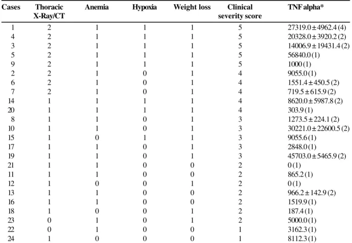
Table 5 shows statistical comparisons between clinical and laboratory variables of TB patients. There were no statistically significant differences between TNF alpha level and the following variables: anemia (p=0.148), fever (p=0.475), night sweats (p=0.859), dyspnea (p=0.514), cough (p=0.678) and sputum (p=0.182). Only the weight loss variable showed significant statistical difference in relation to TNF alpha. The patients with weight loss had higher serum TNF alpha (15468.54 ± 4580.54 pg/mL) than those without weight loss (2904.98 ± 1367.89 pg/mL) (p<0.05). The patients with abnormal chest X-rays had higher levels of TNF alpha (15328.48 ± 4602.19 pg/mL) in relation to the patients with normal chest X-ray (3353.18 ± 1495.29 pg/mL) (p<0.05). There was no difference between TNF alpha and sedimentation rate
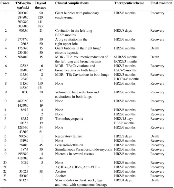
(p=0.848), tissue destruction (p=0.149), multidrug-resistant TB (p=0.695) and clinical survival (p=0.352). The serum TNF alpha level was higher in patients with a positive PPD skin test when compared to those with a negative PPD test (p<0.05). TB treatment reduced significantly the serum TNF alpha values (p<0.05). A positive correlation was found between treatment duration and acid-fast bacilli (AFB) absence (r=0.717; p<0.05). In patients with successive TNF alpha measurements, a clinical improvement was always observed when serum TNF alpha levels dropped (patients 3, 7, 10, 14 and 19).
On the other hand, many patients without a decrease in serum TNF alpha levels presented clinical problems during their clinical evolution. For instance, patients 1 and 4 had pulmonary giant bubbles, patient 6 (MDR-TB) developed pulmonary cavitations, and patient 8 remained AFB positive in sputum until the fifth month of TB therapy.
The highest serum TNF alpha levels (> 15 x normal) were observed in 6 patients with severe TB: patient 1 (pulmonary TB with bubbles), patient 3 (pulmonary TB with cavitations), patient 4 (pulmonary TB with bubbles), patient 5 (pulmonary - MDR TB), patient 10 (pulmonary - interstitial TB) and patient 19 (disseminated TB). We found that high serum TNF alpha levels were associated with TB clinical forms having increased tissue destruction (shown by image methods). In three patients (15, 16 and 17), with pleural-pulmonary TB, high serum levels of TNF alpha were noted. In these patients the concomitant TNF alpha level in pleural fluid was undetectable.
The serum TNF alpha levels had good discriminant power in identifying patients with Tuberculosis (n=24) from control individuals (n=13). The control group upper confidence interval of 95% (UCI 95%) was 618.1 pg/mL. This value was used to categorize the continuous data. The ROC curve showed that the TNF alpha test was reliable (area=0.908 ± 0.049, and p < 0.0001).
Discussion
We studied 24 patients with various TB clinical forms. We tried to show a possible role of TNF alpha as a cytotoxic agent, mainly when high serum levels were observed (patients 1, 3, 5, 4, 10, 14 and 19). The presence of TNF alpha in the peripheral blood is a consequence of cytokine leakage to the serum. This cytokine is produced by tissues and inflammatory cells through monocyte-macrophage activation after M.
tuberculosis stimulation [18].
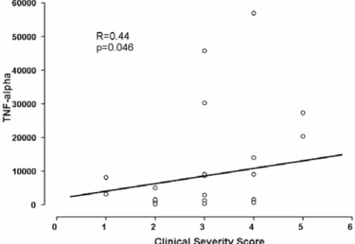
The groups of patients presented illustrate the TNF alpha role in the various TB clinical forms. There was a marked difference of serum TNF alpha levels between the TB group and the control group. When the cases were classified by clinical severity score, we observed a positive correlation between clinical severity and serum TNF alpha levels. The subgroup analysis also showed interesting results. The MDR-TB cases (patients 5, 6, 7) presented severe pulmonary lesions with increased tissue destruction. In this group, the serum TNF alpha level ranged from a slight elevation (patients 6 and 7), to a very high level (patient 5).
Table 1. Demographic characteristics of subject control group and TNF alpha levels.
Figure 1. Positive correlation between serum TNF-alpha levels and the clinical severity score.
Control Age Sex TNF alpha
(pg/mL)
1 27 F 1,415
2 35 F 0.49
3 21 M 8
4 40 M 45
5 40 F 314
6 48 F 598
7 41 F 389
8 50 F 314
9 37 F 133
10 32 F 15
11 35 F 45
12 31 M 1,141
Table 2. Demographic and clinical characteristics of patients with tuberculosis and TNF alpha levels.
On the other hand, in three patients the serum TNF alpha was undetectable: patient 9 (pulmonary with cavitations TB), patient 12 (pulmonary TB), and patient 21 (miliary TB). Patient 21 had a negative PPD skin test. We speculate that in these patients the immune response to the infectious agent could have been very weak. In another two patients, normal serum TNF alpha levels were detected: patient 18 (pleural + lymphatic TB) and patient 20 (lymphatic + pulmonary TB). These patients had normal serum TNF alpha values in spite of the presence of extensive lymphadenopathy with necrosis detected using image methods. We suppose that there can be a compartmentalization of the immune response in some patients with lymphatic TB. Patients 1, 3, 4, and 5 showed the highest serum TNF alpha levels and they also had increased pulmonary tissue destruction. We suggest that TNF alpha may have a direct role in this phenomenon.
We quantified TNF alpha successively in 10 patients and observed that, when serum TNF alpha levels decreased during clinical evolution (patients 3, 7, 10, 14 and 19), a clinical improvement was always noted. In contrast, when serum TNF alpha levels did not decrease during the evolution, a bad clinical course was noted. These results indicate that the serum TNF alpha level may be a good marker to predict the TB patient’s clinical evolution. Ribeiro-Rodrigues et al. [19] showed that decreasesin sputum IFN-γ, TNF alpha, and IL-8 closely parallel andeven precede mycobacterial clearance, and they concluded that these cytokines may be markers of disease activity and inflammation in TB.
In this study we also found a positive correlation between serum TNF alpha levels and weight loss, abnormal chest X-Rays and positive PPD skin tests. The positive correlation between weight loss and serum TNF alpha levels was also observed in other studies. Bossola et al. [20] showed that
Cases Age Sex Clinical form Symptoms length TB diagnosis TNF alpha*
1 40 M Pulmonary 12 months AFB† S + C‡ in sputum 27319.0 ± 4962.4 (4)
2 28 F Pulmonary 2 weeks AFB S + C in sputum 9055.0 (1)
3 50 M Pulmonary 6 months AFB S + C in BAL§ 14006.9 ± 19431.4 (2)
4 18 M Pulmonary 2 months AFB S + C in sputum 20328.0 ± 3920.2 (2)
5 20 F Pulmonary 4 months AFB S + C in sputum 56840.0 (1)
6 20 F Pulmonary 2 months AFB S + C in sputum 1551.4 ± 450.5 (2)
7 31 F Pulmonary 12 months AFB S + C in sputum 719.5 ± 615.9 (2)
8 27 M Pulmonary 6 months AFB S + C in sputum 1273.5 ± 224.1 (2)
9 47 M Pulmonary 12 months AFB S + C in sputum 0 ¦ (1)
10 61 M Pulmonary 6 months Lung biopsy 30221.0 ± 22600.5 (2)
11 72 M Pulmonary 2 months AFB S + C in BAL 865.2 (1)
12 19 M Pulmonary 4 months AFB S + C in BAL 0 (1)
13 24 M Pulmonary 2 months AFB S + C in sputum 966.2 ± 142.9 (2)
14 62 M Pulmonary 2 weeks AFB S + C in BAL 8620.0 ± 5987.8 (2)
15 32 M Pleural–pulmonary 2 months AFB S + C in pleural 9055.6 (1) fluid
16 20 M Pleural-pulmonary 1 month AFB S + C in pleural 1519.9 (1)
biopsy
17 19 M Pleural-pulmonary + 5 months AFB S + C in pleural 2848.0 (1)
pericardial biopsy
18 21 M Pleural + lymphatic 2 months AFB S + C in 187.4 (1)
lymph node biopsy
19 24 M Disseminated ¶ 2 months AFB S + C in 45703.0 ± 5465.9 (2)
lymph node biopsy
20 21 F Lymphatic + 3 months AFB S + C in BAL and 303.9 (1)
pulmonary lymph node biopsy
21 32 F Miliary 15 days AFB C in sputum 0 (1)
22 50 M Peritoneal 18 months AFB C in ascitic fluid 3162.3 (1)
+ peritoneal biopsy
23 23 M Peritoneal 4 months Peritoneal biopsy 5000.0 (1)
24 30 M Cutaneous + 12 months AFB S + C in sputum 8112.3 (1)
pulmonary and skin abscess
Table 3. Severity data and evolution aspects of patients with tuberculosis and levels of TNF-alpha.
serum TNF alpha concentrations were significantly higher in patients with cancer and severe weight loss when compared with patients having cancer and low weight loss. Cakir et al. [21]speculated that serum TNF alpha levels and leptin might be responsible for the weight loss in pulmonary tuberculosis patients. Another study also correlated the chronic administration of TNF alpha with weight loss in rats [22].
We observed that the patients with abnormal chest X-rays had higher serum levels of TNF alpha. Tsao et al. showed that the bronchoalveolar lavage fluid levels of TNF alpha, IL-1β and IL-6 were all significantly higher in patients with large cavities than in patients without cavities or with smaller cavities [23,24] These authors also showed that the relative excess of both TNF alpha and IL-1β, associated with soluble
Cases TNF-alpha Days of Clinical complications Therapeutic scheme Final evolution
(pg/mL) therapy
1 20000.0 90 Giant bubbles with pulmonary HRZ/6 months Recovery
28480.0 120 emphysema
30398.0 141 30398.0 183
2 9055.0 21 Cavitation in the left lung HRZ/8 days Recovery
ESZ/6 months
3 27747.0 30 A big cavitation in the HRZ/6 months Recovery
266.8 60 right upper lobe
4 17556.0 15 Giant bubbles in the right lung/ HRZ/6 months Death 23100.0 30 chronic hypoxia
5 56840.0 15 MDR - TB*, volumetric reduction of SHRZ/9 months Death the left lung and bronchiectasis ECRZ/3 months
6 1232.8 6 MDR - TB; Cavitations and HRZ/2 months Recovery
1870.0 42 bronchiectasis in both lungs ESC+/6 months
7 1155.0 2 MDR - TB; Cavitations in both lungs HRZ/3 months Recovery
284.0 21 IHCCA/6 months
8 1115.0 150 None HRZ/6 months Recovery
1432.0 171
9 1000 30 Volumetric lung reduction and HRZ/6 months Recovery
cavitations in both lungs
10 46202.0 12 None HRZ/6 months Recovery
14240.0 19
11 865.2 4 None HRZ/6 months Recovery
12 0 2 None HRZ/6 months Recovery
13 865.2 15 Thrombocytopenia HRZ/15 days Recovery
1067.3 19 EES/6 months
14 12854.0 34 None HRZ/6 months Recovery
4386.0 61
15 9055.6 1 Respiratory failure HRZ/2 days Death
16 1519.9 1 None HRZ/6 months Recovery
17 2848.0 45 Pericardial effusion HRZ/6 months Recovery
18 187.4 30 Simultaneous Paracoccidioido-mycosis HRZ/6 months Recovery
19 49568.0 4 Necrosis in several tissues HRZ/6 months Recovery
41838.0 44
20 303.9 5 None HRZ/6 months Recovery
21 0 1 AgHBs+, AgHBe+, Anti-VHC+ HRZ/6 months Recovery
22 3162.3 38 Ascites HRZ/6 months Recovery
23 5000.0 1 Ascites HRZ/6 months Recovery
24 8112.3 1 Skin nodules in chest, neck, legs HRZ/4 days Death
and head with spontaneous leakage
Table 4. Distribution of the Tuberculosis patients by clinical severity score and serum TNF-alpha levels.
TNF alpha-receptor secretion imbalance, could have a correlation with the pulmonary cavities in patients having active TB [24]. However, there were few studies in medical literature about the correlation between serum TNF alpha levels and abnormal X-Rays, and further studies are necessary to understand this phenomenon. We also found that the serum TNF alpha level was elevated in patients with a positive PPD skin test. Chu et al. [25] observed that the number of cells staining for TNF alpha and IL-1 in the skin of six normal individuals during the PPD – induced reaction persisted at elevated levels [25]. The authors speculated that TNF alpha might play an important role in the development of delayed-type hypersensitivity in man. Therefore, we added the information that a high serum TNF alpha level occurs in patients with positive PPD skin tests, and this phenomenon suggests a possible correlation between this cytokine serum level and its tissue action.
We described three cases with pleural-pulmonary TB (15, 16 and 17) in which a high serum TNF alpha level was observed without a concomitant increase of TNF alpha in the pleural fluid. Vankayalapati et al. [26] reported that IL-18 and IFN gamma concentrations are higher in the pleural fluid of TB patients than in patients with other diseases. In another study, pleural fluid TNF alpha levels and pleural fluid/serum TNF alpha were higher in tuberculous effusions than in other
exudates, but their diagnostic value appears to be poorer than that of ADA [27]. Our result differs from these studies [26,27] and we did not find a clear explanation. We speculate that there was a barrier separating the pleural fluid and the host’s immune response due to pleural fibrosis present in patient 16. We observed that in three cases, 1, 19 and 14, the serum TNF alpha level increased after the treatment of TB began. However, we can not state whether there was concomitant clinical worsening in these cases. Bekker et al. [13]observed worsening of clinical condition in 16 patients during the first 42 days of treatment, with two deaths among those having severe pulmonary TB. All patients had increased serum TNF alpha and lactic acid levels. These authors associated the worsening clinical condition of these patients with the increase of serum TNF alpha levels. Such evidences show the importance of estimating the serum cytokine level at the beginning of TB treatment.
The relation between TNF alpha and TB stands out due to the reactivation of latent TB infection with treatment with tumor necrosis factor (TNF) antagonists [28] in patients with several diseases such as: rheumatoid arthritis [29], juvenile arthritis, ankylosing spondylitis, Crohn’s disease, psoriasis, glomerulonephritis, sarcoidosis and Behcet’s disease. These diseases are characterized by a Th1 type immune response associated with excess generation of TNF-alpha [30].
Cases Thoracic Anemia Hypoxia Weight loss Clinical TNF alpha*
X-Ray/CT severity score
1 2 1 1 1 5 27319.0 ± 4962.4 (4)
4 2 1 1 1 5 20328.0 ± 3920.2 (2)
3 2 1 1 1 5 14006.9 ± 19431.4 (2)
5 2 1 1 1 5 56840.0 (1)
9 2 1 1 1 5 1000 (1)
2 2 1 0 1 4 9055.0 (1)
6 2 1 0 1 4 1551.4 ± 450.5 (2)
7 2 1 0 1 4 719.5 ± 615.9 (2)
14 1 1 1 1 4 8620.0 ± 5987.8 (2)
20 1 1 1 1 4 303.9 (1)
8 1 1 0 1 3 1273.5 ± 224.1 (2)
10 1 1 0 1 3 30221.0 ± 22600.5 (2)
15 1 0 1 1 3 9055.6 (1)
17 1 1 0 1 3 2848.0 (1)
19 1 1 0 1 3 45703.0 ± 5465.9 (2)
21 1 1 0 0 2 0 (1)
11 1 1 0 0 2 865.2 (1)
12 1 0 0 1 2 0 (1)
13 1 1 0 0 2 966.2 ± 142.9 (2)
16 1 1 0 0 2 1519.9 (1)
18 1 0 0 1 2 187.4 (1)
23 0 1 0 1 2 5000.0 (1)
22 0 1 0 0 1 3162.3 (1)
24 1 0 0 0 1 8112.3 (1)
Nowadays there is much knowledge about TNF alpha influences in tissue injury. Inflammatory cells and cytokines cause death by apoptosis both in inflammatory and tissue cells (pneumocytes, fibroblasts, endothelial, etc.). The extrinsic pathway of apoptosis is activated by receptors belonging to the TNF alpha-receptor superfamily. However, Ashkenasi and Dixit [30] state that TNF alpha rarely starts cellular apoptosis, unless cellular protein synthesis is blocked. In contrast, Krammer [31] states that TNF alpha may cause cellular death by apoptosis, overcoming Rel A protein resistance [32]. Apoptotic cell death caused by TNF alpha may be completely or partially blocked by NF-kB and its antiapoptotic proteins.
Ciaramella et al. [33] showed thatMTB-induced apoptosis is inhibitedby a caspase-1 inhibitor(YVAD) and is associated with the maturation of IL-1β. In addition, IL-1β and TNF alpha were producedmassively in the courseof infection, and both were inhibited by YVAD. Keane et al. [34] described the apoptosis of alveolar macrophages infected by M. tuberculosis. Watson et al. [35] found various pulmonary histopathologic alterations in different mice species with acute infection by M.
tuberculosis. They suggested that apoptosis occurred during
granuloma formation after infection by M. tuberculosis, through a FAS/FAS ligand-independent pathway. Taken together, these studies correlate apoptotic cell death with TNF alpha, and they highlight the role of this cytokine in TB cases.
One of the objectives of our study was to show that increased serum TNF alpha was followed by severe clinical TB with high tissue destruction. On the other hand, we observed that a decrease in the serum cytokine level occurred in parallel with patient clinical improvement.We speculate that a reduction of high TNF alpha serum levels may decrease tissue destruction, particularly when the cytokine serum level remains elevated for a long time. TNF alpha periodic measurements through bioassay or Elisa methods could help to understand tuberculous outcome and itshould be included in the routine TB clinical evaluation in the near future. However, in order to establish definitive conclusions more research should be done.Nowadays the treatment of TB is directed specifically to M. tuberculosis elimination. We suggest that TNF alpha immune modulation should be evaluated in
M.tuberculosis therapy in future studies, especially in cases
with very high TNF alpha serum levels.
Table 5. Statistical comparisons of clinical and laboratorial variables related to serum TNF-alpha levels in TB patients.
Variables TNF alpha* p
Patient with TB 13035.54 ± 15948.35 p < 0.01
Control Group 341.3 ± 127.1
Weight loss 1 § 15468.54 ± 4580.54 p < 0.05
Weight loss 0 ¦ 2904.98 ± 1367.89
Thoracic X-Ray 1 15328.48 ± 4602.19 p < 0.05
Thoracic X-Ray 0 3353.18 ± 1495.29
PPD 1 21710 ± 17710 p < 0.05
PPD 0 101.3 ± 101.3
Anemia 1 13592.57 ± 4228.01 p=0.148
Anemia 0 5785.10 ± 2812.07
Fever 1 10799.27 ± 4498.97 p=0.475
Fever 0 16672.10 ± 6521.49
Night sweats 1 11225.22 ± 7789.59 p=0.859
Night sweats 0 12868.47 ± 4306.21
Dyspnea 1 14557.54 ± 5350.22 p=0.514
Dyspnea 0 9703.46 ± 4964.60
Cough 1 10711.0 ± 4916.57 p=0.678
Cough 0 13801.26 ± 5424.93
Sputum 1 6713.95 ± 3083.72 p=0.182
Sputum 0 14782.53 ± 4932.87
VHS 1 16223.23 ± 5634.26 p=0.848
VHS 0 18511.45 ± 9813.49
Tecidual destruction 1 17535.96 ± 5485.07 p=0.149
Tecidual destruction 0 6912.60 ± 4439.29
MR - TB 1 19742.60 ± 18548.71 p=0.695
MR - TB 0 11266.32 ± 3345.66
Recovery 22890.98 ± 11513.85 p=0.352
Death 10026.92 ± 3611.45
References
1. Kindler V., Sappino A.P., Grau G.E., et al. The inducing role of tumor necrosis factor in the development of bactericidal granulomas during BCG infection. Cell 1989;56:731-40. 2. Cunha R.M.C., Kallas E.G., Rodrigues D.S., et al. Interferon-g and
tumour necrosis factor-a production by CD4+ T and CD8 + T lymphocytes in AIDS patients with Tuberculosis. Clin Exper Immunol 1995;140:491-7.
3. Juffermans N.P., Verbon A., van Deventer S.J., et al. Tumor necrosis factor and ýnterleukin-1 inhibitors as markers of disease activity of tuberculosis. Amer J Respir Crit Care Med 1998;157:1328-31.
4. McDermott M.F. TNF and TNFR biology in health and disease. Cell MolecBiol 2001;47:619-35.
5. Park S.H., Bendelac A. CD1-restricted T-cell responses and microbial infection. Nature 2000;406:788-92.
6. Lewinsohn D.M., Briden A.L., Reed S.G., et al. Mycobacterium tuberculosis-reactive CD8+ T lymphocytes: the relative contribution of classical versus nonclassical HLA restriction. J Immunol 2000;165:925-30.
7. Serbina N.V., Liu C.C., Scanga C.A., Flynn J.L. CD8+ CTL from lungs of Mycobacterium tuberculosis-infected mice express perforin in vivo and lyse infected macrophages. J Immunol 2000;165:353-63.
8. D’Andrea A., Aste-Amezaga M., Valiante N.M., et al. Interleukin 10 (IL-10) inhibits human lymphocyte interferon γ-production by suppressing natural killer cell stimulatory factor/IL-12 synthesis in accessory cells. J Exp Med 1993;178:1041-8. 9. Gerosa F., Paganin C., Peritt D., et al. Interleukin-12 primes human
CD4 and CD8 T cell clones for high production of both interferon-gamma and interleukin-10. J Exp Med 1996;183:2559-69.
10. Gerosa F., Nisii C., Righetti S., et al. CD4+ T cell clones producing both interferon-γ and interleukin-10 predominate in bronchoalveolar lavages of active pulmonary tuberculosis patients. Clin Immunol 1999;92:224-34.
11. Seah G.T., Scott G.M., Rook G.A.W. Type 2 cytokine gene activation and its relationship to extent of disease in patients with tuberculosis. J Infect Dis 2000;181:385-9.
12. Steyn A.J.C., Chan J., Mehra V. Recent developments in mycobacterial research. Cur Opin Infect Dis 1999;12:415-24. 13. Bekker L.G., Maartens G., Steyn L., Kaplan G. Selective increase in plasma tumor necrosis factor - a and concomitant clinical deterioration after initiating therapy in patients with severe tuberculosis. J Infect Dis 1998;178:580-4.
14. Silver R.F., Li Q., Ellner J.J. Expression of virulence of Mycobacterium tuberculosis within human monocytes: virulence correlates with intracellular growth and induction of tumor necrosis factor alpha but not with evasion of lymphocyte -dependent monocyte effector functions. Infect Immun 1998;66:1190-9.
15. Dlugovitzky D., Bay M.L., Rateni L., et al. In vitro synthesis of interferon gamma, interleukin 4, transforming growth factor beta, and interleukin 1beta, by peripheral blood mononuclear cells from tuberculosis patients: relationship with severity of pulmonary involvement. Scand J Immunol 1999;49:210-7. 16. Wang C.H., Liu C.Y., Lin, H.C., et al. Increased exhaled nitric
oxide in active pulmonary tuberculosis due to inducible NO synthase upregulation in alveolar macrophages. Europ Resp J 1998;11:809-15.
17. McGee Z.A., Clemens C.M. Effect of bacterial products on tumor necrosis factor production: quantitation in biological fluids or tissues. Meth Enzymol 1994;236:23-31.
18. Barnes P.F., Wizel B. Type 1 cytokines and the pathogenesis of tuberculosis. Amer J Respir Crit Care Med 2000;161:1773-4. 19. Ribeiro-Rodrigues R., Resende-Co, T.; Johnson, J.L., et al. Sputum
cytokine levels in patients with pulmonary tuberculosis as early markers of mycobacterial clearance. Clin Diagn Lab Immunol 2002;9:818-23.
20. Bossola M., Muscaritoli M., Bellantone R. et al. Serum tumour necrosis factor – alpha levels in cancer patients are discontinuous and correlate with weight loss. Europ J Clin Invest 2000;30:1107-12.
21. Cakir B., Yönem A., Güler S., et al. Relation of leptin and tumor necrosis factor alpha to body weight changes in patients with pulmonary tuberculosis. Horm Res 1999;52:279-83. 22. Ling P.R., Schwartz J.H., Bristrian B.R. Mechanisms of host wasting
induced by administration of cytokines in rats. Amer J Physiol 1997;272(3pt1):E333-9.
23. Tsao T.C., Hong J., Huang C., et al. Increase TNF-α, IL-1β and IL-6 levels in the bronchoalveolar lavage fluid with the upregulation of their mRNA in macrophages lavaged from patients with active pulmonary tuberculosis. Tuber Lung Dis 1999;79:279-85.
24. Tsao T.C., Hong, J., Li L.F., et al. Imbalances between tumor necrosis factor-alpha and its soluble receptor forms, and interleukin-1 beta and interleukin-1 receptor antagonist in BAL fluid of cavitary pulmonary tuberculosis. Chest 2000;117:103-9.
25. Chu C.Q., Field M., Andrew E., et al. Detection of cytokines at the site of tuberculin-induced delayed-type hypersensitivity in man. Clin Exp Immunol 1992;90:522-9.
26. Vankayalapati R., Wizel B., Weis, S.E., et al. Production of interleukin-18 in human tuberculosis. J Infect Dis 2000;182:234-9. 27. Tahhan M., Ugurman F., Gozu A., et al. Tumour necrosis
factor-alpha in comparison to adenosine deaminase in tuberculous pleuritis. Respiration 2003;70:270-4.
28. Wallis R.S., Broder M.S., Wong J.Y., et al. Granulomatous infectious diseases associated with Tumor Necrosis Factor antagonists. Clin Infect Dis 2004;38:1261-5.
29. Carmona L., Gomez-Reino J.J., Rodriguez-Valverde V., et al. Effectiveness of recommendations to prevent reactivation of latent tuberculosis infection in patients treated with Tumor Necrosis Factor antagonists. Arthr Rheum 2005;52:1766-72. 30. Ashkenazi A., Dixit V.M. Death receptors: signaling and
modulation. Science 1998;281:1305-8.
31. Krammer P.H. CD95’s deadly mission in the immune system. Nature 2000;407:789-95.
32. Doi T.S., Marino M.W., Takahashi T., et al. Absence of tumor necrosis factor rescues RelA-deficient mice from embryonic lethality. Proc Natl Acad Sci 1999;96:2994-9.
33. Ciaramella A., Cavone A., Santucci M.B., et al. Proinflammatory cytokines in the course of Mycobacterium tuberculosis-induced apoptosis in monocytes/macrophages. J Infect Dis 2002;186:1277-82.
34. Keane J., Balcewicz-Sablinska M.K., Remold H.G., et al. Infection by Mycobacterium tuberculosis promotes human alveolar macrophage apoptosis. Infect Immun 1997;65:298-304. 35. Watson V.E., Hill L.L., Owen-Schaub L.B., et al. Apoptosis in