Hospital de Clínicas de Porto Alegre - Federal University of Rio Grande do Sul, Porto Alegre, Brazil
Mailing address: Nadine Clausell - Serviço de Cardiologia - Hospital de Clínicas de Porto Alegre Rua Ramiro Barcelos, 2350 90035003 Porto Alegre, RS -Brazil - E-mail:[email protected]
Objective - To compare circulating plasma levels of immunoinflammatory markers in patients with known de novo coronary artery disease and patients with postan-gioplasty restenosis.
Methods - Using enzymatic immunoabsorbent assay, we measured plasma levels of soluble interleukin-2 receptosr, tu-mor necrosis factor alpha, and soluble tutu-mor necrosis alpha receptors I and II in 11 patients with restenosis postcoronary angioplasty (restenosis group), in 10 patients with primary atherosclerosis (de novo group) who were referred for coronary angiography because of stable or unstable angina, and in 9 healthy volunteers (control group). Levels of soluble interleukin-2 receptors were significantly higher in the de novo group compared with that in the restenosis and control groups. Levels were also higher in the restenosis group compared with that in the control group. Plasma levels of tumor necrosis alpha and receptor levels were significantly higher in the de novo group compared to with that in the restenosis and control groups, but levels in the restenosis group were not diffe-rent from that in the controls.
Conclusion – Coronary artery disease, either prima-ry or secondaprima-ry to restenosis, is associated with significant immunoinflammatory activity, which can be assessed by examining the extent of circulating plasma levels of in-flammatory markers. Moreover, patients with de novo le-sions appear to have increased inflammatory activity com-pared with patients with restenosis.
Key words: coronary artery disease, inflammation, tumor necrosis factor alpha, interleukin-2
Arq Bras Cardiol, volume 76 (nº 5), 385-9, 2001
Alexandre Schaan de Quadros, Jorge Pinto Ribeiro, Waldomiro Carlos Manfroi, Cristiane Leitão, Karen Ordovás, Letícia Weiss, Nadine Clausell
Porto Alegre, RS - Brazil
Plasma Levels of Immunoinflammatory Markers in
De Novo
Coronary Atherosclerosis and Coronary Restenosis
Postangioplasty
Immunoinflammatory mechanisms are thought to be involved in the pathogenesis of restenosis after coronary balloon angioplasty. Mechanical injury to the vascular wall caused by angioplasty stimulates the production of growth factors and cytokines by inflammatory cells attracted from the peripheral blood to the injury site, and by vascular cells that become activated 1. This “restenosis cascade”
invol-ves cytokines, such as tumor necrosis factor alpha, interleu-kin-1 beta, and interleukin-2, which may contribute to the development of intimal hyperplasia and vascular remode-ling 2. In previous studies, we demonstrated that tumor
ne-crosis alpha expression was increased in coronary resteno-tic lesions retrieved by atherectomy compared with expres-sion in de novo lesions 3. In addition, Pietersma et al. 4
de-monstrated that increased production of interleukin-1 beta by circulating monocytes predicted restenosis.
Although these studies strongly suggest that immu-noinflammatory mechanisms can be pivotal in the develop-ment of restenosis, it remains unclear whether intraplaque events, such as increased cytokine content, are mirrored by increased circulating levels of these biological markers in patients presenting with coronary restenosis. Therefore, in this study we measured levels of tumor necrosis alpha and its soluble receptors I and II, and soluble interleukin-2 re-ceptors in the peripheral blood of patients with coronary restenosis postangioplasty and compared that with levels in patients with de novo coronary lesions and in normal individuals.
Methods
pre-sentation was classified as stable or unstable angina class IB, IIB, or III B according to Braunwald’s criteria 5. Use of
cardiac drugs as prescribed by the attending cardiologists was recorded. Exclusion criteria were: systemic inflammato-ry disease, heart failure, ejection fraction less than 40%, de-compensated diabetes mellitus, current use of antiinflamma-tory (except for aspirin) or immunosupressive drugs, acute myocardial infarction or surgery in the last three months, systemic infections, or neoplasia. To minimize chances of asymptomatic coronary artery disease, the control group was formed by subjects under 35 years of age, with no symp-toms or signs of cardiac disease, absence of risk factors for coronary artery disease, and no significant medical history. They did not undergo coronary angiography. The study protocol was previously approved by the Ethics and Rese-arch Committee of Hospital de Clínicas de Porto Alegre, and we obtained written informed consent from all subjects pri-or to enrollment.
Clinicians used Judkin’s technique to perform corona-ry arteriography on the patients. The presence and severity of coronary angiographic lesions were evaluated from at least three projections by one investigator who was blinded to the clinical characteristics of the patients. Coronary reste-nosis was defined as the recurrence of a 50% stereste-nosis in the site of a previously dilated vessel. Patients in the group of
de novo coronary atherosclerosis had a 70% stenosis in at least one coronary artery. The number of affected vessels was also recorded.
Patients had 20mL of blood drawn from a venous site to an EDTA-containing tube immediately prior to coronary an-giography. Control subjects had blood drawn in a quiet laboratory room. Samples were immediately centrifuged at 2000 rpm for 10 minutes, and the plasma was stored at - 20oC for up to
6 months for future combined analysis. Enzyme-linked immunoabsorbent assays were then run using duplicate samples, to minimize interassay variability, to measure plasma circulating levels of soluble interleukin-2 receptors (sensitivity <6 pg/mL; range 78.1 – 5000 pg/mL), tumor necrosis alpha (sensitivity <4.4 pg/mL; range 15.6 – 1000 pg/mL), soluble recep-tor I of tumor necrosis alpha (sensitivity <3 pg/mL; range 7.8 – 500 pg/mL), soluble receptor II of tumor necrosis alpha (sensitivity <1 pg/mL; range 7.8 – 500 pg/mL) (R&D Systems, Minneapolis). Lipid profile, creatine phosphokinase (CPK), and MB fraction were also measured in all subjects.
Continuous variables are expressed as means ± SD. Tumor necrosis alpha was not normally distributed and is presented as median [range]. Differences among the three groups were analyzed by ANOVA with Scheffé’s procedu-re, except for tumor necrosis alpha, for which the Kruskall-Wallis test was used. Associations among variables were evaluated using Pearson’s correlation coefficient or Spearman’s rank-order correlation coefficient, and differen-ces between groups of patients were analyzed by the Stu-dents t test or Fisher’s exact test for categoric variables. A p value of <0.05 was considered significant for all tests. Line-ar regression models were used to analyze vLine-ariables adjus-ting for baseline clinical differences among groups.
Results
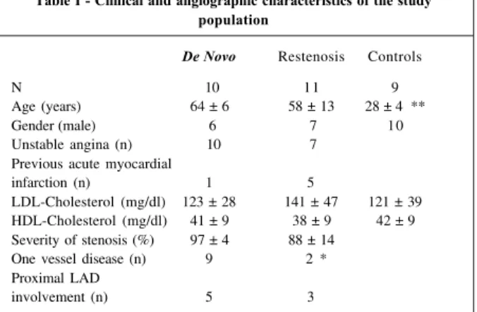
Characteristics of patients and control subjects are shown in the Table I. According to inclusion criteria, normal individuals were significantly younger than both groups of patients. Unstable angina accounted for the totality of cases in the de novo lesions group, but only 7/11 patients in the restenosis group had unstable angina; however, this was not statistically significant. The time from the last episode of an-gina in the group of patients with restenosis was significantly shorter than that of patients with de novo lesions (77±214 versus 124±144 hours; p<0.05). No statistically significant differences occurred regarding the use of cardiovascular drugs, which included beta-blockers, aspirin, nitrates, calcium channel blockers, ticlopidin, or intravenous heparin. The lipid profile was also similar between the two groups of patients, and no change in cardiac enzyme profiles were observed. The mean elapsed time from coronary angioplasty in the group of patients with restenosis was 165±35 days.
Significantly more patients in the de novo group pre-sented with one vessel disease compared with the resteno-sis group (Table I). Other angiographic characteristics, su-ch as proximal left anterior descending artery involvement and severity of stenosis, were not significantly different between the groups.
between tumor necrosis alpha and soluble tumor necrosis alpha receptor II (r= 0.6; p<0.05) were observed. Similarly, soluble receptor levels correlated with each other (r= 0.85; p<0.05). After adjustment for differences in age among groups, results remained unchanged.
Discussion
Atherosclerosis can be considered an immunoinflam-matory process evolving over a number of years according to the presence or absence of risk factors and genetic back-ground 6. Restenosis can be viewed as an accelerated form of
this process with associated wound-healing characteristics 2.
In this study, we demonstrated an enhanced state of inflam-matory activity detected in the plasma of patients presenting with coronary artery disease syndromes secondary to both
de novo or restenotic lesions compared with that in the nor-mal controls.
Patients enrolled in this study constituted a typical group of individuals referred by their attending cardiolo-gists for coronary angiography for known or suspected co-ronary artery disease. No major clinical or laboratory diffe-rences were observed between the two groups of patients, except for an increased number of patients presenting with unstable angina in the group with de novo lesions. Althou-gh this difference did not reach statistical significance, this is in keeping with the notion that, overall, restenotic lesions are less likely to cause unstable syndromes 7. Use of
medi-cations was also similar in both groups at the time of blood sampling, except for two patients receiving heparin in-travenously in the de novo atherosclerosis group.
Atherogenesis can be viewed as an inflammatory pro-cess in which vascular cells can play important roles media-ting several immunoinflammatory mechanisms secondary to endothelial injury. Endothelial cells express adhesion mo-lecules in their surface that will induce T cells and monocy-tes/macrophages to adhere to the vascular endothelial sur-face and subsequently migrate to the subendothelial space. Increased plasma levels of different inflammatory
media-tors, indicative of this inflammatory process, have been identified in established coronary artery disease 8-11. Our
group has also recently demonstrated that patients with stable and unstable angina with significant coronary artery disease by angiography and even patients without flow-li-miting coronary stenosis but presenting with chest pain may have increased plasma levels of adhesion molecules 12. In
fact, Ridker and co-workers 13 have recently shown that
in-creased levels of intracellular adhesion molecule-1 in heal-thy men are predictors of future acute myocardial infarction. In the present study, yet investigating different mar-kers of inflammatory activity, we also showed that patients presenting with stable or unstable angina feature a pattern consistent with an enhanced state of immunoinflammatory activation. In our study, the source of increased levels of the different markers we examined is unclear. Because both vascular and more likely T cells and monocytes present in the atherosclerotic plaque can synthesize tumor necrosis alpha, it can be speculated that this cytokine could be ini-tially formed in the plaque and subsequently released in the circulation. In fact, Rus et al. 14 have recently shown
increa-sed C protein and interleukin-6 levels eluted from human ar-terial wall with atherosclerotic disease. On the other hand, Liuzzo et al. 15 have indicated that an enhanced
immunoin-flammatory response in unstable angina patients could be secondary to nonspecific stimuli. Similarly, soluble interleu-kin-2 receptors, which are derived from activated T cells, could also be produced by intraplaque lymphocytes or by activated circulating T cells. We have previously shown that, compared with primary lesions, restenotic coronary le-sions subsequent to both atherectomy or balloon angiopla-sty feature increased expression of tumor necrosis alpha 3.
This was associated with a higher number of T cells in these lesions. Others have also suggested that an enhanced state of inflammatory activity could be important to the develop-ment of restenosis 4,16,17. Whether these features can be
translated into a clinical situation of patients presenting with coronary syndromes and detected in circulating plas-ma of patients replas-mains unclear. Our study aimed to characte-rize patients with known restenotic lesions or de novo le-sions regarding circulatory levels of different markers of in-flammation. The pattern of tumor necrosis alpha and its soluble receptors and that of soluble interleukin-2 receptors consistently indicated an enhanced inflammatory activity in the group of patients with de novo lesions, when compa-red with that in controls or in patients with restenosis. This is in contrast to our previous immunohistochemical studies on both primary and restenotic plaques. It is possible that differences in the expression of tumor necrosis alpha, which has a known short half-life 18,19, observed in atherectomy
specimens do not have the magnitude to be maintained when clinical studies measuring this peptide in the circula-ting plasma are carried out. In addition, all of the patients in the de novo group from the present study had unstable an-gina compared with 7/11 in the restenosis group. Although this difference was not statistically significant, the known ruptured/fissured plaque associated with unstable angina may have contributed to a more pronounced inflammatory
Table I - Clinical and angiographic characteristics of the study population
De Novo Restenosis Controls
N 10 11 9
Age (years) 64 ± 6 58 ± 13 28 ± 4 **
Gender (male) 6 7 10
Unstable angina (n) 10 7 Previous acute myocardial
infarction (n) 1 5
LDL-Cholesterol (mg/dl) 123 ± 28 141 ± 47 121 ± 39 HDL-Cholesterol (mg/dl) 41 ± 9 38 ± 9 42 ± 9 Severity of stenosis (%) 97 ± 4 88 ± 14
One vessel disease (n) 9 2 * Proximal LAD
involvement (n) 5 3
reaction in this group of patients, offsetting a potential in-creased inflammatory activity expected to be present in pa-tients with restenotic lesions.
Our data should be interpreted in the light of specific study limitations. First, our limited sample size may not sup-port definitive answers, but regardless of the number of stu-dy individuals, our data collectively show a consistent pat-tern of heightened inflammatory activity in the de novo
group as opposed to the restenosis group. Second, we chose a control group that was significantly younger than both other groups. While this may have influenced our ana-lysis, interleukin 2 soluble receptor levels may correlate with age only up to 4 years when the immune system is still be-ing developed 20. Tumor necrosis factor alpha was not
in-fluenced by age in similar studies 21,22. Third, as mentioned
above, our data may not reflect intraplaque events, because only peripheral blood was studied. It can be speculated that blood sampling from the coronary sinus could more appro-priately identify differences occurring inside the atheroscle-rotic plaque. Finally, because coronary syndromes can be complex, it would be interesting to investigate patients pre-senting with clinical syndromes as comparable as possible
(all stable angina or all unstable angina patients with primary and restenotic lesions).
In conclusion, our study reinforces the notion that co-ronary artery disease is associated with a pattern of immu-noinflammatory activity and suggests that this can be as-sessed by measuring plasma levels of tumor necrosis alpha and its receptors and soluble interleukin-2 receptors. Our data also indicate significant increases in circulatory levels of these markers in patients with primary atherosclerosis when compared with that in patients with restenotic lesions or in normal controls. Finally, a moderate degree of immuno-inflammatory activity was shown in patients with resteno-sis as increased levels of soluble interleukin-2 receptors only were observed in this group compared with controls.
Acknowledgements
We are thankful to Dr. Karen Prado and Dr. Jarbas Oli-veira for their support to carry out the immunoabsorbent assays. We are also grateful to Dr. Murilo Foppa for his sug-gestions and help in the initial phases of the study. Statisti-cal support was provided by Ms. Luciana Bertoldi Nucci.
Atero Rest Ctr
Atero Rest Ctr Atero Rest Ctr
Fig. 1 - Individual data for immunoinflammatory markers in patients with de novo coronary lesions (de novo), patients with restenosis (Rest), and controls (Ctr). Groups connected by lines are significantly different. A) Plasma levels of soluble receptor of interleukin-2 (sr-IL2); B) plasma levels of soluble receptor I of tumor necrosis factor alpha (sr I-TNF-a); C) plasma levels of soluble receptor II of tumor necrosis alpha (sr II-TNFa); D) plasma levels of tumor necrosis alpha (TNFa). Data and mean + SD (except for TNF-a).
1. Serrano CV, Ramires JA, Venturinelli M, et al. Coronary angioplasty results in leuko-cyte and platelet activation with adhesion molecule expression. Evidence of in-flammatory responses in coronary angioplasty. J Am Coll Cardiol 1997; 29: 1276-83. 2. Libby P, Schwartz D, Brogi E, Tanaka H, Clinton SK. A cascade model for resteno-sis. A special case of atherosclerosis progression. Circulation 1992; 86(suppl 3): 47-52.
3. Kelso A. Cytokines: structure, function and synthesis. Curr opin immunol 1989; 2: 215-25.
4. Le J, Vilcek J. Tumor necrosis factor and interleukin-1: cytokines with multiple overlapping biological activities. Lab Invest 1987; 56: 234-48.
5. Clausell N, Molossi S, Sett S, Rabinovitch M. In vivo blockade of tumor necrosis factor-alpha in cholesterol-fed rabbits after cardiac transplant inhibits acute co-ronary artery neointimal formation. Circulation 1994; 89: 2768-79. 6. Dinarello CA, Mier JW. Lymphokines. N Engl J Med 1987; 317: 940-5. 7. Rubin LA, Nelson DL. The soluble interleukin-2 receptor: biology, function,
and clinical application. Ann Intern Med 1990; 113: 619-27.
8. Clausell N, de Lima VC, Molossi S, et al. Expression of tumour necrosis factor al-pha and accumulation of fibronectin in coronary artery restenotic lesions retrie-ved by atherectomy. Br Heart J 1995; 73: 534-9.
9. Pietersma A, Kofflard M, de Wit LE, et al. Late lumen loss after coronary angio-plasty is associated with the activation status of circulating phagocytes before treatment. Circulation 1995; 91: 1320-5.
10. Braunwald E. Unstable angina: a classification. Circulation 1989; 80: 410-14. 11. Ross R. Mechanism of disease: atherosclerosis - an inflammatory disease. N Engl
J Med 1999; 340: 115-26.
12. Nobuyoshi M, Kimura T, Oshishi H, et al. Restenosis after percutanous translu-minal coronary angioplasty: pathological observations in 20 patients. J Am Coll Cardiol 1991; 17: 433-9.
13. Blum A, Sclarovsky S, Shohat B. T lymphocyte activation in stable angina pec-toris and after percutaneous transluminal coronary angioplasty. Circulation 1995; 91: 20-2.
14. Schumacher M, Halwachs G, Tatzber F, et al. Increased neopterin in patients with chronic and acute coronary syndromes. J Am Coll Cardiol 1997; 30: 703-7.
Referências
15. Hasdai D, Scheinowitz M, Leibovitz E, Sclarovsky S, Eldar M, Barak V. Increa-sed serum concentrations of interleukin-1b in patients with coronary artery di-sease. Heart1996; 76: 24-8.
16. Neri Serneri GG, Prisco D, Martini F, et al. Acute T-cell activation is detectable in unstable angina. Circulation 1997; 95: 1806-12.
17. Clausell N, Prado K, Ribeiro JP. Increased plasma levels of soluble vascular cel-lular adhesion molecule-1 in patients with chest pain and angiographically nor-mal coronary arteries. Int J Cardiol 1999; 68: 275-80.
18. Ridker PM, Hennekens CH, Roitman Johnson B, Stampfer MJ, Allen J. Plasma concentration of soluble intercellular adhesion molecule 1 and risks of future myocardial infarction in apparently healthy men. Lancet 1998; 351: 88-92. 19. Rus H, Niculescu F. Inflammatory response in unstable angina Circulation 1999;
100: 98.
20. Liuzzo G, Buffon A, Biasucci LM, et al. Enhance inflammatory response to coro-nary angioplasty in patients with severe unstable angina. Circulation 1998; 98: 2370-6.
21. Moreno PR, Bernardi VH, Lopez Cuellar J, et al. Macrophage infiltration predicts restenosis after coronary intervention in patients with unstable angina. Circula-tion 1996; 94: 3098-102.
22. Lima VC, Gottlieb AI, Clausell N, et al. Analysis of atherosclerotic plaques ob-tained by coronary atherectomy: foam cells correlated positively with subse-quent restenosis. Cardiovasc Pathol 1996; 5: 265-9.
23. Packer M. Is tumor necrosis factor an important neurohormonal mechanism in chronic heart failure? Circulation1995; 92: 1379-82.
24. Mueller AR, Platz K, Haak M, et al. The release of cytokines, adhesion molecules, and extracellular matrix parameters during and after reperfusion in human liver transplantation. Transplantation 1996; 62: 1118-26.
25. Rubin LA, Nelson DL. The soluble interleukin-2 receptor: biology, function, and clinical application. Ann Intern Med 1990; 113: 619-27.