Enrique Rassi,s He’ctor Monzh, 4 Macario Castillo,5 Icar Hernhdez, 6
Jaime Ramirez P&z,’
and Jacinto Convit a
A survey of villages in Venezuela near a focus of onchocerciusis
(river blindness) across the border in Brazil has revealed a high prevalence of cases. Future human settEement of the area may well de$end on control of this disease.
The presence of onchocerciasis in Vene- zuela was first observed by Potenza et al. in 1949 (24), but it was not until 1958 that an epidemiologic survey was conducted in the area believed to be affected (22). Since that time the nature and extent of two foci, both of them in the coastal mountain range, have gradually been defined (Figure 1). One, located in the north-central region of the country, has primarily involved the states of Aragua, Carabobo, Miranda, and Guarico, but it has also caused a few sporadic cases in the states of Yaracuy and Cojedes. The other, in northeastern Vene-
zuela, covers portions of the states of Anzokegui, Monagas, and Sucre (13, 17, 22).
These affected areas are fairly populous, the total number of inhabitants being reported at 1,898,827 in 1974. In early June 1974 some 1,628,370 persons were examined for onchocerciasis, and 40,091 were found to be positive (by the Mazzotti test in most instances). Thus the apparent rate of infection was 24.6 per 1,000 population.
The characteristic features of the disease in these endemic areas are (23,17,22):
IAlso appearing in Spanish in BOG Of Sunit Panam. ZStudies conducted by the Pan American Center for Research and Training in Leprosy and Tropical Diseases, Caracas, Venezuela. Dr. Celia P. Motta R. was adviser for the investigation. Also cooperating was the Border Service, Venezuelan Ministry of Health and Social Welfare-Dr. Luis Gonzalez Herrera, Chief Medical Officer.
SAssistant Director, National Institute of Dermatol- ogy; Pan American Center for Research and Training in Leprosy and Tropical Diseases, Caracas, Vene- zuela.
4First Assistant Director, National Institute of Dermatology; Pan American Center, Caracas.
bCommissioner General of Public Health, Federal Territory of Amazonas, Venezuela.
6Medical Officer, Public Health Dermatology Service. State of Apure, Venezuela.
7Entomologist, National Institute of Dermatology: Pan American Center, Caracas.
SDirector, National Institute of Dermatoldgy: Pan American Center, Caracas.
42 PAHO BULLETIN l Vol. Xi, No. I, I977
l.YARACUY STATE 2. CAAABOBO STATE 3. COlELIES STATE
4. ARAGUA STATE 5. GUAM0 STATE
6. MIRANDA STATE 7. ANZOATEGUI STATE
8. MONAGAS STATE 9. SUCRE STATE
61
Known extent of onchocerciaris foci, 1958.
Known extent of onchocerciasis foci ..*... as of 31 October 1974.
Source: National Registry of Patients and Contacts, Department of Public Health Dermatology, Ministry of Public Health and Social Welfare, Caracas, Venezuela.
l Absence of dermatologic lesions; l Absence of lymphatic involvement; l Presence of ocular lesions in some 30
per cent of the cases, with 49.6 per cent of the reported cases involving punctate kera- titis (no sclerosing keratitis observed):
l Low frequency of subcutaneous nod-
ules, which were found in only 23 per cent of the subjects examined at the time control measures were first introduced and are currently very rare;
l Localization of the nodules low on the
body, mainly from the hips downward; and
l Transmission of the Onchocerca vol-
vulus parasite by Simulium metallicum and, to a much lesser degree, by Simulium
exiguum- the only vectors heretofore known in these foci (8,13,15,16).
The two foci described above diminish and fade out toward the south as ecologic conditions change and the two vectors gradually disappear. Neither of the vectors has been found in the vast expanse of the Venezuelan plains farther south, and until very recently no other Venezuelan foci were known,
American Health Organization’s advisory group completed a study in parts of neigh- boring Brazil where new endemic areas of the disease had been found. It was observed in the course of this study that onchocercia- sis was present among Yanomama Indians in the far northern portions of Amazonas State and the Federal Territory of Roraima (10,21)-areas close to the Venezuelan border (Figure 2).
Brazil’s three known foci-near, respec- tively, the Auaris, Surucucu, and Toototobi Missions-are not directly linked to each other. However, many of the Indians’ com- munication routes extend into Venezuela. At Auaris there is a main route used by the local Sanuma and Makiritare which crosses the border and goes northward into the mountains, traveling along the valleys of the Upper Ventuari, Merewari, Canara- cuni, and Upper Caura rivers. Another proceeds west by way of the Padamo and Cunucunuma river valleys. At Surucucu the Aikam-teri Indians have a major travel route that extends west into Venezuela through the mountainous Parima region. And the Xiriano-teri of Toototobi have a seasonal migration route across the moun- tains along the Brazil-Venezuela border which takes them into the valleys of the Upper Orinoco, Ugueto, and Siapa (21).
Only a very small proportion (0.75 per cent) of the predominant man-biting Si- mu&m vectors captured at the Toototobi Mission were found to be naturally infested with 0. volvulus (20). This low index is not compatible with the high infection rate
(606.5 per 1,000 population) observed among the Xiriano-teri residents of this area. The investigators therefore reached the following conclusion:
Since the most heavily parasitized patients come from the area inhabited by the Vene- zuelan Yanomama Indians, we may assume that in some of the following places-the
Upper Ventuari, Upper Merewari, and
Padamo river valleys, the Parima mountain range, and the Upper Orinocovalley-oncho-
cerciasis transmission must be greater, with a higher vector infestation index, and that the Indians of Auaris, Toototobi, and Surucucu come in temporary contact with these more efficient vectors in the course of their seasonal migrations (21).
Mater%& and Methods
On the basis of the foregoing facts and assumptions, an expedition was planned for early 1975 into the region of the Parima mountains and the Upper Orinoco river. The expedition’s itinerary, shown in Figure 2, was as follows: Puerto Ayacucho-Tama- Tama-Parima Mission-Coyowa-teri Mis-
sion-Platanal-Boca de Ocamo-La Es-
meralda-Puerto Ayacucho. In addition, side trips were made by launch from Platanal to the area of the Guajaribos and Pefiascal rapids and from Boca de Ocamo to Boca de Mavaca.
The basic aims of the undertaking were: to detect cases of onchocerciasis in the local populations, and to collect entomologlc specimens for the purpose of identifying man-biting Simuliidae and determining indexes of natural 0. volvulus infestation in these species. Other activities carried out included tuberculin-testing and BCG administration, collection of Phlebotomus
flies, biopsy of dermatologic lesions, and removal of subcutaneous nodules. Specifi- cally, the case-detection work consisted of procuring and examining skin snips from the inhabitants (one each, taken with a razor from the area above the shoulder blade), usually from persons 10 years of age or over; administering the Mazzotti test (50 mg of hetrazan--2,3,9,10); conducting a general dermatologic examination: palpat- ing the skin to detect nodules; and, in every tenth subject, testing for the presence of
Figure 2. Onchocerciasis in southern Venezuela: Preliminary investigation of possible foci in the Federal Territory of Amazonas, April 1975.
APURE STATE
ff BOLIVAR h STATI
/,
‘-‘--x
..
.,
A
. . . .‘...,,,,
,,..,
‘,.,
.’
;.’ )
.’
. ..I
,..., .___.,.
..’ ‘x
f’...‘..,.,
i j ,A...
__..
. . . i
:
( i...
. . .._.
. . . . _,
,,,
‘..,
A~ n
PUERTO AYbCUCHO _/-.._/"--'-ATURES UEPAR
/:
.I; FEDERAL .’
iOMAROA
. r&F--
FEDERAL
\
t
MA TERRITORY
OF RORAIMA
TOOTOTORI xillana.tsri
. LOCALITIES WITH ONGHOCERCIASIS
BRAZIL 0 LOCALITIES WITHOUT ONCHOCERClASlS @ LOCALITIES IN BRAZIL WITH ONCHOCERCIASIS 44
obtained are described in the sections that from one hour to five days on foot (Figure
follow. 3). Several of the individuals examined
were from these other settlements. Parima Mission
Description Results
The Parima Mission is located near the Brazil-Venezuela border (Z050’N, 64O ZO’W). It is set in a valley near a small stream at an altitude of about 850 meters. The rainy season begins in May and ends in September. About 200 Yanomamas, from the Niayoba-teri group, live near the mission. In addition, there are some 15 other settlements with a total population of about 1,140 located nearby at distances of
Skin snips were obtained from 104 persons, of whom 30 (28.8 per cent) proved to be positive (Table 1). The differences between the sexes were not statistically significant. The average number of micro- filariae per unstained biopsy specimen was 14.4; counts with Giemsa-stained specimens were higher, averaging 21.8 per slide. Two of the individuals tested were children, four and five years old respectively, who had
Figure 3. Distribution of the Yanomama Indian groups studied in the Federal Territory of Amazonas, Venezuela, April 1975 (see Annex on page 46).
0
No. Groups with onchocerciasis Groups not studiedBroups apparently without onchocerciasis
(negative findings]
BOCA OE OCAMO
..l.-- -.m._-
46 PAHO BULLETIN l Vol. XI, No. I, 1977
ANNEX TO FIGURE 3
Yanomama Groups Living in the Area Studied
Mission visited and Indian Distance away
nearby Indian groups populations from missions
PCZTilW2 1) Niayoba-teri (0) 2) Vainama-teri (0) 3) Pablo-teri (a)
4) Mayubaaeri (4 groups) (0) 5) Ysha-teri (4 groups) (0) 6) MLnapa.teri (several groups) 7) Shitali
Subtotal 200 60 100 300 200 200 80 1,140
Coyowa-teri Mtscion 8) Coyowa-teri (0) 9) Motowa-teri 10) Tolobo-reri (0) 11) Tacowa-teri 12) Adajubayaca-teri (0) 13) Yulucalewa-teri 14) Ijiliha-teri (0) 15) Shimuwa-teri (Zgroups) 16) Fubiramao-teri 17) Coyoshiwa-teri 18) Obowaca-teri
19) Mayo-teri (several groups)
Subtotal 85 95 65 125 60 60 85 80 60 80 60 250 1,105 Platanal
20) Majecoto-teri (0) 21) Chachano-teri (0) 22) Conoboredue-teri (o) 23) Nabtawe-teri 24) Kashorawa-teri 25) J asubue-teri 26) J ijiramawa-teri 27) u arocoao-teri 28) Yesidueari
Subtotal 120 20 150 60 110 180 50 150 100 940 MaVUCU
29) Patano-teri (3 groups) (0) 30) Pisahi-teri (5 groups) (0) 31) Mono-teri
32) Masiquidue-teri
33) Moeropoi-teri and other groups
226
200 60 88 200
34) Macorima-teri 60
35) Ohyari-teri 60
36) Carawaieri 60
Subtotal 954
OClZ?7W 37) Iiiavi-teri 38) Vitocayo-teri 39) Sibari-teri
Subtotal
168 60 70
298
Total 4,437
Near the mission l/2 hour on foot 1 hour on foot 8 hours on foot 6 hours on foot
Between 12 hours and 3 days on foot 24 hours on foot
Near the mission 4 hours on foot 5 hours on foot 8 hours on foot 5 hours on foot 6 hours on foot 5 hours on foot 18 hours on foot 12 hours on foot 8 hourson foot 10 hours on foot 24 hours on foot
Near the mission 20 min. by boat 90 min. by boat 90 min. by boat 2 hours by boat 5 hours by boat 6 hours by boat 8 hours by boat 12 hours by boat
2 days on foot
Near the mission Near the mission 1 hour by boat Upper Mavaca. 4 to 12
hours by boat
1 hour from Mavaca in the Manaviche Gorge Between Mavaca and Platanal Between Mavaca and Platanal
Near the mission 1 hour by boat
Upper Ocamo, 5 hours by boat
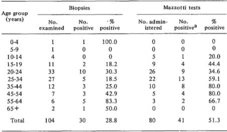
Table I. Biopsy and Mazzotti test results obtained from members of the Niayoba-teri and Mayuba-teri tribes at Parima, Federal Territory
of Amazonas, Venezuela (April 1975).
Age group (wars)
Biopsies Mazzotti tests
NO. NO. .% No. admin- No. %
examined positive positive istered positivea positive
o-4 1 1 100.0 0 0 0
5-9 1 0 0 0 0 0
10-14 4 0 0 5 1 20.0
15-19 11 2 18.2 9 4 44.4
20-24 33 10 30.3 26 9 34.6
25-34 27 5 18.5 22 13 59.1
35-44 12 3 25.0 10 8 80.0
45-54 7 3 42.9 5 4 80.0
55-64 6 5 83.3 3 2 66.7
65+ 2 1 50.0 0 0 0
Total 104 30 28.8 80 41 51.3
aWeak positive responses (l-i-) to the Mazzotti test (such as pruritis without patent scratching lesions or infiltration of the tissues, erythema, and edema) were not included in these figures as positive findings.
been found on palpation to have nodules. The younger one turned out, in fact, to be positive. About eight hours away by foot (10 minutes by light aircraft) a Mayuba-teri settlement was found at an altitude of about 950 meters. It was only possible to obtain skin snips from nine individuals, three of whom were positive.
The Mazzotti test was given to 80 of the people who provided biopsy material, including some who had positive biopsies. The results (Table 1) were clearly positive in 41 instances (51.3 per cent). The positive responses included itching lesions, ery- thema, edema, and conjunctival reaction - this last being the most frequent and patent manifestation.
In the dermatologic examination, chronic dermatitis and “elephant skin” attributable to onchocerciasis were observed in three cases. Other findings included one case of ichthyosis and several of pyodermatitis. In addition, 13 onchocercal nodules were
found on 12 patients, 10 (76.9 per cent) of them being localized on the scalp, two in the pelvic region, and one on the leg.
Coyowa-teri Mission
Description
The Coyowa-teri Mission is located in the foothills of the Parima mountains to the south of the Parima Mission (Figure 3) on a bank of the Orinoquito river at an altitude of about 250 meters. The river is between 20 and 30 meters wide at this point.
48 PAHO BULLETIN l Vol. XI, No. I, I977
Results
Skin snips were taken from 45 individ- uals, not all of whom belonged to the Coyowa-teri group. As Table 2 shows, 33 of these subjects (73.3 per cent) showed positive findings. The average microfilaria count was 18.8 per unstained slide and 24.0 per Giemsa-stained slide. Significant levels of infection were observed among young people (including the five-to-nine-year bracket), and the positivity rate among subjects over 20 years of age was nearly 100 per cent.
Of the 12 individuals with negative bi- opsies, seven had a clearly positive response in the Mazzotti test. Addition of these seven cases to the 33 detected earlier gave an overall infection rate of 88.9 per cent.
All the 40 patients above (33 plus the additional seven positive by the Mazzotti test) were examined dermatologically. Cuta- neous manifestations of the disease were very evident and included some features previously described only in certain foci in Africa. Significant inguinal and femoral adenopathy was observed in eight subjects. In another four, including an eight-year- old boy, involvement of the inguinal and
Table 2. Biopsy results obtained from Yanomama Indians near the Coyowa-teri Mission,
Amazonas, Venezuela (April 1975).
Age group No. No. %
(wars) examined positive positive
o-4 3 1 33.3
5-9 6 4 66.7
LO-14 2 1 50.0
15-19 6 3 50.0
20-24 1 1 100.0
25-34 17 14 82.4
35-44 6 5 83.3
45-54 1 1 100.0
55-64 2 2 100.0
65+ 1 1 100.0
Total 45 33 73.3
femoral lymph nodes was most impressive, the inflammatory process affecting the tegument. These latter cases showed the feature described by British authors in Africa as “hanging groin.” A total of 12 persons had obvious inguinal involvement.
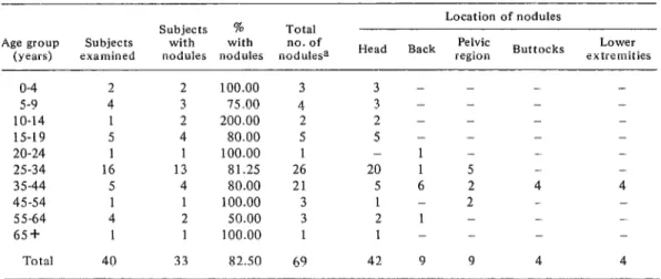
Chronic papular dermatitis attributable to onchocerciasis was also found, together with “elephant skin,” in 12 of the subjects (Figures 4,5). Two of these 12, as well as three others, also had acute skin lesions in the form of erythematous plaques, which were rounded, banded, or, in one case, ir- regular in shape, bearing a clear resem- blance to lesions of dimorphous or lepro- matous leprosy (Figures 6,7). In sum, chronic and/or acute skin lesions were observed in 15 persons, or 37.5 per cent of the patients under study. In addition, a total of 69 nodules were found, either by palpation or by visual inspection, in 33 of the patients (Table 3, Figure 8).
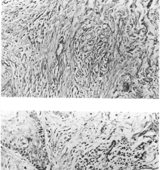
The histopathologic findings can be seen in Figure 9. The first frame (A) shows an H & E-stained section with perivascular in- filtration by eosinophils, macrophages, and lymphoid cells. This tissue was obtained from a papillomatous lesion on the back of a 21-year-old male. The subject had severe papillomatous and irregular erythematous lesions, generalized chronic dermatitis, “elephant skin” on the buttocks, and ingui- nal adenopathy. Figure 9-B likewise shows a perivascular infiltrate composed of eosino- phils, macrophages, and lymphoid cells. The biopsy came from an erythematous plaque on the shoulder-blade region of a 35-year-old male who had several such plaques, chronic generalized dermatitis, and inguinal adenopathy. Figure 9-C shows fragmentation of elastic fibers and, in some places, their actual disappearance from the dermis. This tissue was taken from an erythematous plaque located on the lumbar region of a 32-year-old male who had many irregular erythematous lesions and plaques
Fii
4. A Coyowa-teri case of generalized and marked chxuuic dermatitis with exten-
sive involvement of elastic fibers approaching the condition descrii
by writers in Africa
as “elephant skin.”
50
PAHO BULLETIN
lVol. Xl, No. I, I977
Figum 6. A well-defined erythematous plaque in another Coyowa-teri subject. Similar
lesions, corresponding to the acute phase of cutaneous involvement, were observed in
five cases.
Figum 8. Nodules on the scalp of an eight-yealcold Coyma-teri
boy. The swellings are
confluent.
Platanal
workers
putting
up a building
for
the
Malaria
Service, missionaries,
mission per-
sonnel and their families,
etc.
Description
Results
Platanal
(2O23’N, 64O55’W) is situated
a
short
distance
from
the Upper
Orinoco
river
at an altitude
of about
180 meters
(Figure
2). Two settlements
of Yanomamas
located 10 to 15 minutes
upriver
from the
mission
by boat-one
of 120 individuals
belonging
to the Majecoto-teri
group
and
the other of 20 belonging
to the Chachano-
teri -were
investigated.
From
there
the
study went on to include
some of the 150
members
of the Conoboredue-teri
group
living
about 90 minutes
farther
upstream.
Six other settlements
are located along this
waterway
between
the
Guajaribos
and
Peiiascal
rapids.
Al together,
about
9Ob
Indians were found living in these nine set-
tlements. Additional
subjects included
some
50-odd persons living
in the vicinity
of the
mission
post: a nurse,
malaria
personnel,
It was only possible to take biopsies from
36 individuals
in the Majecoto-teri,
Cha-
chano-teri,
and Conoboredue-teri
groups,
since most of the residents were away from
their communal
homes. Ten of the biopsies
(27.8 per cent) were positive.
The material
showed an average of nine microfilariae
per
unstained
slide,
but most of them were
found in skin snips obtained
from two sick
members
of the Conoboredue-teri
group.
Biopsies were also taken from 30 of the 50
Platanal
residents
not belonging
to the
Yanomama
groups, most of whom had only
been in the area a short time. These latter
specimens
yielded
generally
negative
re-
sul ts.
52 PAHO BULLETIN l Vol. XI, No. 1, 1977
Table 3. Number and distribution of nodules found on members of the Coyowa-teri tribe, Coyowa-teri Mission, Amazonas, Venezuela (April 1975).
Location of nodules
Subjects % Total
Age group Subjects with with no. of PelViC Lower
(wars) examined nodules nodules nodulesa Head Back region Buttocks extremities
o-4 2
5-9 4
10-14 1
IS-19 5
20-24 1
25-34 16
35-44 5
45-54 1
55-64 4
65+ 1
Total 40
2 3 2 4 1 13 4 1 2 1 33
100.00 3 3 - - - -
75.00 4 3 - - - -
200.00 2 2 - - - -
80.00 5 5 - - - -
100.00 1 - 1 - - -
81.25 26 20 1 5 - -
80.00 21 5 6 2 4 4
100.00 3 1 - 2 -
50.00 3 2 1 - - -
100.00 1 1 - - - -
82.50 69 42 9 9 4 4
apercentage of nodules located on the head: 62.3 % .
to read the results in nine cases, but of these,
eight were positive. The test was also given to the 30 non-Yanomama residents with negative biopsies. Twenty-nine of them responded negatively, but one woman, who had resided in the region of the Upper Ventuari, gave an intense positive response, including pruritus, erythema, and edema of the face and arms.
No cutaneous lesions definitely attrib- utable to onchocerciasis were observed in the dermatologic examination.
Ocular lesions were the most significant and surprising finding at Platanal (Figures 10-13). Four individuals residing in this general area were found to have lesions of major importance, including sclerosing keratitis. In one instance the condition had progressed over a three-year period to total blindness; in another the sight of the right eye had been lost; and in a third there was significant impairment of vision. The fourth subject, a boy only four years of age, was found to have a 1.0 x 0.5 cm opaque area on his left cornea. In addition to these four cases, one of bilateral keratitis was found in an Indian girl visitor from the Upper Ventuari region (Figures 14, 15).
Boca de Mavaca
There is a mission post at the juncture of the Upper Orinoco and Mavaca rivers (2O25’N, 65O15’W) about two hours down- stream by boat from Platanal. Some 250 members of the Pisashi-teri and Mono-teri groups were found living in three com- munes near the mission.
Biopsies obtained from 40 of the subjects gave negative results, but specimens from two additional Patano-teri visitors con- tained microfilariae.
The Mazzotti test was given to all 42 individuals examined, and 15 had positive responses. In one case this response included severe facial edema.
Dermatologic examination failed to re- veal any cutaneous lesions or onchocercotic nodules, but it should be mentioned that this phase of the investigation was con- ducted in the jungle under very difficult conditions.
54
PAHO BULLETIN lVi-d. XI, No. 1, 1977
Figura 10. Lesions of keratitis and bilateral cataracts involving loss of sight in the
right eye of a 50 yearold Majecoto-teri male (Platanal).
Figure 12. Closeup of lesions in right eye of patient in Fii
11.
56
PAHO BULLETIN lVol. XI, No. I, I977
Figure! 14. Ocular lesions of an Upper Ventuari Indian girl with bilateral keratitis.
Boca de Ocamo
The Ocamo Mission (Z045’N, 65O15’W) is situated near the confluence of the Ocamo and Upper Orinoco rivers about two hours downstream from Boca de Mavaca. Biopsies and Mazzotti tests were performed on over 100 Indians in this area, and the results were negative in all instances.
Tama-Tama
The mission post serving this area (Figure 2) is on the Upper Orinoco near the con- fluence of the Tama-Tama river (3”15’N, 65O45’W). Some 250 Piaroa Indians (not belonging to the Yanomama Nation) were living at the mission at the time of the inves- tigation. Biopsies were done on 38 subjects and Mazzotti tests were performed on over 100, all with negative results.
Shulium Vectors
The predominant species of man-biting Simulium flies identified in the foci under study were Simulium pintoi (at the Parima and Coyowa-teri Missions) and S. amazo- nicum (at Platanal). S. amazonicum was the predominant man-biting species found at Boca de Ocamo and Tama-Tama.
Eight per cent of the S. pintoi collected at Coyowa-teri Mission were found to be infected with 0. volvulus; of 100 females dissected, two had an infective form of the parasite in the head and six had develop- mental (sausage) forms in the thorax (Fig- ure 16-A). In addition, one of the 400 S.
amazonicum species captured at Platanal was found to have an infective form of 0. VOZVUZUS in the head (Figure 16-B). Hence S. pintoi and S. amazonicum were revealed as new vectors of onchocerciasis in Vene- zuela.
Discussion
The objectives of this study were:
l To confirm the presence of oncho-
cerciasis in at least part of the Venezuelan territory inhabited by Yanomama Indians (21) and to determine the prevalence of the disease among the affected groups:
l To determine the intensity of infection
and the severity of the clinical picture;
l To identify the predominant species of
man-biting Simuliidae in the existing foci and ascertain the infestation index in these species; and
l To train personnel assigned to the
study with a view to their effective partici- pation in a future control program.
The results in terms of these objectives are reported on the pages that follow.
Disease Prevalence
Biopsies obtained near the Parima Mis- sion showed high rates of infection among members of the two groups tested, the Niayoba-teri (28.8 per cent positivity) and the Mayuba-teri (33.3 per cent positivity). To the south, in the Parima foothills, members of the Coyowa-teri group showed a much higher rate of positivity (73.3 per cent). Considerably west and somewhat south of these two areas, between Platanal and the Guajaribos rapids of the Upper Orinoco, small settlements of Majecoto- teri, Chachano-teri, and Conoboredue-teri yielded an overall positivity rate of 27.8 per cent.
58
PAHO BULLETIN lVol. XI, No. 1, 1977
Table 4. Synopsis of findings.
Place=
Parima
Mayuba-teri Mission Coyowa-teri Mission Platanal
Boca de Mavaca Total
Biopsy and/or Mazzotti test No. examined No. positive y0 positive
104 45 43.3
9 3 33.3
45 40 88.9
36 18 50.0
42 15 35.7
236 121 51.3
‘Does not include Tama-Tama or Boca de Ocamo (178 people), which are outside the affected area.
In general, when the figures reflect the
total positive findings from persons showing a positive response either on biopsy or in the Mazzotti test-which is considered to be specific for the disease (3) -then the overall prevalences become higher (43.3 per cent at Parima, 88.9 per cent at Coyowa-teri Mission, 50.0 per cent at Platanal, and 35.7 per cent at Boca de Mavaca), for a general average of 51.3 (Table 4). Neither the biop- sies nor the Mazzotti tests yielded positive results farther north at Boca de Ocamo (among Yanomamas belonging to the Ifiavi- teri group) or farther west among the Piaroa Indians at Tama-Tama.
‘Intensity of Infection
The average number of microfilariae found per unstained biopsy specimen was 14.4 at Parima, 18.8 at Coyowa-teri Mis- sion, and 9.3 at Platanal. It should be noted, however, that a disproportionate share of the Platanal microfilariae were found in specimens from the Conoboredue- teri group. The other subjects from the Platanal area (members of the Majecoto-teri and Chachano-teri groups) yielded an average of only 3.0 microfilariae per un- stained specimen, even though it was in this series of subjects that the severe ocular
lesions, supposedly onchocercal, were ob- served.
Giemsa-stained slides yielded higher aver- age microfilaria counts at both Parima and Coyowa-teri Missions, the respective aver- age figures being 21.8 and 24.0 per biopsy. By way of comparison, a previous survey in a northern Venezuela focus showed a much lower average count: in all, 71 microfilariae were found in 93 biopsy specimens from 31 patients, for an average count of 0.76 per specimen (19).
Another matter to consider in evaluating the intensity of infection is the extent to which young children were affected (12). Although it was not possible to make a systematic study in this regard, several cases were observed in children in the four-to- nine age bracket: three four-year olds-one at Parima Mission, another in Platanal, and the third at Coyowa-teri Mission-plus four other Coyowa-teri children between five and nine years of age.
Severity of Clinical Symptoms
60 PAHO BULLETIN l Vol. XI, No. 1, I977
of lymphatic involvement; and the fre- quency of onchocercal nodules (12).
Four residents with significant ocular lesions were observed at Platanal. One sub- ject had bilateral blindness (complete loss of vision, developed over a three-year period); another had lost the use of one eye; and two others, including a boy four years of age, had less advanced ocular lesions. However, the microfilaria counts were relatively low among the population groups in which the lesions were found, and thus the oncho- cereal etiology of these lesions has not been confirmed.
Significant cutaneous lesions were ob- served at Coyowa-teri Mission. These in- cluded both acute lesions (multiple ery- thematous plaques) and chronic ones (ex- tensive or generalized papular dermatitis with “elephant skin”), often accompanied by severe involvement of the femoral and inguinal lymph nodes. Several of these cases had led to a “hanging groin” picture of the sort previously reported only in Africa (I, 3.
Onchocercotic nodules were found in 33 of the 40 Coyowa-teri residents who tested positively for onchocerciasis (Table 3), although one of the 33, a four-year-old boy, was negative on biopsy. This proportion, 82.5 per cent, was considerably higher than that found in Parima, where 12 out of 45 subjects (26.7 per cent) had a total of 13 nodules. An even lower frequency was observed at Platanal, where the findings were limited to a single nodule on one of the nine subjects examined.
.
Vector S$ecies
The predominant man-biting Simuliidae species near Parima Mission (altitude 850 meters) and at Coyowa-teri Mission (altitude 250 meters) was S. pintoi. S. ama-
zonicum was found to be the predominant
de Mavaca foci and also at Boca de Ocamo and Tama-Tama.
Infestation Index
The infective and developmental (sau- sage) forms of 0. volvulus found in eight of 100 unstained S. pintoi specimens collected at Coyowa-teri Mission clearly demonstrate the role of this species as vector of the disease. It should also be noted that S. $&ztoi was the predominant man-biting simulid found in the onchocerciasis focus at Auaris in Brazil’s Federal Territory of Roraima. This focus was previously described on the basis of work done in 1974 (21). At that time it was not possible to determine the natural rate of vector infestation at Auaris, since no developmental stages of the parasite were found in the 178 specimens examined. Nevertheless, it may be assumed that the natural infestation index was far lower at Auaris than at Coyowa-teri Mission.
Only one infective (final) form of 0. volvulus was found in 400 S. amazonicum females captured on the banks of the Orinoco at Platanal. By comparison, a slightly higher index of S. amazonicum
infestation (0.75 per cent) was previously found in flies captured along the Toototobi river in Brazil’s Amazonas State (20).
Personnel Training
Personnel from the Department of Public Health Dermatology, National Institute of Dermatology, and three auxiliaries ap- pointed by the administration of the Feder- al Territory of Amazonas to be responsible for carrying out the programs of the Public Health Dermatology Service, participated alternately in the work of the investigation.
Conclusions
analysis of the disease foci in Venezuela. The traditional foci in the central and eastern parts of the country-both located mainly in the coastal highlands (23, 22)- fade out as they approach the southern parts of Monagas and Guarico states. This is at- tributed to changing ecologic conditions beyond the base of the highlands, together with disappearance of S. metallicurn and S. exiguum, the two local vectors of the disease. To the south the great plains of Venezuela extend for hundreds of miles, crisscrossed periodically by the rivers of the Orinoco basin. Eventually the Amazon jungle begins, rising finally into the upper headwaters of these rivers, which is where the Yanomama groups reside.
This situation in terms of geography and the vectors suggests that there is no epide- miologic relationship between these tradi- tional Venezuelan foci in the coastal highlands and the newly discovered focus in the Amazon region. In other words, the latter appears to be indigenous to the tropical forest area shared by Venezuela and Brazil-in the same way, in turn, that the Western Hemisphere foci appear to be indigenous and epidemiologically unrelated to those in Africa. In regard to this latter point, Mazzotti and others have felt-and Duke has recently confirmed (X6)-that the African foci are quite distinct in their etiology from those in Guatemala and along the Venezuelan coast.
Direct observation of the Amazon focus and analysis of its epidemiologic and clinical features likewise confirm that it is distinct from the coastal foci. The high positivity rates found at Coyowa-teri and Parima Missions and at Platanal contrast sharply with the much lower rates recorded in the traditional foci in the north, where, before the initiation of control measures, response to the Mazzotti test usually ranged between 3 and 10 per cent, rarely exceeding the latter figure.
The presence of severe skin lesions in 37.5
per cent of the Coyowa-teri subjects and of significant lymphatic involvement in 26.7 per cent (including four cases of “hanging groin”) also provide a marked contrast to the absence of such lesions in the coastal foci. Nor have the latter areas ever reported the progressive sclerosing keratitis which was observed at Platanal.
Similarly, before control measures were adopted in the coastal foci, onchocercal nodules were found in some 23 per cent of the patients examined-a proportion far smaller than the 82.5 per cent observed among the Coyowa-teri subjects.
The same kind of evidence that prompted the search for onchocerciasis in the Parima mountains, at Coyowa-teri Mission, and along the Upper Orinoco (21) now suggests that the Amazon focus must extend farther north toward the headwaters of the Ven- tuari, Merewari, Canaracuni, and Caura rivers and farther west toward the source of the Padamo.
Quite apart from the personal conse- quences suffered, the transmission of 0. volvulus with such a high infestation index among small groups of primitive Indians scattered in a vast rain forest is a matter of real epidemiologic concern; the Indians, who are identified with the local environ- ment, act as the reservoir of infection, and secondary cases have already occurred in missionaries in Brazil and Venezuela
(1OJl).
The area in question is of particular im- portance because its development is called for in the national plan. Given the wide distribution-far beyond the zone investi- gated-of S. amazonicum and S. pintoi,
which were found to be naturally infected by 0. volvulus at Platanal and Coyowa-teri Mission, the implications are serious.
62 PAHO BULLETIN l Vol. XI, No. 1, I977
contains numerous Yanomama groups and a total of about 4,300 inhabitants, about 50 per cent of whom could be affected.
Anthropologist Napoleon Chagnon, who has been studying the population dynamics and other features of the Yanomamas since 1965, has reported (personal communica- tion) that the various groups are becoming increasingly numerous, some more than others, in the wake of declining infant mortality. Moreover, they are spreading out, since as soon as a settlement reaches a
population of about ZOO, some of its people will move away and establish a new one.
Thus it is considered essential that a broad control program, with adequate human and material resources, be estab- lished in the affected area to combat oncho- cerciasis through case-finding and treat- ment of patients. If this is not done, the disease will frighten away settlers and a sizable portion of the Federal Territory of Amazonas will remain blocked to progress.
ACRNOWLEDGMEiVTS
The authors would like to express their appre- ciation to the following individuals and their institutions without whose cooperation it would not have been possible to complete the research described: Dr. Hector Acevedo Zuleta, Governor
of the Federal Territory of Amazonas; Dr. Albert0 Anzart Mantilla, Amazonas Commis- sioner of Public Health; Monsignor Ciaccarelli, Bishop of Puerto Ayacucho; and Dr. Luis Gon- zllez Herrera, Chief Medical Officer, Border Service, Venezuelan Ministry of Health and Social Welfare.
They also wish to thank, in chronological order of their assistance, Dr. Jaime Cardenas, Chief, Amazonas Rural Endemic Disease Serv- ice; Walter Mood, pilot of the “Wings of
Deliverance”; Missionary Jacob0 V. Toewa and members of his family: Jose Temple: personnel of the facility at Tama-Tama; Missionary Wallys Jank and members of his family at Parima;
Missionaries Cecil and Wilfred Neese and
anthropologist Raymond Hames at Coyowa-teri Mission: Father Jesus Gonzalez and Brother Franc0 at Platanal; Father Bis, Sister Felicita, and Dr. Napoleon Chagnon at Boca de Ocamo; and Sister Nora at Boca de Mavaca.
Special mention should also be made of the work done during the study by the National Supervisor of Rural Programs, Juan B. Oviedo: by Inspector Felipe Perez; and by the entomo- logic assistant, Alirio RamIrez.
SUMMARY
The discovery of onchocerciasis (river blind- ness) at sites in Brazil near the Venezuelan border prompted a survey of the Venezuelan area most likely to be affected. The localities investigated-small, scattered settlements of Yanomama Indians deep in the jungle-are situated in Venezuela’s Federal Territory of Amazonas near the Parima mountains and the Upper Orinoco river.
Several cases of disease were observed in children four to nine years of age. Two Simulium flies (S. pintoi and S. amazonicum) appear to be the predominant vectors of Onchocerca volvulus in the area surveyed.
Severe ocular lesions, including one case of total blindness, were observed at Platanal, but it is not certain that they were caused by onchocer- ciasis. On the other hand, numerous significant dermatologic lesions definitely attributable to the disease were found at the Coyowa-teri Mission. Several of these cases presented a “hanging groin” picture of the sort previously reported only in Africa. Nodules were found in
82.5 per cent of the infected Coyowa-teri subjects.
Epidemiologic analysis of this Venezuelan Amazon focus suggests that it is unrelated to the other traditional Venezuelan foci near the coast. At the same time, there are indications that the Amazon focus extends farther to the north and west, into the headwaters of several Orinoco tributaries not included in the present survey.
The area in question is scheduled for develop- ment under the national plan. The wide distri- bution of the two Simulium vectors of the disease is cause for concern. It is essential that a broad control program be established in the focal area, lest it become blocked to human settlement.
REFER.ENCEs
(I) Anderson, J., and H. Fuglsang. Clinical Aspects of Onchocerciasis in Uganda and Yemen Arab Republic Compared with a Rain Forest and Savanna Focus in Cameroon. Document
WHO/ONCH0/73.102. Geneva, World Health
Organization, 1973.
(2) Burch, T. S. Prurito producido por el hetrazan coma una prueba de diagnostico para la oncocercosis. Rev Co1 Med Guatemala 2(l): 53-57, 1951.
(3) Castellazzi, Z., F. Hernando, andE. Rassi. Respuesta al test de Mazzotti (test de hetrazan) en poblaciones no endemicas de oncocercosis. (To be published.)
(4) Chagnon, N. A., J. V. Neel, L. Weitkamp, H. Gershowitz, and M. Ayres. The influence of cultural factors on the demography and pattern of gene flow from the Makiritare to the Yanomama Indians. Am J Phys Anthropol32(3): 339-349, 1970.
(5) Duke, B.O.L. Onchocerca-Simulium com- plexes. VI. Experimental studies on the transmis- sion of Venezuelan and West African strain of 0. volvulus by S. metallicurn and S. exiguum in Venezuela. Ann Trap Med PaTasitol 64(4):421- 431, 1970.
(6) Duke. B.O.L. Report on a Visit to Venezuela for the Purpose of Investigating Some Aspects of the Transmission of Onchocerciasis. 1971. (Mimeographed booklet.)
(7) Duke, B.O.L., and J Anderson. Oncho- cerciasis and its treatment. TTofiical Doctor 2(3), 1972.
(8) Lewis, D. J. Los simzilidos y su relacio’n con la oncocercosis en el norte de Venezuela. Report presented to the Ministry of Health and
Social Welfare of Venezuela, March-June 1961. (9) Mazzotti, L. Posibilidad de utilizar coma medio de diagndstico auxiliar en la oncocercosis las reacciones alergicas consecutivas a la adminis- tracidn de1 hetrazan. Rev Znst Salud Enf Trap (Mexico City) 9(3):235-237, 1948.
(10) Moraes, M.A.P., H. Fraiha, and G. M. Chaves. Onchocerciasis in Brazil. Bull PAHO 7(4):50-56, 1973. Also published in Portuguese in Bol Of Sanit Panam 76(1):48-54, 1974.
(11) Nelson, C. S., and F.R.N. Pester. The identification of infective filarial larvae in Simuliidae. Bull WHO 27:473-481, 1962.
(12) Ovazza, M. Evaluacidn de m&odos y t&z- nicas relativas al par&it0 y a la enfermedad para inuestigacidn masiva de la oncocercosis. Docu-
ment WHO/ONCH0/66.48. Geneva, World
,Health Organization, 1966.
(13) Peiialver, L. M., J. Convit, A. Rivas, E. Rassi, et al. Estado actual de la oncocercosis en Venezuela. Paper presented at the VII Congress of Tropical Medicine and Malariology (Rio de Janeiro, September 1963).
(14) Potenza, L., R. Febres Cordero, and P. T. Anduze. Nuevo foco endemic0 de oncocer- cosis en el mundo. Bol Med Caracas 1(8):263-285, 1949.
(15) Ramirez Perez, J ., Distribution geografi- cay revision taxondmica de 10s simfilidos (Dipte- ra Nematocera) de Venezuela con description de 10 especies nuevas. Acta Biol Vener 7(3):271-372, 1971.
64 PAHO BULLETIN . Vol. XI, No. I, I977
(17) Rassi, E. Epidemiologia y control de la oncocercosis en Venezuela. Bol Des-m Sanit (Caracas) 14(1-4):44-57, 1971-1972.
(18) Rassi, E., and E. Gonzalez. Comparacion de la sensibilidad de1 test de Mazzotti y de la biopsia cutanea en el foco de oncocercosis de Guanaguana, Venezuela, 1973-1974. Paper pre- sented at the XV Annual Meeting on Dermato-
leprology (Caracas, November 1973). To be published.
(19) Rassi, E., H. Monzbn, M. E. Pinardi, and 0. Sanchez. Basqueda de microfilarias en orina, sangre y esputo en foco de oncocercosis de baja prevalencia (Venezuela, 1975). (To be published.)
(20) Rassi, E., N. Lacerda, J. A. Guaimaraes, M. A. Vulcano, J. R. Perez, and A. Ramirez. Preliminary report on a new vector of onchocer- ciasis in the Americas: Simulium amazonicum (Goeldi, Lutz, 1910 and 1917). Bull PAHO 9(l): 10-12, 1975. Also published in Spanish in Bol Of
Sanit Panam 79(2):136-138, 1975.
(21) Rassi, E., N. Lacerda, and A. Guaima- raes. Study of the area affected by onchocerciasis in Brazil: Survey of local residents. Bull PAHO 10(1):33-45, 1976.