O
O
ABSTRACT
RESUMO
COMPARATIVE IN VITRO STUDY OF ROOT ROUGHNESS
AFTER INSTRUMENTATION WITH ULTRASONIC AND
DIAMOND TIP SONIC SCALER
ESTUDO COMPARATIVO IN VITRO DA RUGOSIDADE RADICULAR APÓS
INSTRUMENTAÇÃO COM ULTRASSOM E PONTAS SÔNICAS DIAMANTADAS
Fernanda Vieira RIBEIRO1, Renato Correa Viana CASARIN1, Francisco Humberto NOCITI JÚNIOR2,
Enilson Antônio SALLUM2, Antonio Wilson SALLUM3, Márcio Zaffalon CASATI4
1- DDS, fellowship, Department of Prosthodontics and Periodontics, Division of Periodontics, Piracicaba Dental School, University of Campinas - UNICAMP.
2- DDS, MS, PhD, Associate Professor, Department of Prosthodontics and Periodontics, Division of Periodontics, Piracicaba Dental School, University of Campinas - UNICAMP.
3- DDS, MS, PhD, Titular Professor, Department of Prosthodontics and Periodontics, Division of Periodontics, Piracicaba Dental School, University of Campinas - UNICAMP.
4- DDS, MS, PhD, Assistant Professor, Department of Prosthodontics and Periodontics, Division of Periodontics, Piracicaba Dental School, University of Campinas - UNICAMP.
Corresponding address: Márcio Zaffalon Casati (corresponding author) - e-mail: [email protected] - Av. Limeira 901 - Areião
Caixa Postal: 052 - Cep.: 13414-903 - Piracicaba - S.P. - Brazil / Fone: 00 55 19 3412-5301/ Fax: 00 55 19 3412-5218
Received: June 06, 2005 - Modification: October 20, 2005 - Accepted: February 23, 2006
bjective: The purpose of this study was to evaluate the root surface roughness after instrumentation with hand curette and diamond-coated sonic and universal ultrasonic tips. Materials and Methods: Forty root surfaces of human teeth were randomly assigned to four treatment groups: control group (without instrumentation), curette instrumentation, ultrasonic instrumentation with universal tip and sonic instrumentation with diamond-coated tip. Each sample was instrumented with fifteen strokes. Before and after instrumentation, surface roughness was measured. In addition, the root surface topography was examined after treatment under the scanning electron microscope. Results: Significant statistical differences (p<0.05) were observed when comparing the control group (0.48±0.07mm) to the treated groups (hand 1.246±0.279mm, ultrasonic -1.468±0.177mm and sonic instrumentation - 1.576±0.20mm). The highest roughness was produced by diamond-coated sonic tip and by ultrasonic universal tip (p>0.05). Conclusion: The diamond-coated tip with sonic scaler instrumentation and ultrasonic instrumentation produce similar root surface roughness, higher than curette instrumentation.
Uniterms: Dental calculus; Dental plaque; Instrumentation; Dental scaling; Root planning; Microscopy electron scanning.
bjetivo: O objetivo do presente estudo foi avaliar a rugosidade radicular obtida após instrumentação por aparelho sônico com pontas diamantadas, curetas e ultrassom. Material e Métodos: Quarenta superfícies radiculares, devidamente polidas e incluídas em resina acrílica, foram dividas em 4 grupos de tratamento: grupo controle (sem instrumentação) e instrumentação com cureta Gracey 5/6, ultrassom ou aparelho sônico com ponta diamantada. Em cada amostra foram realizados 15 movimentos de raspagem. Antes e após esta instrumentação foi utilizado um rugosímetro para a medição da rugosidade radicular. Além disso, a topografia da superfície radicular foi avaliada após o tratamento com microscopia eletrônica de varredura. Resultados: Diferenças estatisticamente significantes (p<0.05) foram observadas ao se comparar o grupo controle (0.48±0.07mm) aos grupos tratados (cureta - 1.246±0.279mm, ultrassom - 1.468±0.177mm e aparelho sônico com ponta diamantada - 1.576±0.20mm). As maiores rugosidades foram produzidas pela ponta sônica diamantada e ponta universal de ultrassom (p>0.05). Conclusão: A instrumentação sônica com pontas diamantadas promove uma rugosidade radicular equivalente à instrumentação com ultrassom, sendo essa rugosidade superior àquela apresentada pela instrumentação manual.
INTRODUCTION
The removal of dental plaque, calculus, and altered cementum by scaling and root planing is fundamental in periodontal treatment19. However, complete removal of
subgingival calculus with hand or machine instruments is difficult to achieve, even when a surgical approach is used3.
The ultimate objective of all root treatment procedures is to render the treated root surface biologically compatible with host periodontal tissues8.
The use of ultrasonic and sonic scalers in periodontal therapy has been studied since the 1950s. These instruments have shown many advantages such as reduced instrumentation time spent per tooth5 and better accessibility
in furcation defects7. Recently, many tip designs for ultrasonic
and sonic scalers have been modified to provide better access and instrumentation6,15.
Diamond-coated sonic inserts improve access to furcations, and reduce the average treatment time1,14,15,16,17.
Despite the advantages described, many studies have shown that diamond-coated sonic inserts removed more tooth structure than conventional sonic scaler inserts13,14,15,18. These
observations suggest that the diamond-coated sonic scaler tips can damage the root surface if improperly handled.13
The ideal goal of periodontal instrumentation is to effectively remove plaque and calculus without causing root surface damage. Studies evaluating differences in root surface alterations due to hand, sonic, and ultrasonic instruments are inconclusive24,28. Furthermore, there are few studies regarding
root surface roughness caused by diamond-coated sonic scaler tips after instrumentation.
Therefore, the aim of this in vitro study was to evaluate the root roughness caused by diamond-coated sonic instrument tips, hand curette and ultrasonic universal tips.
MATERIAL AND METHODS
Collection of Experimental Sample
Forty mandibular and maxillary premolars extracted for orthodontics reasons were selected for this study. All teeth were extracted after written informed consent of the patients (Resolution no. 196/96 from the National Health Council, Brazilia, DF, 10/03/1996). After extraction, teeth were rinsed with water for approximately 60 seconds and placed in 10% formalin.
Selection Criteria
All teeth had to meet the following criteria: intact root surface, caries free, negative history of periodontal involvement, clean and free of gross soft and hard debris and unaltered by extraction procedure. The final selection was made at 4x magnification through a stereomicroscope and teeth with excessive root concavities or convexities were excluded.
Mounting Procedure
The crowns of the teeth were removed and each root was mounted in a 2cm high plastic tube filled with acrylic resin (Jet Classico, São Paulo – SP, Brazil) with one root face exposed. Before the instrumentation, roots were polished to reach a similar roughness for all samples. The mounted teeth were numbered from 1 to 40 and randomly assigned to one of the four study groups. To avoid reading location errors a 3x3mm area in each root was delimitated as the reading area.
Pre instrumentation roughness reading
Surface roughness was measured with a surface roughness measuring instrument (Surf-Corder SE 1200 Kosaka Laboratory Ltd.) at 0.1mm/sec reading speed following the ANSI standard. Each root received 6 roughness readings (3 parallel and 3 perpendicular to the scaling) to determine a mean roughness for each tooth.
Root Scaling
The root surfaces were treated by the same operator using one of the following instruments: 1) Gracey hand curettes 5/ 6 (Gracey curette 5/6, Hu Friedy, Chicago, USA), 2) ultrasonic scaler (Dabi Profi III–Bios, Dabi Atlante, Ribeirão Preto, SP, Brazil) -power setting at medium with universal tip (9Q, Dabi Atlante, Ribeirão Preto, SP, Brazil) and 3) sonic scaler (Sonicborden2000N®, KaVo, Biberach, Germany) with
diamond-coated sonic instrument tip (Sonicflex® rootplaner,
KaVo, Biberach, Germany). The control group has not received any treatment.
Groups treated with curettes received 15 apical to coronal strokes, parallel to the axis of the tooth. The curette was resharpened with a sharpening stone(Arkansas stone No.6A, Hu-Friedy, Leimen, Germany) after each five strokes. The groups treated with ultrasonic and sonic scalers received 15 apical to coronal strokes with an inclination of zero degree of the tip9.
Post instrumentation roughness evaluations
A roughness reading (Surf-Corder SE 1200 Kosaka Laboratory Ltd.) was performed again on all treated roots to determine a mean roughness for each treated root surface. Four samples of each group were selected for scanning electron microscopy (SEM) (JEOL JSM-T330A, Japan) with a magnification of 100X. The images acquired were used for descriptive analysis.
Statistical analysis
Differences in roughness means after instrumentation were evaluated by analysis of variance (ANOVA) and by the Tukey test (a=0.05).
RESULTS
Roughness
smoothness in root surfaces due to the standardized preparation.
All treated groups showed a significant increase in roughness (p<0.05) compared to the control group (0.485 ±0.076mm). Significant statistical differences were found when hand instrumentation was compared (1.246±0.279mm) to ultrasonic (1.468±0.177mm) and sonic (1.576±0.20mm) treatments (p<0.05). The diamond-coated sonic tips created the roughest surface; however, this roughness was not significantly different when compared with roughness created by the ultrasonic scaler (Table 1).
Microscopy Descriptive Analysis
The SEM images showed a smooth and polished root surface in the control specimens (Figure 1). After instrumentation, differences could be observed in the surface topography of treated groups. All treated surfaces showed an irregular aspect, different from non scaled root surfaces. The treated surfaces after instrumentation, independently of the experimental group, revealed that dental tissue was removed along the entire instrumentation stroke (Figure 2, 3, 4).
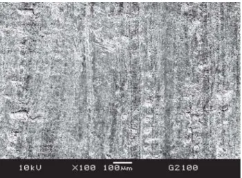
Hand curette instrumentation produced the smoothest surfaces among the treated groups. In this group, grooves were observed following the same direction of the scaling movements and less roughness was found when compared to ultrasonic and sonic groups (Figure 2).
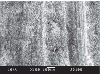
The surfaces after instrumentation with the ultrasonic group showed the presence of deeper sulcus and a rough surface (Figure 3). In the ultrasonic group and sonic scaler with diamond-coated tips group, irregular scratching was found in all surfaces (Figures 3, 4). Both of these instruments produced uneven surfaces marked with scratches due to the vibrating movements of machine scalers (Figures 3, 4).
The group instrumented by sonic scaler with diamond-coated tips also showed a rough surface caused by scaling (Figure 4). The diamond coating caused an irregular aspect because of the grinding action. The diamond splinters give the tool a multitude of edges, and every individual cutting grain forms part of the multifaceted tool, which leaves a characteristic roughness, as illustrated by SEM images (Figure 4).
DISCUSSION
According to the present study, the roughness reading and the SEM examinations showed that all treated groups presented a significant increase in roughness compared with the control group and demonstrated that the diamond-coated sonic tip and ultrasonic universal tip caused increased roughness when compared to hand curettes.
Group Treatment Mean
G1 (n=10) Control 0.485 ± 0.076 C
G2 (n=10) Curette 1.246 ± 0.279 B
G3 (n=10) Ultrasonic scaler/universal tip 1.468 0.177 A
G4 (n=10) Sonic scaler/diamond-coated tip 1.576 ± 0.204 A
TABLE 1- Comparison of all control and treated groups showing mean values and standard deviation of roughness after instrumentation
Mean values followed by different letters exhibited statistical difference (p<0.05)
FIGURE 1- Micrograph of not scaled root surface. Note the root surface smoothness. Original magnification 100x
Previous studies have evaluated differences regarding the roughness produced by sonic, ultrasonic and hand instruments10,24. However, the angulation and design of
instrument tip, sharpness of the working edge, the length of time the instrument is in contact with the root, and the cumulative number of strokes have impact on the degree of root damage and this situation can be explained by the lack of standardization.
Furthermore, the Roughness Loss of Tooth Substance Index (RLTSI) has been used by some studies10,12,24, but the
loss of tooth substance of a specific instrument cannot be directly correlated with its produced roughness13,30 and a
separate evaluation of tooth substance loss and surface roughness produced is necessary30. Therefore, considering
all these variables in previous studies, it is difficult to come to a conclusion regarding the method of instrumentation that causes the least amount of root surface alterations.
In the present study, differences in surface roughness have been found among different instruments, although it
remains to be determined whether these differences are of clinical significance. To understand the issue of roughness created after debridement and the success of periodontal treatment, different aspects have to be distinguished: supragingival or subgingival roughness and supragingival plaque control during healing.
Studies have demonstrated that the most important prerequisite for healing after periodontal treatment is a root surface free of plaque and calculus29. Mierau25 (1984) and
Quirynen and Bollen27 (1995) have clarified that
supragingival rough surfaces subsequent to professional instrumentation can promote plaque formation and contribute to bacterial adhesion. Supragingival surface roughness and surface irregularities increase the surface area, promote bacterial colonization, plaque formation and thereby can compromise daily plaque removal20,23.
Concerning subgingival roughness, some studies demonstrated that changes over subgingival root topography did not interfere with the response to periodontal treatment4,28. Rosenberg and Ash28 (1974) did not find that
the different instruments had a significant effect on histologically assessed healing. Khatiblou and Ghodossi11
(1983) have reported that periodontal healing following flap surgery occurs regardless of whether the subgingival root surface is rough or smooth. These results were confirmed by Oberholzer and Rateitschak26 (1996), who have found no
difference in pocket reduction and clinical attachment gain after creating rough or smooth surfaces during a flap operation. This indicates that subgingival roughness does not interfere with healing if there is a good supragingival plaque control. In an animal experiment, subgingival roughness following surgery, without supragingival plaque control during healing, favored plaque retention and colonization21. Leknes, et al.22 (1996) demonstrated that
roughness resulting from subgingival instrumentation significantly influenced the subgingival microbial colonization. Then, a smooth root surface may be advantageous near the gingival margin, since a smooth surface is less likely to accumulate plaque than a rough surface.
Therefore, for clinical application, it can be assumed that a meticulous scaling and root planing procedure during initial cause-related therapy should be performed30 and the
long-term success of this treatment is dependent on the quality of the maintenance therapy2.
Although there are many advantages of using power-driven scalers and diamond-coated sonic tips instead of hand curettes1,14,15,16,17, the present study showed that
diamond-coated sonic tips produced rougher root surface than curettes. Even though a clinical evaluation has not been conducted in the current investigation, according to the findings of this study and based on the in vivo evidences2,13,20,21,23,27, it can be suggested that caution should
be important when utilizing this instrument and that a higher standard of supragingival oral hygiene of the patient can be required. More studies are needed to clarify the influence of diamond-coated sonic tips on root surface roughness.
FIGURE 3- Micrograph of root surface instrumented by ultrasonic scaler. Note the rougher surface. Original magnification 100x
CONCLUSION
Within the limits of the present study, it can be concluded that diamond-coated sonic tips and ultrasonic universal tips produce a similar roughness surface that is higher than that produced by hand curettes.
ACKNOWLEDGMENTS
The authors would like to thank to Dr Gláucia Bovi Ambrosano Department of Social Dentistry, UNICAMP -for the statistical analysis support and to Dr Marcelo Giannini - Department of Restorative Dentistry, UNICAMP - who supplied the surface roughness measuring instrument for the execution of this study (Process 51234 – FAPESP).
REFERENCES
1- Auplish G, Needleman IG, Moles DR, Newman HN. Diamond-coated sonic tips are more efficient for open debridement of molar furcations. A comparative manikin study J Clin Periodontol. 2000;27(5):302-7.
2- Axelsson P, Lindhe J. The significance of maintenance care in the treatment of periodontal disease. J Clin Periodontol. 1981;8:281-94.
3- Caffesse RG, Sweeney PL, Smith BA. Scaling and root planning with and without periodontal flap Surgery. J Clin Periodontol. 1986;13:205-10.
4- Chapple IL, Walmsley AD, Saxby MS, Moscrop H. Effect of instrument power setting during ultrasonic scaling upon treatment outcome. J Periodontol. 1995;66(9):756-60.
5- Copulos TA, Low SB, Walker CB, Trebilcock YY, Hefti AF. Comparative analysis between a modified ultrasonic tip and hand instruments on clinical parameters of periodontal disease. J Periodontol. 1993;64(8):694-700.
6- Dragoo M. A clinical evaluation of hand and ultrasonic instruments on subgigival debridment. Part I. With unmodified and modified ultrasonic inserts. Int J Periodontics Restorative Dent. 1992;12:311-23.
7- Drisko CL, Cochran DL, Blieden T, Bouwsma OJ, Cohen RE, Damoulis P, et al. Research, Science and Therapy Committee of the American Academy of Periodontology. Position paper: sonic and ultrasonic scalers in periodontics. Research, Science and Therapy Committee of the American Academy of Periodontology. J Periodontol. 2000;71(11):1792-801.
8- Eschler BM, Rapley JW. Mechanical and chemical root preparation in vitro: efficiency of plaque and calculus removal. J Periodontol. 1991;62(12):755-60.
9- Flemmig TF, Petersilka GJ, Mehl A, Hickel R, Klaiber B. The effect of working parameters on root substance removal using a piezoelectric ultrasonic scaler in vitro. J Clin Periodontol. 1998;25(2):158-63.
10- Jotikasthira NE, Lie T, Leknes KN. Comparative in vitro studies of sonic, ultrasonic and reciprocating scaling instruments. J Clin Periodontol. 1992;19(8):560-9.
11- Khatiblou FA, Ghodossi A. Root surface smoothness or roughness in periodontal treatment. A clinical study. J Periodontol. 1983;54:365-7 .
12- Kishida M, Sato S, Ito K. Effects of a new ultrasonic scaler on .broblast attachment
to root surfaces: a scanning electron microscopy analysis. J Periodontal Res. 2004; 39:111-9.
13- Kocher T, Fanghanel J, Sawaf H, Lits R. Substance loss caused by scaling with different sonic scaler inserts – an in vitro study. J Clin Periodontol. 2001;28(1):9-15.
14- Kocher T, Gutshe C, Plagmann HC. Instrumentation of furcation with modified sonic scaler inserts: study on manikins, part I. J Clin Periodontol. 1998;25(5):388-93.
15- Kocher T, Plagmann HC. The diamond-coated sonic scaler tip. Part II: Loss of substance and alteration of root surface texture after different scaling modalities. Int J Periodontics Restorative Dent. 1997;17(5):484-93.
16- Kocher T, Plagmann HC. Root debridement of single-rooted teeth with a diamond-coated sonic scaler inserts during flap surgery – a pilot study. J Clin Periodontol. 1999;26(4):201-5.
17- Kocher T, Plagmann HC. Root debridement of molars with furcation involvement using diamond-coated sonic scaler inserts during flap surgery – a pilot study. J Clin Periodontol. 1999;26(8):525-30.
18- Kocher T, Tersic-Orth, Plagmann HC. Instrumentation of furcation with modified sonic scaler inserts: study on manikins, part II. J Clin Periodontol. 1998;25(6):451-6.
19- Lang NP. Indications and rationale for non-surgical periodontal therapy. Int Dent J. 1983;33(2):127-36.
20- Leknes KN, Lie T. Influence of polishing procedures on sonic scaling root surface roughness. J Periodontol. 1991;62:659-62.
21- Leknes KN, Lie T, Boe OE, Selvig KA. A correlation study of inflammatory cell mobilization in response to subgingival microbial colonization. J Periodontol. 1997;68:67-72.
22- Leknes KN, Lie T, Wikesjo UM, Boe OE, Selvig KA. Influence of tooth instrumentation roughness on gingival tissue reactions. J Periodontol. 1996;67(3):197-204.
23- Leknes KN, Lie T, Wikesjo UM, Bogle GC, Selvig KA. Influence of tooth instrumentation roughness on subgingival microbial colonization. J Periodontol. 1994;65:303-08.
24- Lie T, Leknes KN. Evaluation of the effect on root surfaces of air turbine scalers and ultrasonic instrumentation. J Periodontol. 1985;56(9):522-31.
25- Mierau HD. Relations between plaque formation, tooth surface roughness and self-cleaning. Dtsch Zahnarztl Z. 1984;39(9):691-8.
26- Oberholzer R, Rateitschak KH. Root cleaning or root smoothing. An in vivo study. J Clin Periodontol. 1996;23(4):326-30.
27- Quirynen M, Bollen CM. The influence of surface roughness and surface-free energy on supra- and subgingival plaque formation in man. A review of the literature. J Clin Periodontol. 1995;22(1):1-14.
29- Rosling B, Nyman S, Lindhe J, Jern B. The healing potential of the periodontal tissues following different techniques of periodontal surgery in plaque-free dentitions. A 2-year clinical study. J Clin Periodontol. 1976;3(4):233-50.