Durability of the adhesive cementation to the dentin substract
Durabilidade da cimentação adesiva ao substrato dentinário
Rodivan BRAZ1
Márcia de Almeida DURÃO1 Gabriela Luna Santana GOMES1 Fabio Barbosa DE SOUZA2 Eliane Alves LIMA1
1 Universidade de Pernambuco, Departamento de Odontologia. Av. Gal. Newton Cavalcanti, 1650, 54753-220, Camaragibe, PE, Brasil. Correspondên-ABSTRACT
Objective
The aim of this study was to evaluate, in vitro, the union stability of resin cements to the dental substract through microtensile bond strength (µTBS) analysis and scanning electron microscopy (SEM).
Methods
Fifty-four third human molars, stored in water for a short (24 hours) and long period of time (1 year) presented a lat oclusal supericial dentin. The teeth were distributed in six different groups: G1- Panavia F2.0/Kuraray; G2- RelyXUnicem/3M ESPE; G3- G-Cem/GC; G4- Biscem/ Bisco; G5- Panavia F2.0/Kuraray without pre-treatment and G6- Multilink Sprint/Ivoclar-Vivadent which were adhered to its respective indirect resin composite restoration, (G1- Clearil AP-X/Kuraray; G2- Filtek Z350/3M ESPE; G3- Gradia Direct X™/GC; G4- Aelite™/ Bisco; G5- Clearil AP-X/ Kuraray; G6- Tetric Ceram/ Ivoclar-Vivadent). The resin blocks were cemented and the sticks were obtained by tooth, with an area of adhesive interface of 0,8mm² (±0,2).
Results
The mean values, submitted to Mann-Whitney and Kruskal-Wallis tests (α = 5%) were in MPa after 24 hours: G1 = 9.66 (A), G2 = 13.37 (A); G3 = 15.89 (A); G5 = 4.18 (B); G6 = 11.01 (A) and after 1 year: G1 = 9.75 (A), G2 = 11.73 (A); G3 = 20.10 (B); G5 = 6.80 (A); G6 = 21.09 (B). All G4 group presented pretest failures.
Conclusion
During the one year period, with the exception of BisCem, the self-adhesive resin cements were a favorable alternative for the adhesive cementation, standing out among these, the G-Cem and Multilink Sprint.
Indexing terms: Dentin. Resin cements. Tensile strength.
RESUMO
Objetivo
Avaliar, in vitro, a estabilidade da união de cimentos resinosos ao substrato dental através da resistência de união e análise em microscopia eletrônica de varredura (MEV).
Métodos
Cinquenta e quatro terceiros molares humanos, armazenados em água por um curto (24 horas) e longo período de tempo (1 ano), tiveram a face oclusal removida expondo a superfície dentinária. Os dentes foram distribuídos em seis grupos distintos: G1- Panavia F2.0 / Kuraray; G2- RelyX Unicem / 3M ESPE; G3- G-Cem / GC; G4- BisCem / Bisco; G5- Panavia F2.0 / Kuraray sem pré-tratamento e G6- Multilink Sprint / Ivoclar-Vivadent que foram aderidas ao seu respectivo restaurações de resina composta indireta, (G1- Clearil AP-X / Kuraray; G2- Filtek Z350 / 3M ESPE; G3- Gradia Direct X ™ / GC; G4- Aelite ™ / Bisco; G5- Clearil AP-X / Kuraray; G6- Tetric Ceram / Vivadent Ivoclar-). Os blocos de resina foram cimentados e foram obtidos palitos, com área de interface adesiva de 0,8mm² (± 0,2).
Resultados
Os valores médios, submetidos à Mann-Whitney e Kruskal-Wallis testes (α = 5%) foram em MPa após 24 horas: G1 = 9,66 (A), G2 = 13,37 (A); G3 = 15,89 (A); G5 = 4,18 (B); G6 = 11,01 (A) e depois de 1 ano: G1 = 9,75 (A), G2 = 11,73 (A); G3 = 20,10 (B); G5 = 6,80 (A); G6 = 21,09 (B). O grupo todo G4 apresentou falhas pré-teste.
Conclusão
Durante o período de um ano de investigação, os cimentos de resinosos auto-adesivos, exceto o Bis Cem, eram uma alternativa favorável para a cimentação adesiva, destacando-se entre estes o G-Cem e Multilink Sprint.
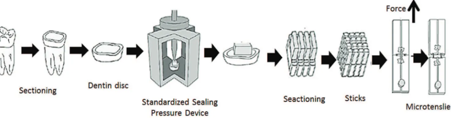
human third molars were cleaned, disinfected (0.5% ChloramineT, 24h), and stored in distilled water (4˚C). Each tooth was individually fixed to a sectioning machine (Elquip, São Paulo, Brazil) and sectioned perpendicular to its longitudinal axis using a flexible diamond disc (Extec, Einfield) under cooling to obtain a flat dentin area of medium depth. Forty-eight dentin discs of 3 mm thick were used for the µTBS test, and six discs for SEM evaluation. A smear layer was produced on this surface using water abrasive papers number 180, 400 and 600, for 30s each under continuous cooling, at 300 rpm in a polishing machine (Risitec, São Paulo, Brazil). The dentin discs were randomly divided into 6 groups, according to the cementing strategy (Table 2). The aim of G5 was to test and evaluate the potential for self-adhesiveness of the self-etching cement Panavia F2.0, thus the dentin surface received no treatment.
Restorations with 5.5x5.5x2mm of restorative materials described in Table 3 were performed with the aid of a two-piece matrix. The resin blocks were photoactivated for 40s on each side (VIP Junior/Bisco-600mW/cm²). The restoration surface in contact with the dentin was roughened with a diamond point, and then submitted to an ultrasonic bath in distilled water for 10min. The resin blocks were cemented onto the dentin in accordance with the respective manufacturer’s recommendations. The pressure exerted on the restoration was standardized at 40g/mm2 using a device specially
developed for this purpose (Figure 1).
The specimens were stored in distilled water at 37˚C for 24h. Half of the specimens were stored for 24h, while the other half were stored for 1 year. Using a diamond disk (Extec, Einield–thickness=0.3mm), the tooth-restoration set was sectioned vertically, in perpendicular direction to the adhesive interface, to obtain 16 test specimens (sticks), with an adhesive interface area standardized at 0.8mm2. The number
of specimens obtained for each tooth varies due to differences in the dentin surface area.
To perform the µTBS test, a fast-drying adhesive (Super Bonder Gel/Loctite, São Paulo, Brazil) was used to attach each stick to the two sides of the device for the microtensile test, which was attached to a universal test machine (KRATOS/São Paulo, Brazil). The test was conducted in two parts: after 24h and 1 year after the luting procedure of composite resin blocks. The maximum load at rupture of each test specimen was recorded. The cross sectional bond area of the stick
INTRODUCTION
The clinical success of indirect restorations is attributed to the effective union between the mineralized tissues and luting systems, as deined by the combination of resin cement and adhesive agent. The differences in composition of resin cements and amounts of monomers diluents in functional groups and in the percentage of iller particles produce a signiicant variation in the commercial products properties1-2.
Currently, the resin luting agents can be classiied as total-etching, self-etching and self-adhesives, according to the need of pretreatment of the tooth surface3.
Self-adhesive cements have been indicated and used for yielding less sensitivity and reduced postoperative sensitivity4-5.
According to the manufacturers, the functional monomers are able to chemically bind calcium from hydroxyapatite, which demineralize and iniltrates the dental substrate resulting in a micromechanical retention of the restoration6.
Adhesion degradation occurs via enzymatic attack of the collagen ibers not protected by the adhesive and dissolution of the resin components iniltrated into the dentin matrix. When acid etching agents are used to remove the smear layer, resulting in demineralization of the dentin surface, there is a risk that, the adhesive does not fully enclose the collagen ibers, which remain exposed7.
Such ibers become susceptible to the action of proteolytic enzymes present in the human dentin - endogenous metalloproteinases (MMPs) - resulting in the dissolution of the hybrid layer. The dissolution of the adhesive components can occur if there is an incomplete polymerization of the adhesive on the hybrid layer due to the presence of residual water in dentin during its application.
The main problem of adhesive restorations is their limited longevity. Several studies evaluate the bond strength of self-adhesive resin cements to the dental substrate in the short term. However, will the bond strength of self-adhesive resin cements remain stable in the long term? The analysis of this parameter is useful for a better understanding of the technology of self-adhesive luting.
The null hypothesis is that the bond strength of self-adhesive cements to dentin is similar to conventional cement, both initially and on the long term.
METHODS
Cement / Lots Composition Resin / Batch Composition Manufacturer
Kit Panávia F2.0 (61166)
ED Primer 2.0 - ED Primer II A: HEMA, DP, 5-NMSA, water, accelerator. ED Primer B: 5-NMSA, gas, water, benzene sodium
sulinateED Primer 2.0Panavia F2.0 -
Base: hydrophobic aliphatic and aromatic dimethacrylate; aromatic sulinate sodium
N, N-diethanol-p-toluidine, sodium luoride, functionalized glass, barium silaniizado. Catalyst: MDP dimethacrylate
aromatic and aliphatic hydrophobic, hydrophilic dimethacrylate, silanized silica, photoinitiator, dibenzoyl peroxide. (70.8%
iller particles, particles of 2μ.
Clearil AP-X (01191A)
Barium silanized crystal, silanized colloidal silica, silanized silica, Bis-GMA, TEGDMA,
di-camphorquinone.
Kuraray, Osaka, Japan
RelyX Unicem (265704)
Powder: glass illers, silica, calcium hydroxide, self-curing initiators, pigments, light-curing initiators (iller load 72% wt, particle size < 9.5 μm) Liquid: methacrylated phosphoric esters, dimethacrylates, acetate, stabilizers,
self-curing initiators, light-self-curing initiators
Filtek Z350 (7KU) Filler particles, bisphenol A polyethylene glycol diether dimethacrylate, diurethane dimethacrylate, bisphenol A di-glycidyl
ether dimethacrylate, triethylene glycol dimethacrylate and pigment. 3M-Espe, Seefeld, Germany
G-CEM (0702171)
Powder: luoroaluminosilicate glass, initiator, pigment Liquid: 4-MET, phosphoric acid ester monomer, water,
UDMA, dimethacrylate, silica powder, initiator, stabilizer
Gradia DirectX (0705101)
UDMA (15-30% \ 0; aluminum luoride silicate glass (30-40%),
silica powder (10-20%); prepolymerized loading (20-30%); dimethacrylate (0-5%); camphorquinone (<1%) G-Cem Tokyo, Japan Biscem
(0700006829) phosphate(base), tetraethylene glycolBis (hydroxyethyl methacrylate) (070008486)Aelite
Bis-EMA (15-40%), TEGDMA (3% -7); glass particles (50-90%), amorphous
silica (1-20%)
Bisco, Schaumburg, IL USA
Multilink Sprint (k25953)
Dimethacrylates and acidicmonomers. The inorganic illers arebarium glass, ytterbium triluorideand silicon dioxide. The mean
particlesize is 5 μm. The total volume oinorganic illers is approx. 48%
Tetric Ceram (k00802)
Urethane dimethacrylate,
Bis-GMA, TEGDMA. Inorganic iller: glass,
barium, ytterbium triluoride, aluminum
luorosilicate glass and barium, highly dispersed silicon dioxide and mixed
oxides spheroidal
Ivoclar Vivadent Schaan, Liechtenstein
Table 1. Resin cements and resin composites used in this study.
was measured with a digital pachymeter (Digimess, São Paulo, Brazil). The transformation of the unit into load/ area values (MPa) was carried out subsequently. Figure 1 shows the experimental stages of this methodology. The machine was calibrated so that the load to be applied to the dentin bond line/restorative material occurred at a speed of 0.5mm/min. The bond strength values of the tested specimens (sticks) were grouped per tooth as the arithmetic mean. As a consequence, each tooth was considered as a statistical unit. The data with reference to the bond area, bond strength and standard deviation received descriptive statistics and inferential treatment at a level of signiicance of α =0.05 (5%).
Table 2. The Protocol of use.
Group Product Dentin Pre-Treatment Cimentation
G1 Panavia F2.0(Kurarya)
Prophylaxis with pumice and water, mix the ED Primer II (Dispense 1 drop of Liquid A and Liquid B; apply on the tooth structure and wait 30 s, remove the excess with paper towels, dry
with slight air jet
Apply Clearil Ceramic Primer on the piece restorer with microbrush and remove excess; dispense equal amounts of paste A and B of the
resin cement; manipulate for 20s, cementing the restoration under pressure 40g/mm² for 60s, 37°C; remove excesses, light curing each
interface and the occlusal for 20s, apply the Oxiguard II for 3min. and then remove material
with cotton pellet and water jet.
G2 RelyX Unicem(3M ESPE) No pre-treatment
Activate the capsule for 4s in the metallic device that comes with the kit; manipulate in
mechanical mill for 15s, apply the cement to dentin through the applier; cementing under pressure 40g/mm² for 60s, 37°C; to remove excesses; light cure each interface and occlusal
for 20s.
G3 GCem(GC) pumice and waterProphylaxis with
Agitate the capsule manually to homogenize the material, press the plunger until the level of the main body, and immediately places a capsule in the applicator and activate only once; take the crusher and rotate for 10s, remove immediately
and put the crusher on the applicator; perform 2 clicks and prepare the capsule for the application; apply the cement to dentin and cementing restorative piece under pressure of 40g/mm² for 4min, 37°C; remove excess of the cement, light curing each interface and occlusal
surface for 10s.
G4 BisCem (Bisco) pumice and waterProphylaxis with
Dispense small amount of the material; dovetail the applicator; apply the cement to the inner surface of the restoration, cementing the part
under pressure 40g/mm² and removing the excess, immediate photoactivation of each
interface and occlusal for 20s.
G5 Panavia F2.0(Kurarya) No pre-treatment
Apply Clearil Ceramic Primer on the piece restorer with microbrush and remove excess; dispense equal amounts of paste A and B of the
resin cement; manipulate for 20s, cementing the restoration under pressure 40g/mm² for 60s, 37°C; remove excesses, light curing each interface and the occlusal surface for 20s, apply
the Oxiguard II for 3min. and then remove material with cotton pellet and water jet.
G6 (Ivoclar Vivadent)Multlink Sprint pumice and waterProphylaxis with
Apply cement, cement the restoring under pressure 40g/mm²; remove excess; cover interfaces with oxygen blocking (Aqua gel / Kley
Hertz SA); immediate photoactivation of each adhesive interface and occlusal for 20s, rinse
thoroughly to remove the blocker.
degrees of acetone. After dehydration, the specimens were submitted to a drying process through exchange with liquid CO2 by sublimation at the critical point, at a certain temperature and pressure (Balzers CPD-020 Critical Point drier, Linchenstein). The specimens were attached to metal stubs with double-faced carbon tape and gold-sputter coated using a vacuum metalizing
RESULTS
The µTBS means and standard deviations by groups are shown in Table 3. Analyzing the resin cements, the Kruskal-Wallis test revealed that after 24h (p=0.022), the use of G5, Panavia without any dentin pre-treatment, resulted in lower adhesion in comparison to the other groups. After 1 year of storage, the same test indicated signiicant statistical superiority for groups 3 and 6 (p=0.033) compared to the other cements studied. The Mann-Whitney test (p>0.05) indicated no statistically signiicant differences when comparing the averages of 24h and 1 year of storage in distilled
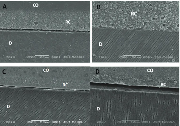
water. All specimens of G4 suffered failures before being submitted to the mechanical tests. In SEM it was possible to observe an homogeneous cement layer in G1, but no signs of hybrid layer formation were detectable (Figure 2A). Discontinuities were present along the interface with dentin and porosities could be noticed within the cement layer (G2), the hybrid layer was thin to undetectable and no tags were formed (Figure 2B). Good continuity could be seen between the cement layer and the dental substrate (Figure 2C), with no evidence of hybrid layer formation. Discontinuities were present along the interface with dentin. No detectable signs of hybrid layer formation were observed (Figure 2D).
Figure 1. Schematic drawing of Tooth Preparation and Microtensile Bond Test.
Table 3. Mean (standard deviation) bond strength (MPa) of resin cements to dentin.
Statistical Analysis Groups p - Value
24 hours
G1 G2 G3 G5 G6
Mean 9.66 (2.22)A 13.37 (4.09)A 15.89 (5.57)A 4.18 (1.99)B 11.01 (4.02)A p = 0.022*
1 year
DISCUSSION
At 24h of this study, Panavia (G1) achieved a capacity of adhesion similar to that found by Goracci et al.4
The primer Panavia consists of three amphiphilic monomer (HEMA, MDP and 5-NMSA) with low molecular weight, the latter being responsible for the dentin demineralization. According to Yoshida et al.8, the hydroxyapatite remaining
after the product application may serve as a receptor for the chemical interaction with functional monomers, and subsequently contribute to the adhesive performance in addition to micro-mechanical hybridization.
In this study, after 24 hours, the Panavia F2.0 (G1), which is used in combination with the ED primer 2.0, showed union capacity numerically similar to that reported by Goracci et al.4. The primer of Panavia F2.0 consists of
three amphiphilic monomers (HEMA, MDP and 5-NMSA) with low molecular weight, the latter being responsible for
the demineralization of the dentin. According to Hiraishi et al.3, through the microscopic observation of the treated
dentin surface and the measurement of its permeability, it is possible to establish that the ED primer 2.0 is able to provide a moderate demineralization with dissolution of the layer and smear plugs. In 2007, Al-Assaf et al.11 showed that
the extent of demineralization provided by this material is 51.99% of the dentin and the thickness of the hybrid layer is 0,95μm.
In the short term, Panavia F2.0 without prior primer application (G5) had statistically signiicant lower bond strength when compared to G1, which followed the manufacturer's recommendations.
According to Monticelli et al.9, although the
mechanism of adhesion seemed similar for all the self-adhesive cements, these materials are still relatively new and detailed information on its composition and adhesive property are limited. Still according to the same authors,
these cements contain phosphoric acid methacrylate, which has the ability to react with the hydroxyapatite. This ester is not only able to decalcify hydroxyapatite, but may also chemically adhere to it allowing a micromechanical retention10. However, in Monticelli et al.9, no evidence of
demineralization and iniltration in the dentin was found for the self-adhesive resin cements evaluated in this study.
In a study by Mazzitelli6, a 30-days analysis of the
BisCem one μTBS 2.4 MPa obtained 68% of failure in the pretest, while the present study obtained a 100% failure rate in the pretest. However, the application of 5min of autopolimerization and then 40s of photoactivation may have enhanced numerically the union capacity of this cement. In this study, the light curing immediately after cementation of their respective indirect restorative materials (composite), may have contributed to the occurrence of 100% of pre-testing failures. This can be explained by the limited penetration ability of the cement into the tooth structure when polymerization is performed immediately.
The RelyX Unicem may set the reaction in two ways. The free radical polymerization can be initiated by exposure to visible light or redox mechanism. There is also an acid-base reaction between the metal ions of the non-silanized glass particles luorine aluminum silicate and the phosphate radical of methacrylate generated by the water produced during the neutralization reaction of the phosphate monomer, that is important for its long-term stability regarding bond strength to dentin11.
Among the cements tested in this study, the GCem showed the greatest capacity of adhesion to dentin. For Mazzitelli et al.6, the presence of water in the chemical
composition of this cements could explain the results obtained since it is important to promote the ionization of existing acidic monomers, thus reducing the necessary time for the interaction with the dental substrate. Furthermore, the presence of the acid functional monomer 4-META in the composition, although a high molecular weight particle, could also have contributed to the chemical reaction with the dentin.
This monomer is capable of adhering to the calcium ions of the apatite through a reaction of chelation9. Yoshida et al.8 found that the application of
4-META hydroxyapatite for 30 min increased the carbon bonds energy in comparison with the application for 30s. In the same year Abo, Uno and Sano evidenced that an adhesive containing the 4-META produced similar performance to dentin when compared to one containing MDP12. This may indicate that the potential
formation of the hybrid layer is practically the same for these two components.
Thus, differences in the chemical composition of these cements are indeed able to provide different results for bond strength. A long period of storage simulating oral conditions is necessary to evaluate dental composites. However, to simulate the time of permanence in the oral cavity expected for these materials, a storage time of ive to ten years would be necessary. This time interval is unfeasible, since every year new materials are commercially available. Thus, evaluation results for a ten-year period were not relevant. The storage in water for 1 year does not decrease the bond strength of self-adhesive resin cements to dentin. Conversely, the humidity conditions can change the bond strength to dentin; however, these results are product-dependent13.
Acid neutralization in cement is a key factor for its hydrophobicity and to guarantee that it remains intact in a humid environment. Acid materials retain some degree of hydrophilicity that leaves them prone to capture water, expand and degrade their structural matrices14. For cements
RelyXUnicem and G-Cem, a work published by Han et al.15 conirm an increase in their pH within 48h following
photoactivation.
A study of the shear strength of resin cements demonstrated highly divergent behavior to dentin. While some of the self-adhesive resin cements had similar values of bond strength to the conventional cement Panavia, others showed lower values before and after aging, in the long term16. Not all self-adhesive resin cements can be a valid
alternative to conventional cement for binding silica based glass-ceramic to dentin17. Regarding the Multilink Sprint,
although no statistical difference was showed for the two periods studied, there was a numerical increase in adhesive resistance after 1 year of storage. However, the lack of information provided by the manufacturer with respect to the monomers that compose this cement do not allow a further discussion to justify the result. We can argue, though, that the acid neutralization of RelyXUnicem and G-Cem must have contributed to their adhesive stability.
CONCLUSION
strength values to dentin, standing out among the other resin cements evaluated in the same period. In the long-term, the self-adhesive resin cements demonstrated to be stable with respect to bond strength, presenting itself as a favorable alternative.
ACKNOWLEDGEMENTS
FACEPE and CNPq through the scholarship.
Collaborators
R BRAZ, advisor. FB SOUZA, he worked on the methodology and SEM analysis. GLS GOMES, she worked on the methodology and SEM analysis. MA Marcia Durão - Monitoring methodology and was responsible for preparing the paper and its submission to the RGO. EA LIMA, worked on the methodology and collaboration in preparing the article.
REFERENCES
1. Fonseca RG, Cruz CAS, Adabo G L. The inluence of chemical activation on hardness of dual-curing resin cements. Braz. Oral Res. 2004;18(3):228-32. doi: 10.1590/S1806-83242004000300009
2. Aguiar TR, Di Francescantonio M, Bedran-Russo AK, Giannini, M. Inorganic composition and iller particles morphology of conventional and self-adhesive resin cements by SEM/ EDX. Microsc Res Tech. 2012;75(10):1348-52 doi: 10.1002/ jemt.22073.
3. Hiraishi N, Yiu CKY, King NM, Tay FR. Effect of pulpal pressure on the microtensile bond strength of luting resin cements to human dentin. Dent Mater. 2009;25(1):58-66. doi: 10.1016/j. dental.2008.05.005
4. Goracci C, Cury AH, Cantoro A, Papacchini F, Tay FR, Ferrari M. Microtensile bond strength and interfacial properties of self-etching and self-adhesive resin cements used to lute composite onlays under different seating forces. J Adhes Dent. 2006;8(5):327-35.
5. Christensen GJ. Should resin cements be used for every cementation? J Am Dent Assoc. 2007;138(6):817-9. doi: 10.14219/jada.archive.2007.0271
6. Mazzitelli C, Monticelli F, Osório R, Casucci A, Toledano M, Ferrari M. Effect of simulated pulpal pressure on self-adhesive cements bonding to dentin. Dent Mater. 2008;24(9):1156-63. doi: 10.1016/j.dental.2008.01.005
7. Carrilho MRO, Carvalho RM, De Góes MF, Di Hipólito V, Geraldeli S, Tay F R, et al. Chlorhexidine preserves dentin bond in vitro. J Dent Res. 2007;86(1):90-4.
8. Yoshida Y, Nagakane K, Fukuda R, Nakayama Y, Okazaki M, Shintani H, et al. Comparative study on adhesive performance of functional monomers. J Dent Res. 2004;83(6):454-8. doi: 10.1177/154405910408300604
9. Monticelli F, Osorio R, Mazzitelli C, Ferrari M, Toledano M. Limited decalciication/diffusion of self-adhesive
cements into dentin. J Dent Res. 2008;87(10):974-9. doi: 10.1177/154405910808701012
10. Fu B, Sun X, Qian W, Shen Y, Chen R, Hannig M. Evidence of chemical bonding to hydroxyapatite by phosphoric acid esters. Biomaterials. 2005;26(25):5104-10. doi:10.1016/j. biomaterials.2005.01.035
11. Al-Assaf K, Chakmakchi M, Palaghias G, Karanika-Kouma A, Eliades G. Interfacial characteristics of adhesive luting resins and composites with dentine. Dent Mater. 2007;23(7):829-39. doi: 10.1016/j.dental.2006.06.023
12. Abo T, Uno S, Sano H. Comparison of bonding eficacy of an all-in-one adhesive with a self-etching primer system. Eur J Oral Sci. 2004;112(3):286-92. doi: 10.1111/j.1600-0722.2004.00126.x
13. André CB, Aguiar TR, Ayres APA, Ambrosano GMB, Giannini M. Bond strength of self-adhesive resin cements to dry and moist dentin. Braz Oral Res. 2013;27(5):389-95. doi: 10.1590/ S1806-83242013000500002.
14. 3M ESPE. Scientiic update: RelyX Unicem self-Adhesive universal resin cement [text internet]; 2006.
15. Han L, Okamoto A, Fukushima M, Okiji T. Evaluation of physical properties and surface degradation of self-adhesive resin cements. Dent Mater J. 2007;26(6):906-14. doi: 10.4012/ dmj.26.906
16. Sarr M, Mine A, De Munck J, Cardoso Mv, Kane Aw, Vreven J, et al. Immediate bonding effectiveness of contemporary composite cements to dentin. Clin Oral Investig. 2010;14(5):569-77. doi: 10.1007/s00784-009-0327-8
17. Hitz T, Stawarczyk B, Fischer J, Hämmerle CH, Sailer I. Are self-adhesive resin cements a valid alternative to conventional resin cements? A laboratory study of the long-term bond strength. Dent Mater. 2012;28(11):1183-90. doi: 10.1016/j. dental.2012.09.006.