Rev Bras
Cineantropom
Hum
DOI: http://dx.doi.org/10.5007/1980-0037.2017v19n5p585
original article
Acute effects of passive static stretching on
the vastus lateralis muscle architecture of
healthy young men
Efeitos agudos do alongamento estático passivo sobre
a arquitetura muscular do vasto lateral de jovens
saudáveis
Eurico Peixoto César1
Letícia de Oliveira Teixeira2
Daniel Vieira Braña Côrtes de Souza1
Paulo Sergio Chagas Gomes3
Abstract – he aim of the study was to investigate the acute efects of passive static stretching (PSS) on the fascicle length (FL) and fascicle angle (FA) of the vastus later-alis muscle (VL) in two diferent joint positions. Twelve physically active men (26.9 ± 7.5 years, 178.6 ± 7.0 cm, and 82.5 ± 16.8 kg) were placed in the prone position for the acquisition of ultrasound images (US) of VL, registered with extended and totally lexed knee up to the heel contact with the gluteus, before and after a PSS routine comprised of three 30-s repetitions maintained in the maximal discomfort position as reported by the participant. Results of the paired t-test indicated an increase in FL (16.2%; p = 0.012) and reduction in FA (15.5%; p = 0.003) in pre vs. post stretching comparisons for the extended knee position. here was also a signiicant increase in FL (34%; p = 0.0001) and reduction in FA (25%; p = 0.0007) when compared the extended knee vs. lexed knee positions. here were no signiicant diferences in muscle architecture variables for the lexed knee position. he results showed high and moderate correlation of FL and FA for the extended (r = -0.89 and r = -0.74) and lexed knee (r = -0.76 and r = -0.78) position, pre and post stretching, respectively. It was concluded that the static stretching acutely afects the vastus lateralis muscle architecture only in the extended knee position, but not in the lexed knee position.
Key words: Muscle stretching exercises; Skeletal muscle; Ultrasonography.
Resumo – O objetivo do estudo foi veriicar os efeitos agudos do alongamento estático passivo (AEP) sobre o comprimento (CF) e ângulo do fascículo (AF) do músculo vasto lateral (VL) em duas diferentes posições articulares. Doze homens (26,9 ± 7,5 anos; 178,6 ± 7,0 cm; e 82,5 ± 16,8 kg), isicamente ativos foram posicionados em decúbito ventral para aquisição de imagens de ultrassonograia (US) do VL, registradas com joelho estendido e totalmente lexionado, até o conato do calcanhar com o glúteo, antes e após uma rotina de AEP composta por três repetições de 30 s com manutenção da posição no limite de desconforto relatado pelo participante.O teste t de Student para amostras pareadas indicou aumento no CF (16,2%; p = 0,012) e redução no AF (15,5%; p = 0,003) nas comparações pré vs. após alongamento na posição com o joelho estendido. Também houve aumento signiicativo do CF (34%; p = 0,0001) e redução do AF (25%; p = 0,0007) na comparação entre as posições de joelho estendido vs. lexionado. Não foram encontradas diferenças signiicativas nas variáveis da arquite-tura muscular investigadas na posição com o joelho lexionado. Os resultados apontaram para correlação alta e moderada do CF e AF com joelho estendido (r = -0,89 e r = -0,74) e joelho lexionado (r = -0,76 e r = -0,78), pré e após alongamento, respectivamente.
Concluiu-se que o alongamento estático afeta de forma aguda a arquitetura muscular
1 “Presidente Antônio Carlos” Uni-versity. Barbacena, MG. Brazil
2 Gama Filho University. Rio de Janeiro, RJ. Brazil.
3 State University of Rio de Janeiro. Rio de Janeiro, RJ. Brazil.
Received: March 23, 2017
INTRODUCTION
Static stretching contributes to the reduction of passive stifness and to increase of range of motion (ROM)1. Changes in the muscle-tendon
mechanical properties are of great inluence to increase the ROM of a joint, being the efect attributed to the structure extensibility, including sarcomeres, aponeuroses and connective tissues2. Among the diferent
types of stretching, static stretching one is one of the most commonly used, where muscles are extended to the point of discomfort and maintained for a certain period, with 30 seconds of insistence time repeated 3 to 4 times to increase ROM and reduce passive stifness3.
Although acute changes in muscle-tendon properties and passive stifness are well documented1,4 after static stretching exercises, little is
known about their efects on muscle architecture variables, such as fasci-cle length (FL) and fascifasci-cle angle (FA). FA is the angle formed between the insertion of the fascicle and the internal muscle aponeurosis and FL is the distance from the junction point of the fascicle with the external aponeurosis5. hese muscular architecture variables drastically afect the
functional characteristics of a muscle and the force production capacity6,
since FA is related to the amount of contractile tissue per unit of muscle area. hus, in hypertrophied muscles, FA is signiicantly increased, and higher FL is associated with higher muscle contraction rate5.
Changes in muscle architecture caused by conventional strength training in trained youth and adults are already well documented, such as increased cross-sectional area (CSA) and FA, but with less apparent modiications in FL7-9. However, little is known about the isolated acute
efect of static stretching on muscle architecture. Most studies indicate alterations generated in muscle architecture during stretching exercises and not the acute efect generated after this routine10. Moreover, the few
studies that veriied the acute efect of static stretching on muscle archi-tecture showed controversial results, possibly due to the diferent methods adopted and muscles tested11-13.
In the study conducted by Morse et al.1, no signiicant changes were
observed in FL and FA immediately after an extensive routine of ive 1-min repetitions of static stretching in the triceps sural muscle. However, muscle architecture variables were measured during dorsilexion movement and not at rest. In contrast, Sá et al.13 measured FL and FA of the vastus lateralis
and femoral biceps muscles before, immediately after three 30 s repetitions of static stretching and 10 min after three sets of strength exercises pre-ceded by the stretching routine. he authors veriied a signiicant increase in the FL of femoral biceps immediately after the stretching routine, but no alterations in FA and in these same muscle architecture variables of the vastus lateralis muscle. It should be noted that in this study, US measure-ments were performed in only one position (supine position).
According to Lieber and Friden6, muscle architecture and the
dif-ferent positions. In this sense, static stretching provides didif-ferent efects as a function of volume, insistence time in the discomfort position, intensity, joint position and type of muscle group tested. In addition, as suggested by Cè et al.11, possible changes in muscle architecture caused by static stretching
may explain the decline in strength performance presented by some studies, but for a more correct conclusion, diferent joint positions should be tested, since force production varies substantially according to this variable.
In this sense, due to the need to verify the isolated efect of static stretching on the muscle architecture in diferent joint positions, the present study aimed at verifying the acute efect of three 30-s repetitions of passive static stretching (PSS) on muscle architecture variables of the vastus lateralis muscle (VL) at two diferent knee angles.
METHODOLOGICAL PROCEDURES
he present study was approved by the Ethics Research Committee of the Gama Filho University under protocol number 173.786. he involvement of volunteers occurred after signing the informed consent form (ICF), in addition to completing a questionnaire for risk stratiication (Par-Q ), where one or more positive answers to the seven questions in the questionnaire served as exclusion criteria, and a detailed explanation of the objectives and procedures of the present study. hey were informed that at any time they would be free to leave the study. he study was conducted according to the recommendations deined in Resolution 466/2012 of the National Health Council.
Sample
Twelve physically active men (26.9 ± 7.5 years, 178.6 ± 7.0 cm, and 82.5 ± 16.8 kg) visited the laboratory on four diferent occasions. Volunteers were invited through posters ixed in classrooms. Inclusion criteria were: a) participant should be able to touch the heel in the gluteus during the knee lexion movement; b) participant should practice physical exercises three or more times per week; c) participant should not have any type of injury or impairment in the right lower limb that restricted the knee lexion move-ment. he irst visit was aimed at familiarizing with procedures and the completion of the ICF and the questionnaire for risk stratiication. Visits 2 and 3 were used to determine the reliability of US measurements and in the last visit, the experimental procedure was performed. he maximum interval between visits was 72 h.
Instruments
being positioned at the measurement site. Gel was used for the acoustic coupling (Ultrex-gel, Farmativa Indústria e Comércio Ltda).
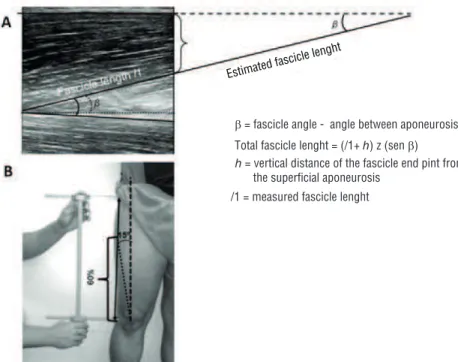
All images were analyzed using public domain software (ImageJ, National Institute of Health, USA, version 1.42). As the used transducer used has 40 mm, an estimation model 14 was used to calculate FL using
the following equation: CF: (l1 + h) / (sen α). Where l1 is the visible FL measured in the image; h is the vertical distance from the end point of l1 to the supericial aponeurosis and α is the angle between the fascicle and the deep aponeurosis. When aponeuroses were not parallel, the angle between the fascicle and the deep aponeurosis was subtracted from the angle of the measured fascicle (Figure 1A).
Procedures
In order to acquire the images, the following procedures were performed: a) with participant standing, a marking was made at the center of the pa-tella and at the point related to the anterior superior iliac crest. From these two points, a straight line was drawn connecting them, thus identifying the line of action of the rectus femoris muscle; b) from the center of the patella, using the straight line drawn for the action of the rectus femoris, a universal goniometer was positioned and 15° lateral was identiied from that point, identifying the line of action of the VL; c) the trochanteric and lateral tibial points were marked to measure the femur length and; d) from the line of action drawn from the VL, a proximal point was marked at 60% of the femur length, where the apparatus for itting the US transducer (Figure 1B) was positioned.
Once demarcations were made, the participant was placed in a ventral decubitus position with the right knee extended and supported on a padded apparatus and the distal part of the thigh wrapped in an inextensible band, ixing the limb and ensuring that there was no hip rotation. hree US im-ages were captured in this position, with an interval of about 1 min between them. hen a passive, slow and gradual mobilization of the knee lexion was performed until there was contact of the heel with the gluteus. In this position, three more US images were recorded. After this procedure, with the subject positioned in the ventral position, three repetitions of passive static stretching were performed to the quadriceps, with a slow and gradual knee lexion until the heel touched the gluteus. hen, hyperextension of the participant’s hip was performed up to the limit of reported discomfort, maintaining this position for 30 s. Subsequently, three US images of the vastus lateralis muscle were captured with extended knee and three with lexed knee. All images were analyzed and the mean results for each posi-tion were used as the value for the statistical calculaposi-tions.
Statistical analysis
he Shapiro-Wilk test showed normal distribution for variables tested. he reliability of the measurement was made by means of the intraclass correla-tion coeicient (ICC parallel method), of the typical measurement error (TME), which according to Hopkins15, is determined by the relationship
between standard deviation of the diferences obtained between measure-ment pairs and the square root of two. Finally, the degree of agreemeasure-ment among measures was veriied, according to Bland and Altman16. he
com-parison between measures before and after the stretching routine for each joint position was made through the Student t test for paired measures. In addition, Pearson’s correlation was used to verify the relationship between FL and FA in the diferent positions tested. Analyses were performed using commercially available software (SPSS 17.0 for Windows®, IBM Corporation, New York, USA, Prism 5.0 for Mac, Graphpad Software, La Jolla, Calif., USA) and signiicance level of 5% was adopted.
RESULTS
he reliability results of FL and FA measurements were based on 10 of the 12 recruited subjects due to problems with the US measurement in two of them, where the diference between the two tests in one section was 69.6% for one subject and 87.8% for the other. he reliability results for FL were R = 0.916, BIAS 4.1 and TME 7.4% and for FA were R = 0.928, BIAS 3.0 and TME 5.3%.
signiicant statistical diference. TMEs were also low with values below 8.0% for both variables. he Bland-Altman graphic representations (Figure 2A and 2B) showed that in both dependent variables used, all measures are within the determined conidence limits (± 1.96 SD) with low associated error (BIAS) and absence of heterocedastic error.
Figure 2. Bland-Altman graphic representations for (A) Fascicle Angle and (B) Fascicle Length. (°) = degrees.
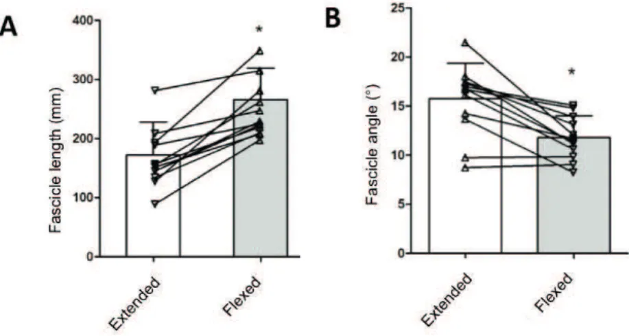
he Student’s t-test for paired measures identiied a signiicant increase in FL (34%, p <0.05) and reduction in FA (25%; P <0.05) in the comparison between extended and lexed knee positions (Figure 3A and 3B).
Figure 3. Comparison of FL (A) and FA (B) at extended and flexed knee positions. Columns represent the means and standard deviations and the symbols connected by rows represent the raw data of each subject tested. * FL (p = 0.0001) and FA (p = 0.0007).
Static stretching promoted a signiicant increase in FL (16.2%, p <0.05) and in FA (15.5%, P <0.05) in the position with knee extended but not lexed (FL , p = 0.430 and FA, p = 0.493) (Figure 4A and 4B). hese results exceed TME and indicate the real efect of the intervention on the study variables.
Figure 4. Effect of passive static stretching on FL (A) and FA (B) for the extended knee position. Columns represent the means and standard deviations and the symbols connected by rows represent the raw data of each subject tested. * FL (p = 0.012) and FA (p = 0.003).
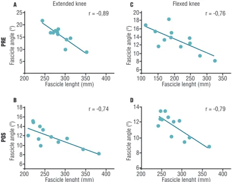
Figure 5. Relationship between FL and FA for extended knee in pre (A) and post stretching (B) conditions and the relationship between FL and FA for flexed knee pre (C) and post stretching (D) conditions. r = Pearson’s correlation coefficient; (°) degrees.
DISCUSSION
studied variables was real. However, no changes were observed in FL and FA in the lexed knee position in the pre and post stretching comparisons.
It is well known that the arrangement of muscle ibers is directly as-sociated with muscle strength production17. FA is associated with CSA
and muscle eiciency, that is, how much force generated in sarcomeres is efectively transferred to aponeurosis. FL is related, in part, to muscle contraction rate7. Some studies have reported the acute efect of static
stretching on muscle architecture1,11-13, but the results were inconsistent
with each other. While some authors observed an increase in FL and a reduction of FA1,12 in the gastrocnemius muscle after 5 1-min repetitions
of static stretching, others only found FL changes for the vastus lateralis muscle, but not for the femoral biceps muscle13 after three 30-s repetitions,
and did not ind any type of alteration for the gastrocnemius muscle after six 45-s repetitions11. Diferences in the amount of stretching exercises
in the protocols to acquire US images, in the joint position in which the measurements were made and in the muscle groups tested, may explain such discrepancies in results.
In the present study, there was an increase in FL with concomitant reduction in FA immediately after static stretching only in the extended knee position, which reinforces some indings in literature1,12. However, no
change was observed for the lexed knee position. A possible explanation for this would be the greater level of passive tension generated in the posi-tion with more stretched muscle, which would remove all the addiposi-tional complacency caused by the stretching routine, being no longer possible to change FL or FA. his hypothesis has already been demonstrated in literature18,19.
Although studies have already investigated the acute efect of static stretching on muscle architecture, a large part of them veriied this dur-ing stretchdur-ing and used unusual volumes (e.g., 5 min) in a speciic group (gastrocnemius) and in only one joint position1,12. Another study that
measured the muscle architecture parameters after static stretching13 had
inluence of strength training performed after stretching, which makes it impossible to know its isolated efect. herefore, the indings of the present study may be important for understanding the isolated efect of usual static stretching routines on the muscle architecture as a function of the joint position in which the muscle is located, in addition to allowing associations with force performance.
Some authors suggest an angle-dependent relationship, showing that only in positions where the muscle is at a shorter length (e.g., extended knee position), deleterious efects on force promoted by stretching are more common. However, when the muscle is positioned at a greater length (e.g., lexed knee), the decrease in strength performance becomes less evident, since more elongated muscle position is able to remove the additional complacency caused by stretching18- 21. his fact may be associated to the
It is important to emphasize that there are few studies that have veri-ied the isolated acute efect of static stretching on the muscle architecture variables studied here. Most of them identiied changes in muscle archi-tecture during stretching rather than after the session. Kato et al.2 veriied
the changes in the medial gastrocnemius muscle architecture during ive 60-s repetitions of static stretching and reported a signiicant but low inversely proportional relation (r2 = 0.46; P <0.001), with the increase of
FL and concomitant reduction in passive torque from the irst to the ifth stretching session. hese results indicate that changes in passive stifness associated with ROM gain after static stretching are strongly associated with increased FL. Corroborating these indings, some studies have pointed out that the efects of stretching on the passive stifness of the muscle-tendon unit occur mainly due to changes in the relative stifness of the muscle and not the tendon10,22,23. Another study sought to identify changes
in the gastrocnemius muscle architecture during stretching and reported a gradual increase in FL and reduction in FA as ROM increased10 from
10° of plantar lexion to 30° of dorsilexion. hese indings corroborate the results of the present study.
FA is the angulation of muscle ibers in relation to the muscle action line5. It has long been evidenced that during muscle contraction, a
rota-tion of the muscle iber occurs, promoting its increase24. he same pattern
occurs for structural adaptations to strength training, which promotes implications for the transfer of the force generated in the muscle iber to the aponeurosis, since, for muscles with the same AST and muscle iber length, a greater fascicle angle will promote lower eiciency in the transmission of force from muscle ibers to aponeurosis25. Associations of
hypertrophy with increase in FA and also with reduction in muscle ef-iciency with concomitant increase in force production due to the increase in the contractile material are pointed out in literature5,7. hus, it may
be advantageous to present lower FA during muscle contraction, as this will imply lower loss of eiciency in the transfer of force from the muscle iber to the tendon. However, there is little evidence for improved force performance after stretching, which is most noticeable at positions where the muscle is at a longer length, near its maximal ROM19.
CONCLUSION
REFERENCES
1. Morse CI, Degens H, Seynnes OR, Maganaris CN, Jones DA. he acute efect of stretching on the passive stifness of the human gastrocnemius muscle tendon unit. J Physiol 2008;586(1):97-106.
2. Kato E, Vieillevoye S, Balestra C, Guissard N, Duchateau J. Acute efect of muscle stretching on the steadiness of sustained submaximal contractions of the plantar lexor muscles. J Appl Physiol 2011;110(2):407-15.
3. Shellock FG, Prentice WE. Warming-up and stretching for improved physical per-formance and prevention of sports-related injuries. Sports Med 1985;2(4):267-78.
4. Herda TJ, Costa PB, Walter AA, Ryan ED, Hoge KM, Kerksick CM, et al. Ef-fects of two modes of static stretching on muscle strength and stifness. Med Sci Sports Exerc 2011;43(9):1777-84.
5. Kawakami Y. he efects of strength training on muscle architecture in humans. International J Sport Health Sci 2005;3:208-17.
6. Lieber RL, Friden J. Clinical signiicance of skeletal muscle architecture. Clin Orthop Relat Res 2001(383):140-51.
7. Aagaard P, Andersen JL, Dyhre-Poulsen P, Lefers AM, Wagner A, Magnus-son SP, et al. A mechanism for increased contractile strength of human pennate muscle in response to strength training: changes in muscle architecture. J Physiol 2001;534(Pt. 2):613-23.
8. Kawakami Y, Abe T, Kuno SY, Fukunaga T. Training-induced changes in muscle architecture and speciic tension. Eur J Appl Physiol 1995;72(1-2):37-43.
9. Ema R, Wakahara T, Miyamoto N, Kanehisa H, Kawakami Y. Inhomogeneous architectural changes of the quadriceps femoris induced by resistance training. Eur J Appl Physiol 2013;113(11):2691-703.
10. Abellaneda S, Guissard N, Duchateau J. he relative lengthening of the myoten-dinous structures in the medial gastrocnemius during passive stretching difers among individuals. J Appl Physiol 2009;106(1):169-77.
11. Ce E, Longo S, Rampichini S, Devoto M, Limonta E, Venturelli M, et al. Stretch-induced changes in tension generation process and stifness are not accompanied by alterations in muscle architecture of the middle and distal portions of the two gastrocnemii. J Electromyogr Kinesiol 2015;25(3):469-78.
12. Nakamura M, Ikezoe T, Takeno Y, Ichihashi N. Acute and prolonged efect of static stretching on the passive stifness of the human gastrocnemius muscle tendon unit in vivo. J Orthop Res 2011;29(11):1759-63.
13. Sa MA, Matta TT, Carneiro SP, Araujo CO, Novaes JS, Oliveira LF. Acute Efects of Diferent Methods of Stretching and Speciic Warm Ups on Muscle Architecture and Strength Performance. J Strength Cond Res 2016;30(8):2324-9
14. Finni T, Ikegawa S, Lepola V, Komi PV. Comparison of force-velocity relationships of vastus lateralis muscle in isokinetic and in stretch-shortening cycle exercises. Acta Physiol Scand 2003;177(4):483-91.
15. Hopkins WG. Measures of reliability in sports medicine and science. Sports Med 2000;30(1):1-15.
16. Bland JM, Altman DG. Statistical methods for assessing agreement between two methods of clinical measurement. Lancet 1986;1(8476):307-10.
17. Lieber RL, Friden J. Functional and clinical signiicance of skeletal muscle archi-tecture. Muscle Nerve 2000;23(11):1647-66.
18. Herda TJ, Cramer JT, Ryan ED, McHugh MP, Stout JR. Acute efects of static ver-sus dynamic stretching on isometric peak torque, electromyography, and mechano-myography of the biceps femoris muscle. J Strength Cond Res 2008;22(3):809-17.
19. McHugh MP, Nesse M. Efect of stretching on strength loss and pain after ec-centric exercise. Med Science Sports Exerc 2008;40(3):566-73.
CORRESPONDING AUTHOR
Eurico Peixoto César
Rodovia MG 338 km 12, Colônia Rodrigo Silva
Barbacena, MG, Brasil CEP: 36.201-143
E-mail: [email protected]; [email protected]
21. Weir DE, Tingley J, Elder GC. Acute passive stretching alters the mechanical properties of human plantar lexors and the optimal angle for maximal voluntary contraction. Eur J Appl Physiol 2005;93(5-6):614-23.
22. Kawakami Y, Kanehisa H, Fukunaga T. he relationship between passive ankle plantar lexion joint torque and gastrocnemius muscle and achilles tendon stif-ness: implications for lexibility. J Orthop Sports Phys her 2008;38(5):269-76.
23. Pasquet B, Carpentier A, Duchateau J. Change in muscle fascicle length inluences the recruitment and discharge rate of motor units during isometric contractions. J Neurophysiol 2005;94(5):3126-33.
24. Fukunaga T, Ichinose Y, Ito M, Kawakami Y, Fukashiro S. Determination of fascicle length and pennation in a contracting human muscle in vivo. J Appl Physiol 1997;82(1):354-8.