181
Criado DAB et al. Aesthetic breast augmentation with hyaluronic acid
Radiol Bras. 2012 Mai/Jun;45(3):181–183
Aesthetic breast augmentation with hyaluronic acid: imaging
findings and implications for radiological assessment
*
Preenchimento estético das mamas com ácido hialurônico: aspectos de imagem e implicações sobre a avaliação radiológica
Divanei Aparecida Bottaro Criado1, Fernanda Del Campo Braojos2, Ulysses dos Santos Torres2,
Marcos Pontes Muniz3
New injectable fillers such as hyaluronic acid have recently been employed as a non-surgical alternative to implants such as silicone for aesthetic breast enhancement. Although their utilization is not yet widespread in Brazil, radiologists should be aware of the imaging findings in this context and of the implications of the presence of this filler for the radiological evaluation in the screening for breast cancer.
Keywords: Breast; Breast implants; Hyaluronic acid; Imaging diagnosis.
Novos preenchedores injetáveis, como o ácido hialurônico, vêm sendo empregados recentemente como alternativa não cirúrgica a implantes como os de silicone para aumento estético das mamas. Embora ainda pouco difundido no Brasil, é importante que o radiologista conheça os achados de imagem nesse contexto e as implicações desse preen-chedor sobre a avaliação radiológica durante o rastreamento do câncer de mama.
Unitermos: Mama; Implantes de mama; Ácido hialurônico; Diagnóstico por imagem.
Abstract
Resumo
* Study developed at Hospital de Base – Faculdade de Medi-cina de São José do Rio Preto (Famerp), São José do Rio Preto, SP, Brazil.
1. Fellow Master Degree in Health Sciences, MD, Radiologist at Hospital de Base – Faculdade de Medicina de São José do Rio Preto (Famerp), São José do Rio Preto, SP, Brazil.
2. MDs, Residents of Radiology and Imaging Diagnosis at Hos-pital de Base – Faculdade de Medicina de São José do Rio Preto (Famerp), São José do Rio Preto, SP, Brazil.
3. PhD, Head of Department of Radiology and Imaging Diag-nosis, MD, Radiologist at Hospital de Base – Faculdade de Medi-cina de São José do Rio Preto (Famerp), São José do Rio Preto, SP, Brazil.
Mailing Address: Dra. Divanei Aparecida Bottaro Criado. Hospi-tal de Base – Faculdade de Medicina de São José do Rio Preto, Serviço de Radiologia e Diagnóstico por Imagem. Avenida Briga-deiro Faria Lima, 5544, Vila São Pedro. São José do Rio Preto, SP, Brazil, 15090-000. E-mail: [email protected]
Received December 16, 2011. Accepted after revision Feb-ruary 27, 2012.
Criado DAB, Braojos FDC, Torres US, Muniz MP. Aesthetic breast augmentation with hyaluronic acid: imaging findings and implications for radiological assessment. Radiol Bras. 2012 Mai/Jun;45(3):181–183.
0100-3984 © Colégio Brasileiro de Radiologia e Diagnóstico por Imagem CASE REPORT
evidence of nodular images. Such findings, in correlation with the patient’s clinical history, were considered compatible with collections of hyaluronic acid. Correlation with mammography on craniocaudal (Fig-ure 2) and mediolateral oblique views dem-onstrated a generalized increase in paren-chymal radiodensity of both breasts. Breast magnetic resonance imaging (Figure 3) demonstrated the presence of hyperintense collections on T2-weighted and hypoin-tense collections on T1-weighted images, with no enhancement following intrave-nous contrast agent injection, strengthen-ing their cystic appearance. None of the imaging methods has demonstrated addi-tional alterations, and BI-RADS® 2 was the classification for both breasts.
DISCUSSION
The feasibility of an easy aesthetic pro-cedure for breast enhancement, with local anesthesia and performed on an outpatient basis as advantages related to the use of hyaluronic acid, are considered attractive by those patients who wish to avoid a sur-gical procedure(2). The aesthetic effects of
this product are considered transitory for its acid (NASHA, nonanimal stabilized
hyalu-ronic acid) approved in Europe in 2006 for purposes of aesthetic breast augmenta-tion(1). In Brazil, this practice is still poorly
disseminated.
The present case report is aimed at de-scribing the radiological findings in this type of breast filling and discussing its implications on the routine radiological breast cancer screening.
CASE REPORT
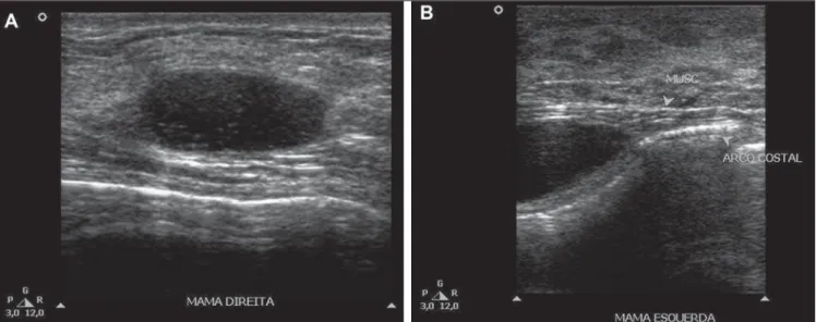
A female, 53-year-old patient was sub-mitted, on an outpatient basis, to a proce-dure of intramammary injection of hyalu-ronic acid-based gel (Macrolane) for breast augmentation for 18 months ago. During the procedure a single point on the skin of each breast was utilized for insertion of the needle. The patient was referred to the au-thors’ institution for investigation of bilat-eral, painless, palpable nodules whose on-set occurred after the procedure. Breast ultrasonography demonstrated the presence of multiple, predominantly anechoic, ovoid, cyst-like collections containing low-amplitude echoes, in intramuscular and intraglandular locations (Figure 1), with no
INTRODUCTION
Aesthetic procedures for breast aug-mentation have become increasingly fre-quent in the last decades, with utilization of different techniques and materials(1).
Although surgical implantation of encap-sulated fillers (such as silicone implants) constitute the most widely adopted tech-nique for breast enhancement, new inject-able non-encapsulated fillers have been available in the market(1). Macrolane™
182
Criado DAB et al. Aesthetic breast augmentation with hyaluronic acid
Radiol Bras. 2012 Mai/Jun;45(3):181–183
Figure 1. Breast ultrasonography demonstrating predominantly anechoic, ovoid, cystic images with low-amplitute echoes in intraglandular (A) and intramus-cular (B) locations.
natural and progressive degradability, and it is expected that its reabsorption occurs over a 12- to 18-month period(1).
Although Macrolane has been approved and is currently being utilized in more than twenty countries, its utilization has not yet been approved by the Food and Drug Ad-ministration in the United States of America(3). Additionally, scarce scientific evidence is reported in the literature about the safety and efficacy of its use, and
long-term prospective studies on this matter are still to be published(1,3–5). However, some
issues about the application of this product have been raised in the literature. The sig-nificant reabsorption of hyaluronic acid (about 50% over 12 months) would lead to the need for additional applications in the future to achieve the desirable aesthetic outcomes, increasing the total cost of the treatment(6) and, similarly to the application
in other sites, it would increase the
fre-quency and risk for development of granu-lomas(4).
On the other hand, recent studies have reported only a minimum rate of hyaluronic acid degradation, with no radiological sign of reabsorption even 24 months after the procedure(5). While an amount between 1
and 5 ml is injected into the face, breasts require from 100 to 150 ml(1), which might
remain in the breast tissue for a still un-known period of time(5). Additionally, the Figure 3. Breast magnetic resonance imaging. Axial sections demonstrating the presence of hyperintense collections on T2-weighted (A) and hypointense collections on T1-weighted (B) images.
183
Criado DAB et al. Aesthetic breast augmentation with hyaluronic acid
Radiol Bras. 2012 Mai/Jun;45(3):181–183 onset of adverse effects related to the prod-uct (development of nodules in 13% and mastalgia in 25% of cases after one year)(7),
which previously were considered as mini-mal, are currently discussed as issues of clinical significance(8) because, in
associa-tion with the decrease in the sensitivity of imaging method, young patients with re-cent painful breast nodules must undergo different supplementary studies and biop-sies to rule out the presence of neoplasia, which represents an additional morbidity(8).
Other complications, such as superficial infections and development of abscesses, have also been described(5).
From the radiological point of view, Macrolane represents a diagnostic chal-lenge, not only for its still recent use, but also for interfering with the images inter-pretation(5). Generally, Macrolane
deter-mines an increase in the breast parenchyma radiodensity at mammography, which may be either generalized or being visualized as multiple radiodense lesions. At ultrasonog-raphy, the finding corresponds to multiple predominantly anechoic collections with internal echoes of variable sizes and echogenicities. At magnetic resonance im-aging, hyaluronic acid collections appear as well delimited areas with hyperintensity on T2-weighted and hypointensity on
T1-weighted images(5). Although in the present
case the assessment of a patient complain-ing of palpable nodules by means of mul-tiple methods has demonstrated only im-ages of well defined cyst-like collections, Macrolane collections sometimes may be involved by fibrotic capsules, assuming a more worrisome radiological appearance(5).
According to Chaput et al.(3), the need
for repeated injections which could cause inflammation of the breast tissue and in-crease in the risk for cancer and develop-ment of nodules; alterations in the breast anatomy which could affect the radiologi-cal interpretation, possibly delaying the breast cancer diagnosis; and the necessity of giving priority to breast cancer screen-ing under the public health point of view, have recently led to the prohibition of us-ing Macrolane for aesthetic breast enhance-ment purposes in France(3). Additionally, in
the United Kingdom, some authors recom-mend that patients submitted to injections of the product undergo long-term follow-up, and also contraindicate its use in pa-tients with personal/family history of breast cancer, previous history of breast cystic or pre-malignant lesions, and family history of ovarian cancer(2).
Considering the relevant impact on the radiological interpretation of images in cases
of patients submitted to breast augmenta-tion with hyaluronic acid, it is important for the radiologist to be familiar with such imaging findings and aware of the clinical history of the patient when interpreting radiological images in these contexts.
REFERENCES
1. McCleave MJ. Is breast augmentation using hyaluronic acid safe? Aesthetic Plast Surg. 2010; 34:65–8.
2. McCleave MJ, Grover R, Jones BM. Breast en-hancement using Macrolane™: a report of com-plications in three patients and a review of this new product. J Plast Reconstr Aesthet Surg. 2010; 63:2108–11.
3. Chaput B, Chavoin JP, Crouzet C, et al. Macrolane is no longer allowed in aesthetic breast augmenta-tion in France. Will this decision extend to the rest of the world? J Plast Reconstr Aesthet Surg. 2012; 65:527–9.
4. Fortea-Sanchis C, Martínez-Ramos D, Alcalde-Sánchez M, et al. Hyaluronic acid breast injections. Potential interferences with mammography. Cir Esp. 2010;88:421–3.
5. Pienaar WE, McWilliams S, Wilding LJ, et al. The imaging features of MACROLANE™ in breast augmentation. Clin Radiol. 2011;66:977–83. 6. Goisis M, Savoldi A, Guareschi M. Is hyaluronic
acid gel a good option for breast augmentation? Aesthetic Plast Surg. 2011;35:134–6.
7. Hedén P, Olenius M, Tengvar M. Macrolane for breast enhancement: 12-month follow-up. Plast Reconstr Surg. 2011;127:850–60.