Arq Neuropsiquiatr 2000;58(3-B):897-900
CEREBELLAR VERMIS HYPOPLASIA
-NON PROGRESSIVE CONGENITAL ATAXIA
CLINICAL AND RADIOLOGICAL FINDINGS IN A PAIR OF SIBLINGS
ISAC BRUCK*, SÉRGIO A. ANTONIUK*, ARNOLFO DE CARVALHO NETO**, ADRIANE SPESSATTO***
ABSTRACT - We describe the clinical and radiological findings of a pair of siblings with cerebellar vermis hypoplasia and compare them with the literature. Both of them present pregnancies and deliveries uneventful and both presented some grade of hypotonia, ataxia, ocular motor abnormalities and mild motor delay and slurred speech. These siblings meet many of the criteria described in non-progressive congenital ataxia in which can occur familial cases with cerebellar atrophy, including vermis hypoplasia. As differential diagnosis we compare them with related syndromes and with Joubert’s syndrome which main radiological finding on MRI is vermis hypoplasia associated with “molar tooth” appearance. The correct answer for these cases will only be possible by molecular genetics.
KEY WORDS: non-progressive congenital ataxia, cerebellar vermis hypoplasia, ocular motor abnormalities.
Hipoplasia do verme cerebelar - ataxia congênita não progressiva: achados clínicos e radiológicos em dois irmãos
RESUMO - Descrevemos e discutimos os achados clínicos e radiológicos de dois irmãos com hipoplasia do verme cerebelar e os comparamos com os relatos da literatura. Ambos apresentaram gestação e parto sem intercorrências, algum grau de hipotonia, ataxia, anormalidades oculares motoras, atraso motor discreto e fala arrastada. Estes irmãos preenchem muitos dos critérios descritos em ataxia congênita não progressiva, nos quais podem ocorrer casos familiais com atrofia cerebelar, incluindo hipoplasia do verme. Como diagnóstico diferencial, nós os comparamos com outras síndromes correlatas e com síndrome de Joubert,cujo achado radiológico característico é a hipoplasia do verme cerebelar associado com aparência de “dente molar” na ressonância magnética do crânio. A correta resposta para estes casos só será possível pela genética molecular.
PALAVRAS-CHAVE: ataxia congênita não progressiva, hipoplasia do verme cerebelar, anormalidades oculares motoras.
In recent decades several sporadic and familial cases of non-progressive congenital ataxia (NPCA) have been published1-15. Steinlin et al.15 reviewing 34 subjects with NPCA found three pairs of siblings but only one child had vermis cerebellar hypoplasia as the only abnormality, since the other siblings had also hemispheres cerebellar hypoplasia or even normal cerebellum. Joubert’s syndrome is a rare autosomal recessive condition and the criteria for the diagnosis has been outlined:
*Professor Assistente em Neurologia Pediátrica, Centro de Neurologia Pediátrica (CENEP), Departamento de Pediatria, Hospital de Clínicas (HC), Universidade Federal do Paraná (UFPR). **Professor Assistente em Radiologia, Departamento de Clínica Médica HC/UFPR; ***Médica Residente em Neurologia Pediátrica, CENEP, Departamento de Pediatria, HC/UFPR. Aceite: 19-maio-2000.
898 Arq Neuropsiquiatr 2000;58(3-B)
irregular breathing in the neonatal period, developmental delay, mental retardation, hypotonia, ocular motor abnormalities, and, occasionally, retinal dystrophy and cystic kidneys associated with cerebellar malformation consisting in vermis hypoplasia or agenesis and abnormalities at the pontomesen-cephalic junction16-19. We describe the clinical and radiological findings of a pair of siblings and compare them with the literature.
CASES
Case 1. FAM (Fig 1) white male, 8 years and 8 months old who had the complain of hypotonia, motor
delay, unsteady gait and eyes deviation in opposite direction to the rotation of the head. All of them improved over the years.
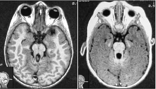
Pregnancy and delivery were uneventful, born at full term, weighing 3 350 g. Started to walk at 2 years, first words at 1 year, sentences at 2 years, with a slurred speech. At the moment he is on the second grade of the elementary school, with difficulty in language, mainly in writing meaningful sentences. Parents are unrelated. On examination macrocephaly was noted (55cm = +2SD). He was right handed, well oriented, good recent and remote memory and speech mildly slurred. Discrete nystagmus was noted on pursuit as the only abnormality of the cranial nerves. There was also mild hypotonia and unbalance with the eyes open and close. He could not perform tandem gait forward and backward. On finger-nose test he was slight dysmetric. The remainder of neurologic examination was normal. Magnetic resonance of the brain (MRI) showed hypoplasia of the posterior vermis and features that resemble the ”molar tooth” appearance (Fig 2). Abdominal ultrasound was normal.
Case 2. MAM (Fig 1) white, male, 2 years and
2 months, the younger brother of Case 1. He was less hypotonic and delayed on motor development than his older brother. When he was 3 to 4 months he used to turn his head, with opposite deviation of the eyes when fixing on an object (the same as his brother).
Pregnancy was uneventful and delivery was by cesarean section, full term, and weighing 3 750 g. When he was 24h of life he had mild hypoglycemia with tremors, that was corrected by glucose IV. He used to thrust his head on the first 30 days of life. At 20 months old he had a febrile seizure lasting 1 minute due to pneumonia. He sat at 7 months, crowed at 12 months, walked at 19 months, started to speak at 12 months and by now he is able to make sentences. On examination he was also macrocephalic (52cm = +2SD) and macrosomic. Mental status was normal for his age and speech was mildly slurred. He had broad base walk and loss the balance with open and close eyes. He is able to kick a ball and do normal finger nose and marionette test. MRI showed posterior cerebellar vermis hypoplasia and “molar tooth” appearance (Fig 2). Abdominal ultrasound was normal.
DISCUSSION
The question that arise in describing these pair of siblings is what is the best diagnosis? A general approach is to call them as NPCA as we mention in the introduction. The incidence is about 0.13 per 1000 children14. Etiology include malformations, congenital infections, syndromal and other hereditary disorders14.
Over recent decades many sporadic and familial case reports have been published1-15. In a
899
Arq Neuropsiquiatr 2000;58(3-B)
recent publication Steinlin et al15 review the charts of 34 cases of NPCA including three pairs of siblings. Age varied from 3 to 29 years (mean age 13,4 years), and 11 were over 18 years. Ataxia in NPCA is a chronic problem, neither deteriorating nor improving significantly. In relation to neuroimaging, the MRI of the three pairs of siblings , findings were discordant in two: in one pair imaging was normal in one subject while the other showed mild cerebellar hypoplasia ( CH), in the other pair one had normal imaging while the other severe CH.
The authors conclusion on pure NPCA is that they present with hypotonia and developmental delay, followed by ataxia. Ataxia is a non-progressive, but persistent symptom. Major problems arise in the majority of these subjects related to cognitive impairment and less to neurologic symptoms. Clinical and neuroimaging findings do not help in defining any subgroups of NPCA and it seems most likely that only progress in the genetics of ataxias will give more information.
At this point we would like to discuss Joubert syndrome, although for the moment is an unlike diagnosis of the siblings, but is always important to mention when we face CH.
Many authors had recently published criteria for the diagnosis of Joubert syndrome, as Sztriha et al.20 and Maria et al.21. The last one included common abnormalities characterized by hypotonia present in all patients, developmental delay including motor, language and adaptative behaviors, and many children are pleasant and friendly. MRI shows “molar tooth” sign in axial plane, deeper than normal posterior interpeduncular fossa, prominent or thickened superior cerebellar peduncules and vermian hypoplasia or dysplasia. Pathologic findings are vermian hypoplasia or dysplasia, elongation of the caudal midbrain tegmentum and marked dysplasia of the caudal medulla. The associated abnormalities are high rounded eyebrows, broad nasal bridge, mild epicanthus, anteverted nostrils, triangular-shaped open mouth with tongue protusion, low-set and coarse ears. Breathing abnormalities are most pronounced in the neonatal period and infancy. Also occur retinal dysplasia, colobomas, nystagmus, strabismus and ptosis. Oculomotor apraxia and microcystic renal disease that can be progressive.
For the description outlined above we can not say that the siblings have Joubert syndrome, and the best that we can say is that they could have a milder expression of the disease, but the answer will be possible only if we have a molecular genetic study done in these patients.
900 Arq Neuropsiquiatr 2000;58(3-B)
Raynes et al22 describe three sisters with Joubert syndrome, two of whom are monozygotic twins with highly discordant phenotypes. While twin A is wheelchair bound, severely retarded, nonverbal, autistic and MRI with “molar tooth” sign, vermian aplasia and absence of the cerebellar hemispheres, twin B is able to walk, run, verbal and MRI with “molar tooth” sign and mild hypoplasia of the inferior cerebellar vermis. Unlike our siblings she shows on ophthalmologic examination diffuse retinal pigmentary changes.
Finally it is important to make differential diagnosis with related conditions such as Arima, Senior Loken and Coach syndromes that also have “molar tooth” signs on axial MRI23. Arima syndrome is an autossomal recessive disorder which consists of vermian hypoplasia, retinopathy, cystic dysplastic kidneys and several patients have Dandy-Walker malformations or occipital encephalocele. Senior Loken syndrome is also an autossomal recessive syndrome, consists of vermian hypoplasia, retinopathy, juvenil onset nephronophthisis and most have mental retardation. Coach syndrome is an autossomal recessive malformation syndrome, consists of vermian hypoplasia, colobomas, nephronophthisis and hepatic fibrosis.
Addendum - After finishing the article we were told by the parents that a children’s cousin, an 1 year old girl, on the mother’s side, from unrelated parents, has the same clinical picture, but with a normal MRI.
REFERENCES
1. Malamud N, Cohen P. Unusual form of cerebellar ataxia with sex linked inheritance. Neurology 1958;8:261-266. 2. Pfeiffer RA, Palm D, Jünemann G, Mandl-Kramer S, Heimann E. Nosology of congenital non-progressive cerebellar
ataxia. Neuropädiatrie 1974;5:91-102.
3. Furman JM, Baloh RW, Chugani H, Waluch V, Bradley WG. Infantile cerebellar atrophy. Ann Neurol 1984;17:399-402. 4. Wichman A, Frank LM, Kelly TE. Autosomal recessive congenital cerebellar hypoplasia. Clin Genet 1985;27:373-282. 5. Tomiwa K, Baraitser M, Wilson J. Dominantly inherited congenital cerebellar ataxia with atrophy of the vermis. Pediatr
Neurol 1987;3:360-362 .
6. Young ID, Moore JR, Tripp JH. Sex-linked recessive congenital ataxia. J Neurol Neurosurg Psychiatry 1987;50:1230-1232.
7. Fenichel GM, Phillips JA. Familial aplasia of the cerebellar vermis. Arch Neurol 1989;46:582-583.
8. Mathews KD, Afifi AK, Hanson JW. Autosomal recessive cerebellar hypoplasia. J Child Neurol 1989;4:189-193. 9. Kornberg AJ, Shield LK. An extended phenotype of an early-onset inherited nonprogressive cerebellar ataxic syndrome. J
Child Neurol, 1991;6:20-23.
10. Rivier F, Echenne B. Dominantly inherited hypoplasia of the vermis. Neuropediatrics 1992;23:206-208.
11. Guzetta F, Mercuri E, Bonanno S, Longo M, Spano M. Autosomal recessive congenital cerebellar atrophy: a clinical and neuropsychological study. Brain Develop 1993;15:439-445.
12. Imamura S, Tachi N, Oya K. Dominantly inherited early-onset non-progressive cerebellar ataxic syndrome. Brain Develop 1993;15:372-376.
13. Al Shawan SA, Bruyn GW, Al Deeb SM. Non-progressive congenital cerebellar hypoplasia. J Neurol Sci, 1995;128:71-77. 14. Esscher E, Flodmark O, Hagberg G, Hagberg B. Non-progressive ataxia:origins, brain pathology and impairments in 78
Swedish children. Dev Med Child Neurol 1996;38:285-296.
15. Steinlin M, Zangger B, Boltshauser E. Non-progressive congenital ataxia with or without cerebellar hypoplasia:a review of 34 subjects. Dev Med Child Neurol 1998;40:148-154.
16. Dooley JM, Laroche GR, Tremblay F, Riding M. Autosomal recessive cerebellar hypoplasia and tapetoretinal degeneration: a new syndrome. Pediatr Neurol 1992;8:232-234.
17. Saraiva JM, Baraitser M. Joubert syndrome: a review. Am J Med Genet 1992;43: 726-731.
18. Maria BL, Hoang KBN, Tusa RJ, et al. “Joubert’s syndrome” revisited: key ocular motor signs with magnetic resonance imaging correlation. J Child Neurol 1997;12:423-430.
19. Shen W-C, Shian W-J, Chen C-C, Chi C-S, Lee S-K, Lee K-R. MRI of Joubert’s syndrome. Eur J Radiol 1994;18:30-33. 20. Sztriha L, Al-Gazali LI, Aithala GR, Nork M. Joubert’s syndrome: new cases and review of clinicopathologic correlation.
Pediatr Neurol 1999:20:274-281
21. Maria BL, Boltshauser E, Palmer SC, Tran TX. Clinical features and revised diagnostic criteria in Joubert syndrome. J Child Neurol 1999;14:583-591.
22. Raynes HR, Shanske A, Goldberg S, Burde R, Rapin I. Joubert syndrome: monozygotic twins with discordant phenotypes. J Child Neurol 1999;14:649-654.