THE DIAGNOSIS OF LEPROSY AMONG PATIENTS WITH SYMPTOMS
OF PERIPHERAL NEUROPATHY WITHOUT CUTANEOUS LESIONS
A FOLLOW-UP STUDY
MICHAEL SKACEL 1, SÉRGIO LUIZ GOMES ANTUNES 2, MÁRCIA MARIA JARDIM RODRIGUES 3, JOSÉ AUGUSTO DA COSTA NERY 4, VÂNIA DA COSTA VALENTIM 5,
RENATA PATRÍCIO BRAGA DE MORAIS 6, EUZENIR NUNES SARNO 7
ABSTRACT - Forty-four patients with neuritic leprosy were individually followed for periods ranging from 4 months to almost 4 years for the purpose of ascertaining the presence and/ or absence of leprosy. The neural symptoms presented were sensory impairment (41), parasthesia (28), nerve enlargement (22), nerve tenderness (20), paresia (20), amyotrophy (8). Leprosy was diagnosed in ten out of the total number of patients studied. Leprosy was confirmed by the appearance of reactional neuritis (4), reversal reaction (2), biopsy of the hypoesthesic area (3) and the appearance of non-reactional cutaneous lesion. We reported an experience in the diagnosis of neuritic leprosy and its most frequent clinical presentation with which clinicians have to be acquainted. We can also state that the clinical follow-up was an effective strategy for the diagnosis of the disease when diagnostic facilities are not available or have not confirmed the diagnosis.
KEY WORDS: leprosy, neuritic leprosy, peripheral neuropathy.
O diagnóstico de hanseníase neural pura entre pacientes com sintomas de neuropatia periférica: acompanhamento clínico
RESUMO - Quarenta e quatro pacientes com sinais de neuropatia periférica foram acompanhados no Ambulatório de Hanseníase da Fundação Oswaldo Cruz por período que variou de 4 meses até 4 anos, com o intuito de confirmar ou afastar o diagnóstico de neuropatia hanseniana. Os sintomas neurológicos apresentados foram hipoestesia (41), parestesia (28), espessamento neural (22), dor nos nervos (20), paresia (20), amiotrofia (8). Dez pacientes dos 44 tiveram o diagnóstico de hanseníase confirmado. A confirmação diagnóstica se deu através da biópsia de áreas hipoestésicas sem lesão dermatológica (3 pacientes), pelo aparecimento de estados reacionais (duas reações reversas e 4 neurites reacionais) e pelo aparecimento de lesão cutânea não reacional característica da forma “borderline” lepromatosa. O acompanhamento clínico regular dos pacientes sem diagnóstico no primeiro exame mostrou ser um método que permitiu o diagnóstico da forma neurítica pura da hanseníase, quando os métodos objetivos de diagnóstico não confirmarem de imediato a doença.
PALAVRAS-CHAVE: hanseníase, neurite, neuropatia periférica.
1“In memoriam”, Professor do Departamento de Neurologia, Universidade do Estado of Rio de Janeiro,
Brasil; 2Doutor em Patologia, Pesquisador Associado do Laboratório de Hanseníase, Instituto Oswaldo Cruz,
Rio de Janeiro, Brasil (IOCruz), Professor de Patologia da Universidade Iguaçu; 3Mestre em Neurologia,
Pesquisadora visitante do Laboratório de Hanseníase, IOCruz; 4Mestre em Dermatologia, Pesquisador Assistente
do Laboratório de Hanseníase, IOCruz; 5Bióloga e técnica do Laboratório de Hanseníase, IOCruz; 6Estagiária de
Pós-Graduação do Laboratório de Hanseníase, IOCruz; 7Doutora em Patologia, Chefe do Departamento de
Medicina Tropical do IOCruz. Aceite: 30-junho-2000.
The main cause of morbidity in leprosy is the peripheral neuropathy1, that is responsible for
the great bulk of disabilities and deformities displayed by many leprosy patients. The nerve lesion is recognized either as a chronic or subacute inflammatory infiltrate, in which either epithelioid cells or M. leprae-glutted macrophages can be present2-4. This infiltrate can occupy the endoneurium, the
perineurium and the epineurium5. As a consequence, there is a progressive impairment of
unmyelinated and myelinated neural fibers6,7 followed by a replacement of the peripheral nerve
parenchyma for fibrous tissue8. Necrotic caseation may also occur in tuberculoid granulomas of the
nerves resulting in its complete destruction by abscesses9. The inflammatory infiltrate of the nerves
may be distinct from the ones in the cutaneous lesions, being multibacillary in the nerves and paucibacillary in the skin10,11.
Leprosy neuropathy may also present without skin lesions12,13, this is known as the neuritic
form of leprosy. The patients with this form of the disease displays only signs and symptoms of sensory impairment, parasthesia, nerve enlargement, nerve pain, and muscle weakness, without dermatological alterations. This poses some difficulties to the leprosy diagnosis, particularly in the services where diagnostic facilities such as bacilloscopy, electroneuromyography and nerve biopsy are not available. Neuritic leprosy has a varied incidence among the total number of cases in an endemic population, ranging from 1% (14), 3% (15), 10,7% (16) to 16%17 of the leprosy patients.
Approaches to the neuritic leprosy found in the literature focus on its clinical characteristics13,16,18,19,
on the value of nerve biopsy on the diagnosis of neuritic leprosy5, on the discrepancy of histological
appearance between dermatological and neurological lesions13.
Neurologists and dermatologists face the demand of an early diagnosis of leprosy neuropathy in order to prevent patients from disability. Being familiar with the peculiar neurological manifestations of this specific peripheral neuropathy renders clinicians capable of disclosing an early diagnosis of leprosy in these patients.
Taking into account these diagnostic difficulties mentioned above, we performed a four-year clinical follow-up of forty-four patients with evidence of peripheral neuropathy trying to ascertain an early leprosy diagnosis on them and to establish the clinical and laboratorial criteria for this diagnosis.
METHOD
Forty-four patients from the Leprosy Outpatient Clinic of the Oswaldo Cruz Institute, suspected of leprosy neuropathy were submitted to a follow-up which ranged from four months to four years. Twenty-nine patients were male and fifteen female. Eighteen were white, 15 were mongrels (mulatto) and seven were black (four patients, not informed). The average age of the patients was 42 years (± 14.1) ranging from nineteen to seventy-eight. This study focused on the leprosy diagnosis as the Souza Araújo outpatient clinic is a leprosy reference center of the Ministry of Health in Brazil to where a great number of patients are referred in order to rule out or confirm the disease. When the presence of leprosy peripheral neuropathy was discarded, the neuropathic patients were usually referred to the Neurology Clinic of the Pedro Ernesto Hospital (Rio de Janeiro State University).
The patients presented in the first visit to the clinic with signs and symptoms of sensory impairment, parasthesia, nerve enlargement, motor involvement and nerve tenderness (Table 1). Only two patients presented with one isolated symptom (sensory impairment and parasthesia), but neither one of them were confirmed to have leprosy. The patients were submitted to routine dermatological and neurological examination. Sensory impairment was evaluated with graded nylon monofilaments20, which is a semi-objective method of determining
the grade of sensory impairment. The examiner inquires the patient on his sensory capacity under stimulation with distinct colored filaments having increasing graded thickness (green, blue, violet, red and black). Voluntary muscle testing (VMT) was employed to evaluate the motor damage. The graded scoring for muscle weakness was from 0 = paralysis to 5 = normal muscle function.
RESULTS
Patients referred to the outpatient service with peripheral neuropathy
The clinical data of the forty-four patients are displayed in Table 1.
Thirty-four out of the 44 patients did not have the diagnosis of leprosy confirmed. Seven out of the thirty-four had the diagnosis of leprosy considered as very unlikely and so were readdressed to the neurological clinic of State University of Rio de Janeiro, where causes other than leprosy could be investigated. The remaining 27 patients in whom the leprosy diagnosis was neither discarded nor confirmed were followed with regular examination in the outpatient service of Oswaldo Cruz Institute for four years. No leprosy diagnosis was confirmed at the end of this follow-up period in these patients. Fifteen patients had an ENMG exam performed and seven were submitted to nerve biopsy. Only one biopsy presented focal perineurial thickening on account of fibrosis.
Patients with confirmed leprosy
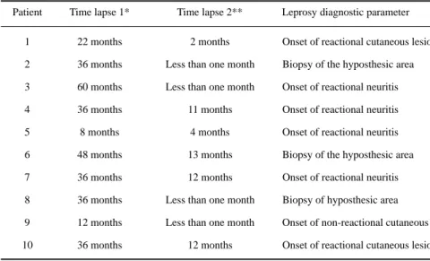
Ten patients had confirmed leprosy disease (Table 2) at diverse time points of the clinical follow-up (Table 4). The average time between the beginning of the symptoms and the first exam was 33 months. The skin smears were negative in all patients in the first exam. Lepromin test was positive in 5 patients; five had negative test and one of the lepromin-negative patient developed borderline lepromatous leprosy (Table 3).
The time lapse between the first exam and confirmation of the diagnosis average 4.25 months, in a time span that ranged from less than one month to twelve months. The nerves Table 1. Clinical data of the 44 patients with symptoms of peripheral neuropathy.
Neural symptoms, exams performed and evolution of the patients Number of patients
Number of patients 44
Touch sensory impairment 41 (93%)
Thermal sensory impairment 18 (44%)
Pain sensory impairment 8 (18%)
Proprioceptive sensory impairment 0
Parasthesia 28 (63%)
Nerve enlargement 22 (50%)
Nerve tenderness 20 (45%)
Paresia 20 (45%)
Amyotrophy 8 (18%)
ENMG performed 15 (34%)
Biopsies of skin hyposthesic lesions 12 (27,2%)
Nerve biopsies performed 7 (15,9%)
Patients clinically discarded to have leprosy 7 (15%)
Patients followed for four years in the study without confirmation of leprosy diagnosis 27 (61%)
Patients with confirmed leprosy along the folow-up 10 (22,7%)
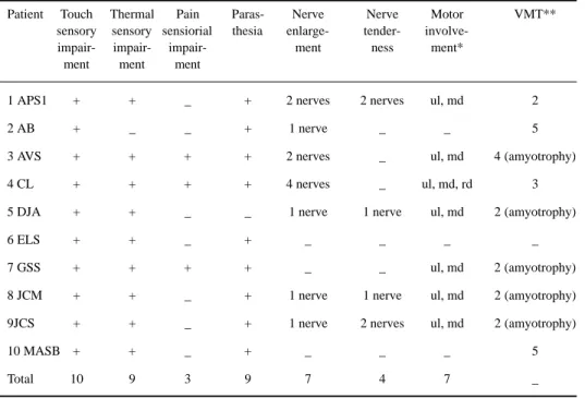
Table 2. Clinical data of the leprosy-confirmed patients (Data of the first exam).
Patient Touch Thermal Pain Paras- Nerve Nerve Motor VMT** sensory sensory sensiorial thesia enlarge- tender-
involve-impair- impair- impair- ment ness ment* ment ment ment
1 APS1 + + _ + 2 nerves 2 nerves ul, md 2
2 AB + _ _ + 1 nerve _ _ 5
3 AVS + + + + 2 nerves _ ul, md 4 (amyotrophy)
4 CL + + + + 4 nerves _ ul, md, rd 3
5 DJA + + _ _ 1 nerve 1 nerve ul, md 2 (amyotrophy)
6 ELS + + _ + _ _ _ _
7 GSS + + + + _ _ ul, md 2 (amyotrophy)
8 JCM + + _ + 1 nerve 1 nerve ul, md 2 (amyotrophy)
9JCS + + _ + 1 nerve 2 nerves ul, md 2 (amyotrophy)
10 MASB + + _ + _ _ _ 5
Total 10 9 3 9 7 4 7 _
*nerves corresponding to the group of muscles involved; **VMT: voluntary muscle test.
Table 3. Laboratory data of the leprosy-confirmed patients.
Patient Lepromin (mm) HPD (skin) ENMG Nerve biopsy
1 10 RR ND Focal perineurium
enlargement
2 0 BT ND ND
3 0 ND MNM Normal
4 0 ND MNM Normal
5 0 Normal MNM ND
6 0 BL MNS ND
7 5 Normal ND Normal
8 4 BT ND ND
9 15 BT ND ND
10 3 RR ND ND
which most frequently presented enlargement in this study were in order of frequency, the ulnar, the auricular, and the radial. The groups of muscles most frequently involved were the intrinsic muscles of the hand and the preferential muscles affected were the ones innervated by the ulnar and by the median nerve.
Each neurological alteration was never found as an isolated clinical manifestation, but association of sensory impairment with another symptom was allways present. Therefore, sensory impairment together with parasthesia (9 patients), with motor involvement (8) or with nerve enlargement (7 patients) and with nerve tenderness (4) were observed.
Only seven in twenty-two patients with nerve enlargement from the total number of patients selected for this study had confirmed leprosy in the follow-up period.
Four out of the ten patients with confirmed leprosy were submitted to electroneuromyographic study before the definitive diagnosis (Table 3). Patient 3 showed absence of sensitive response of the left and right ulnar and sural nerves, a decreased velocity of sensitive conduction of the left median, a decrease of motor conduction of the left fibular nerve and absence of motor response of the left ulnar nerve. Patient 4 exhibited absence of sensitive response of the median and sural nerves, a decreased velocity of sensitive conduction in the right radial and left ulnar, and the velocity of motor conduction was decreased in the right and left median, ulnar and fibular nerves, Patient 5 showed decreased motor and sensitive velocity of the median and ulnar nerve. No sensitive response in the radial, ulnar e median nerves. These electroneuromyographic findings characterized mononeuropathies multiplex cases in three patients. Patient 6 had only absence of sensitive response of the sural nerve, characterizing a mononeuropathy simplex.
Four leprosy patients had been submitted to nerve biopsy. The histopathological appearances of these patients’ nerves were normal except only one who showed a slight and focal thickening of the perineurial layer. No acid-fast bacilli were found on the histopathological sections with the Wade staining. Search for mycobacteria in the nerves with immunohistochemical staining and with polymerase chain reaction were not performed. The nerve biopsies performed in few patients did not allow the prompt elucidation of the neuropathic picture of the patients in this study.
Table 4. Time lapses* and leprosy diagnostic parameter.
Patient Time lapse 1* Time lapse 2** Leprosy diagnostic parameter
1 22 months 2 months Onset of reactional cutaneous lesion
2 36 months Less than one month Biopsy of the hyposthesic area
3 60 months Less than one month Onset of reactional neuritis
4 36 months 11 months Onset of reactional neuritis
5 8 months 4 months Onset of reactional neuritis
6 48 months 13 months Biopsy of the hyposthesic area
7 36 months 12 months Onset of reactional neuritis
8 36 months Less than one month Biopsy of hyposthesic area
9 12 months Less than one month Onset of non-reactional cutaneous lesion
10 36 months 12 months Onset of reactional cutaneous lesion
DISCUSSION
A great practical difficulty posed by neural leprosy without cutaneous manifestations is its diagnosis, specially if electroneuromyography and nerve biopsy21 are not routinely carried out in a
leprosy service.
All of the confirmed leprosy patients of this study presented with local sensory impairment as the main complaint at the first exam. However, a detailed neurological examination detected a much more spread affection of the peripheral nervous system represented by scattered sensory impairment, motor deficit, nerve enlargement, and parasthesia . All of these signs were present on sites far from the ones of the original complaint, characterizing a spread affection of the peripheral nervous system. This evidence indicates that leprosy is not essentially a dermatological disease with complicating neurological manifestations22. In fact, whatever the earliest dermatological manifestation may be,
either a restricted cutaneous region with sensory impairment or an isolated and small macular lesion, there can be a difuse affection of the peripheral nerves. This can only be detected by a careful neurological and electrophysiological examination. The neurological examination, particularly of the nociceptive sensory function requires specific training and this expertise is not usually available in non-neurological outpatient medical services. It is important that dermatologists be aware that a local sensory disturbance or a single skin lesion in leprosy does not mean that the disease is restricted to the site of its cutaneous manifestation.
Touch sensory impairment was the most frequent symptom observed, in the 10 leprosy patients, followed closely by thermal hyposthesia in 9 patients. Sensory impairment was reported by Kaur et al23 as the most frequent symptom found among the neuritic leprosy patients, parasthesia however,
was present in 25% of the patients with neuritic leprosy in this author´s report. Kaur et al23 also
found an association of sensory symptoms with motor symptoms in 33% of patients. In our study the motor involvement was detected in 8 out of 10 (80%) leprosy patients, however two patients exhibited exclusively sensory impairment together with parasthesia at the first exam. Mahajan et al24 found motor deficit as the predominant symptom in 66 out of 179 patients and parasthesia was
the second most frequent clinical manifestation. The high incidence of motor deficit in the leprosy patients of the present study may be caused by the careful search of weakness of the intrinsic muscles of the hands or feet which may pass unnoted.
Only seven patients in twenty-two with nerve enlargement from the whole group of forty-four patients were diagnosed as leprosy. This finding indicates that nerve enlargement may not be specific of leprosy. Nerve enlargement can also be found in other neurological conditions and in normal individuals whose intense physical work may render nerves susceptible to trauma causing fibrosis of the wrapping epineurium, specially at entrapment susceptible points21.
In the follow-up performed in this investigation, the patients presented neurological symptoms of a peripheral neuropathy and were clinically followed until leprosy diagnosis was stated by the appearance of characteristic evidences. In four patients, a typical picture of leprosy reactional neuritis with nerve tenderness and worsening of nerve damage grounded the leprosy diagnosis as the patients remained with no cutaneous lesions. The histopathological examination of the anaesthetic skin without dermatological alteration contributed to the diagnosis, in three patients, therefore, it is recommended that this procedure be performed whenever possible as an aid to the diagnosis.
The onset of the reactional episodes was the most frequent occurrence which allowed the confirmation of leprosy; reactional neuritis and reversal reaction with new cutaneous lesions were the most frequent types of reaction. Srinivasan and Rao25 and Becx-Bleumink and Manetze26 reported
The appearance of skin lesions in the evolution of neuritic leprosy was also reported by Pannikar et al18 who detected the development of skin lesions in 4 out of 17 patients with neuritic
leprosy. Talwar et al16 studied 62 cases of neuritic leprosy and reported the appearance of skin
lesions in 5 patients submitted to dapsone monotherapy and in 3 patients treated with rifampicin and dapsone. An opposite result was shown by Mahajan et al.24, who studied 179 cases of neuritic
leprosy but found no skin lesions developed during the MDT. Girdhar17, in his review, comments a
Kaur’s unpublished observation in which 14 out of 40 patients observed over three and half years presented the appearance of skin lesions along this period. This author considered the skin lesions to be a manifestation of reversal reaction as they appeared along the MDT17. The cases of the present
study were not under treatment but only under regular monitoring when the reactional or non-reactional skin lesions appeared.
The length of the follow-up of the patients (four years) proved to be a good strategy for achieving the diagnosis of leprosy etiology in the neuropathy cases as electroneuromyography and nerve biopsy were not routinely performed in the service during the lapse of time of this study. Whenever a diagnostic doubt persists after the performance of electroneuromyography, nerve biopsy, search for M. leprae antigens with immunohistochemistry and for M leprae DNA in the nerves with polymerase chain reaction, follow-up with regular bacilloscopic and neurological examination for evaluation of progressive nerve damage will be the only alternative strategy for neuritic leprosy diagnosis. The maximal time lapse of 13 months from the patients’ first exam to the confirmation of the diagnosis shows that at least for this period the follow-up should not be interrupted.
Biopsy of the cutaneous region presenting sensory impairment contributed to the diagnosis of three leprosy cases. The presence of a granulomatous tuberculoid infiltrate in a nerve branch of a hyposthesic normal-looking skin is a possible evidence that leprosy affects first the peripheral nervous system, followed by the affection of the cutaneous compartment. Fite27 states, _“To the
histopathologist, leprosy is allways neural”. Based on this statement, exclusively neural leprosy can be found and also instead, there is no leprosy with exclusive cutaneous manifestations.
In the present study the nerve biopsy was performed in a small number of cases and did not contribute to confirm the diagnosis of leprosy on the followed patients. Perhaps a study using semi-thin section would likely be of additional help, however nerve biopsy at the time of this study was not performed as a routine in our clinic. This contrasts with the reports in the literature which show the value of nerve biopsy in neuritic leprosy diagnosis when larger number of cases are analysed. Kaur et al23 stated that 35 out of 37 enlarged nerves of suspected leprosy patients which were biopsied were
affected by the leprosy infiltrates and these were more frequently of the lepromatous type. Only five enlarged nerves did not display inflammatory infiltration, but three of these five showed bacilli. Chimelli et al5 also found a decisive role of nerve biopsy for neuritic leprosy in 15 out of 53 patients.
The electroneuromyographic pattern of leprosy neuropathy described in the literature is the impairment of conduction of nerve impulse28 and decreased amplitude of sensory-motor potentials29.
Tzourio et al.30 reported the absence of correlation between neurological symptoms and
electroneurographic studies in leprosy patients. Unfortunately, in this study the electroneu-romyography was performed in only few patients and only three had the leprosy diagnosis confirmed, so that any evaluation on the contribution of this method to the diagnosis was not possible.
REFERENCES
1. Bryceson A, Pfaltzgraff RE. Complications due to nerve damage. In Medicine in the tropics: leprosy. 3Ed. Edinburgh: Churchill Livingstone, 1990:133-151.
2. Job CK, Desikan KB. Pathologic changes and their distribution in peripheral nerves in lepromatous leprosy. Int I Lepr 1968;36:257-270.
3. Dastur DK, Pandya SS Antia, NH. Nerves in the arm in leprosy II. Pathology, pathogenesis and clinical correlation. Int J Lepr 1970;38:30-48.
4. Dastur DK, Ramamohan Y, Shah JS. Ultrastructure of lepromatous nerves. Neural pathogenesis in leprosy. Ind J Lepr 1973;4:47-80.
5. Chimelli L, Freitas M, Nascimento O. Values of nerve biopsy in the diagnosis and follow-up of leprosy: the role of vascular lesions and usefulness of nerve studies in the detection of persistent bacilli. J Neurol 1997;244:318-323.
6. Gibbels E, Henke-Lubke 0, Klingmüller G. Unmyelinated nerve fibres in leprosy. A qualitative and quantitative study of sural nerve biopsies in 2 cases of lepromatous leprosy. Lepr Rev 1988;59:153-162.
7. Dastur DK, Razzak ZA. Degeneration and regeneration in teased nerve fibers I. Leprous neuritis. Acta Neuropathol 1971;18:286-298.
8. Junqueira LCU, Montes CS, Neto EA, Barros C, Tedesco-Marques AJ. The collagen of permanently damaged nerves in human leprosy. Int J Lepr 1980;48:291-297.
9. Saxena U, Ramesh V, Misra RS, MukherjeeA. Giant nerve abscesses in leprosy. Clin Exp Dermatol 1990;15:349-351. 10. Nilsen R , Mengistu G, Reddy BB. The role of nerve biopsies in the diagnosis and management of leprosy. Lepr Rev
1989;69:18-32.
11. Srinivasan H, Rao KS, Iyer CGS. Discrepancy in the histopathological features of leprosy lesions in the skin and peripheral nerve. Lepr India 1982;54:275-286.
12. Jenkins D, Papp K, Jakubovic HR, Shiffman N. Leprotic involvement of peripheral nerves in the absence of skin lesions. Case report and literature review. J Am Acad Dermatol 1990;23:1023-1026.
13. Uplekar MW, Antia NH. Clinical and histopathological observations on pure neuritic leprosy. Indian J Lepr 1986;58:513-521.
14. Mafoyane NA, Jacyk WK, Lotz BP. Primary neuritic leprosy in a black South African. Lepr Rev 1992;63:177-281. 15. Dongre VV, Ganapati R, Chulawala RG. A study of mono-neuritic lesions in a leprosy clinic. Lepr India 1976;48:132-137. 16. Talwar S, Jha PK, Tiwari VD. Neuritic leprosy: epidemiology and therapeutic responsiveness. Lepr Rev 1992;63:263-268. 17. Girdhar BK. Neuritic leprosy. Ind J Lepr 1996;68:35-42.
18. Pannikar VK, Arunthathi S, Chacko CJ, Fritischi EP. A clinico-pathological study of primary neuritic leprosy. Lepr India 1983;55:212-221.
19. Mishra B, Mkherjee A, Girdhar A, Husain S, Malaviya BN. Neuritic leprosy: further progression and significance. Acta Leprol 1995;9:187-194.
20. Ramu G. Sensory testing at field level. In: Antia NH, Shetty VP: The peripheral nerve in leprosy and other neuropathies. Oxford; Oxford University Press, 1997;37-42.
21. Uplekar MW. Neural manifestations and differential diagnosis in leprosy. In Antia NH, Shetty VP. The peripheral nerve in leprosy and other neuropathies. Calcuta: Oxford University Press, 1997:19-28.
22. Antia NH, Mehta L, Shetty V. Clinical electrophysiological, quantitative, histological and ultrastructural studies of the index branch of the radial cutaneous nerve in leprosy: preliminary report Int J Lepr 1975;43:106-113.
23. Kaur G, Girdhar BK, Girdhar A. A clinical, immunological and histological study of neuritic leprosy patients. Int J Lepr 1991;59:385-391.
24. Mahajan PM, Jogaikar DG, Mehta JM. A study of pure neuritic leprosy: clinical picture. Ind J Lepr 1996;68:137-141. 25. Srinivasan H, Rao KS, Shanmugam N. Steroid therapy in recent “quiet nerve paralysis”. Lepr India 1982;54:412-419. 26. Becx-Bleumink M, Manetje DBWT. The management of nerve damage in the leprosy control services. Lepr Rev
1990;61:1-11.
27. Fite GL. The pathology and pathogenesis of leprosy. Ann N Y Acad Sci 1951;54:28-33.
28. Mc Leod JG, Hargrave JC, Booth GC, Gye RS, Barron SA. Nerve conduction studies in leprosy. Int J Lepr 1975;43:21-31. 29. DeFaria CR, Silva IM. Electromyographic diagnosis of leprosy. Arq Neuropsiquiatr 1990;48:403-413.