81 Work performed in the Hospital de Clínicas, Medical Sciences School
of the State University of Campinas – UNICAMP
Work presented in the XI Brazilian Congress of Adult, Pediatric and Neonatal Intensive Care in June 2004, Curitiba - PR.
1 – Post Graduate student of the Surgery Department, Medical Sciences School of the State University of Campinas
2 - Professor of the Surgery Department, Medical Sciences School of the State University of Campinas
3 – Assistant Professor Cardiac Surgery Service, Medical Sciences School of the State University of Campinas
4 - Assistant Professor, Cardiac Surgery Service, Medical Sciences School of the State University of Campinas
Correspondence address: Marcos Mello Moreira. Rua Conceição 233 sala 810, Centro,
13010-916 Campinas, SP, Brazil. Tel Fax: +55-19-3233-2969. E-mail: [email protected]
Marcos Mello MOREIRA1, Renato G. G. TERZI2, Reinaldo W. VIEIRA3, Orlando PETRUCCI JR.4
Braz J Cardiovasc Surg 2005; 20(1): 81-84
CASE REPORT
Article receiveid in September, 2004 Article accepted in January, 2005
RBCCV 44205-734
Abstract
This report presents data on late dead space fraction (fDlate) of a patient submitted to surgical pulmonary embolectomy. Pulmonary thromboembolism (PTE) was diagnosed by echo-Doppler ultrasound of the lower limbs, lung scintigraphy, computerized helical tomography and angiography. The fDlate was calculated based on volumetric capnography as well as on arterial blood gases according to ERIKSSON et al. [1]. Preoperative fDlate value was 0.16, and was considered positive for the diagnosis of PTE, because it was higher than the cut-off point of 0.12. Postoperative fDlate
value was - 0.04, which was below 0.12 and was characterized as negative. The agreement of fDlate with the image results confirms the validity of this new, noninvasive diagnostic tool.
Descriptors: Pulmonary embolism. Pulmonary gas exchange. Capnography.
Resumo
Este relato de caso apresenta os resultados da fDlate (fração tardia de espaço morto) em um paciente submetido a embolectomia por tromboembolismo pulmonar (TEP). O TEP foi diagnosticado por ultrassonografia ecodoppler de membros inferiores, cintilografia pulmonar, tomografia helicoidal computadorizada e arteriografia pulmonar. O cálculo da fDlate se baseou na capnografia volumétrica e na gasometria arterial de acordo com ERIKSSON et al.[1]. A fDlate pré-operatória foi de 0,16 e foi considerada positiva por estar acima do valor de corte de 0,12. A fDlate pós-operatória foi de - 0,04, um valor inferior ao valor de corte de 0,12e foi caracterizada como negativa. A correlação da fDlate com os resultados de imagem confirma a validade desta nova ferramenta diagnóstica não-invasiva.
Descritores: Embolia pulmonar. Troca gasosa pulmonar. Capnografia.
Fração tardia do espaço morto (fDlate) antes e após embolectomia pulmonar
82
MOREIRA, MM ET AL - Late dead space fraction (fDlate) before and after pulmonary embolectomy
Braz J Cardiovasc Surg 2005; 20(1): 81-84
INTRODUCTION
It is known that unexpected deaths can occur due to pulmonary thromboembolism (PTE) and that anticoagulation is frequently effective in reducing the possibility of a new embolic event and death. For this reason, in patients with suspicion of PTE noninvasive and disposable methods incorporated as part of the bedside evaluation are desirable. Bedside techniques developed to evaluate patients with PTE, are based on some respiratory parameter derived from the alveolar dead space. But, these variables have some limitations because of the difficulties in differentiating patients with PTE and other chronic obstructive pulmonary diseases (COPD). To overcome these difficulties, ERIKSSON et al. [1] described a graphic method to extrapolate the arterial-alveolar gradient (P(a-etCO2) to a late effective expiration. They named this variation the late dead space fraction
(fDlate). The authors performed a study of 38 patients
suspected as suffering PTE and observed the fDlate greater than 0.12 in normal people, while patients with COPD had a
fDlate of less than 0.12. In this report it was possible to correlate the imaging results with fDlate before and after surgical thromboembolectomy.
CASE REPORT
This report is on a 69-year-old man, who arrived in the intensive care unit from a secondary hospital, where he had been hospitalized for one week diagnosed with dyspnea, palpitations and a dry cough. He presented with an arrhythmic pulse, tachycardia and slight hepatomegaly in the physical examination. Measurement of arterial blood gases revealed significant hypoxemia (PaO2 = 48.8 mmHg) and hypocapnia (PaCO2= 31mmHg).
Atrial fibrillation with a deviation of the axis to the right (+ 60 degrees) was evidenced by electrocardiography. An echocardiogram demonstrated a moderate increase of the right ventricle and pulmonary artery hypertension (systolic pressure assessed at 76 mmHg). With the earlier diagnostic hypothesis of PTE, anticoagulation was initiated using non-fractionated heparin. Patient was maintained under oxygen-therapy using a Venturi mask.
Ventilation/perfusion lung scintigraphy showed emboli in multiple regions (Figure 1). The computerized helicoidal tomograph (CHT) confirmed the presence of thrombi in the right and left pulmonary arteries as far as the posterior segmental arteries. Echo-Doppler ultrasound of the lower limbs confirmed the diagnosis of deep venous thrombosis to the left.
Volumetric capnography using a respiratory profile monitor CO2MO Plus 8100® (Dixtal/Novametrix) was made. The PetCO2 was recorded for a period of three minutes in
room air. During this period, a sample of arterial blood on ambient air was drawn for gasometric analysis. fDlate was calculated after the determination of the late PetCO2, extrapolated for 15% of the total pulmonary capacity (TPC), according to ERIKSSON et al.[1], whose mathematical expression is as follows:
Fig. 1 - Pulmonary perfusion scintigraphy in the preoperative period (A) with fDlate of 0.16 (positive for PTE) and six days after pulmonary thromboembolectomy (B) when the fDlate fell to -0.04 (negative for PTE) [1].
A
B
As the evolution was over more than two weeks and the patient was clinically and hemodynamically stability, extensive chronic embolia was suspected and chemical thrombolysis was contra-indicated. Thus, surgical embolectomy was scheduled and the patient was assessed by hemodynamic study. A coronary cineangiography demonstrated normal coronary arteries and ventriculography showed an ejection fraction of 69%. The pulmonary artery
fDlate= PaCO2 – Pet(15% CPT)CO2 PaCO2
Where:
PaCO2 is the partial CO2 pressure in the arterial blood;
Pet(15%TPC) CO2 is the partial CO2 pressure in the expired air extrapolated for 15% of the TPC.
TPC (total pulmonary capacity), obtained from previously published tables and based on the age, weight and height of the patient [2].
83 presented significant bulging of its trunk, presence of
amputation of the peripheral branches suggestive of multiple chronic pulmonary emboli. This image is suggestive of thrombi in the inferior lobar artery of the left lung and hypoperfusion of the middle right pulmonary lobe.
Operative technique
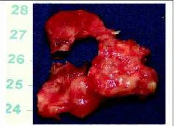
Operative intervention was performed with the help of cardiopulmonary bypass at deep hypothermia and total circulation arrest. Left pulmonary arteriotomy was performed with removal of thrombi and, on the right, thrombi up to the segmental branches were removed (Figure 2), followed by arterial repair and re-warming of the patient. The anatomical-pathological examination evidenced thrombotic substance in several different states. The patient was discharged on the sixth postoperative day. Before release, he was submitted to another pulmonary perfusion scintigram (Figure 1) and after the patient gave written permission, he was submitted to a volumetric capnography to calculate the fDlate. The obtained value was –0.04 which was below 0.12, thus, characterized as negative. In room air (FiO2= 0.21), to PaO2 was normalized at 88.1 mmHg.
The opening of vascular routes by the removal of the thrombotic substance resulted in a significant improvement of the alveolar dead space, which resulted in a reduction of the fDlate. Concomitantly, there was a significant clinical evolution due to the improvement of the hypoxemia that had been observed in the preoperative period.
This present case unequivocally demonstrates the reverse of the fDlate by surgical pulmonary thromboembolectomy and confirms an earlier observation where the reverse was achieved by chemical thrombolysis [6].
Sudden death can occur due to PTE, anticoagulation is frequently effective in reducing this incidence and also new embolic phenomena. Therefore, diagnostic methods by imaging (scintigraphy and CHT) are required every time that there is clinical suspicion of PTE. In our institution, examinations by imaging are negative in more than 40% of the cases where PTE is suspected [5]. On the other side, in developing countries, imaging examinations are not always available. So, noninvasive methods which may exclude the possibility of PTE would considerably reduce the number of patients unnecessarily submitted to lung scintigraphy or to CHT, even in hospitals where these examinations are available. Noninvasive examinations could also be used in small hospitals where these resources of imaging are not available, aiming at selecting patients who need to be transferred to better equipped institutions for a more detailed investigation.
In conclusion, in this report we presented the case of a patient diagnosed as having PTE confirmed by imaging and with a positive fDlate, which became negative after surgical pulmonary thromboembolectomy, validating this new variation as a noninvasive diagnostic tool. If more wide-ranging studies, associated or not to the evaluation of the D dimmer confirm the reliability of fDlate, it is possible to envisage that, in the near future, the decision of using anticoagulation or not in a patient with suspicion of PTE can be exclusively based on noninvasive methods.
COMMENTS
ERIKSSON et al. [1], OLSSON et al. [3] and ANDERSON et al. [4] applying the cutoff value for fDlate of 0.12, obtained good sensitivity and specificity with this method, indicating it to be considerably effective in distinguishing patients with PTE from patients with COPD.
Results previously published of our institution [5] revealed a mean of 0.25 ± 0.17 in 21 patients with confirmed diagnosis of PTE and of 0.02 ± 0.19 in 25 patients with diagnosis confirmed by imaging as negative.
Fig. 2 - Thrombus removed from the pulmonary artery (scale in centimeters)
BIBLIOGRAPHIC REFERENCES
1. Eriksson L, Wollmer P, Olsson CG, Albrechtsson U, Larusdottir H, Nilsson R et al. Diagnosis of pulmonary embolism based upon alveolar dead space analysis. Chest. 1989;96(2):357-62.
2. Grimby G, Söderholm B. Spirometric studies in normal subjects. III static lung volumes and maximum voluntary ventilation in adults with a note on physical fitness. Acta Med Scand. 1963;173:199-205.
MOREIRA, MM ET AL - Late dead space fraction (fDlate) before and after pulmonary embolectomy
84
5. Mello MM, Terzi RGG. Triagem não-invasiva para a exclusão diagnóstica de pacientes com suspeita de tromboembolismo pulmonar (TEP). Rev Bras Terap Intens. 2004;16:124-9.
6. Thys F, Elamly A, Marion E, Roeseler J, Janssens P, El Gariani A et al. PaCO2/ETCO2 gradient: early indicator of thrombolysis
efficacy in a massive pulmonary embolism. Resuscitation. 2001;49(1):105-8.
3. Olsson K, Jonson B, Olsson CG, Wollmer P. Diagnosis of pulmonary embolism by measurement of alveolar dead space. J Intern Med. 1998;244(3):199-207.
4. Anderson JT, Owings JT, Goodnight JE. Bedside noninvasive detection acute pulmonary embolism in critically ill surgical patients. Arch Surg. 1999;134(8):869-75.
MOREIRA, MM ET AL - Late dead space fraction (fDlate) before and after pulmonary embolectomy
![Fig. 1 - Pulmonary perfusion scintigraphy in the preoperative period (A) with fDlate of 0.16 (positive for PTE) and six days after pulmonary thromboembolectomy (B) when the fDlate fell to -0.04 (negative for PTE) [1].](https://thumb-eu.123doks.com/thumbv2/123dok_br/15418489.589157/2.918.480.815.324.635/pulmonary-perfusion-scintigraphy-preoperative-positive-pulmonary-thromboembolectomy-negative.webp)