Editorial
ECHOCARDIOGRAPHY AND DIASTOLIC FUNCTION
Rubens S. THEVENARD
atients with heart defects frequently present with signs and symptoms of systolic function failure. However, recent studies have demonstrated that the abnormalities of diastolic function have an equally significant role in the development of heart failure [1,2]. Currently it is accepted that between 30-50% of patients with clinical heart failure present normal systolic function, pointing to the diastolic dysfunction as being responsible for the physiopathologic abnormalities. This fact corroborates the previously described hypothesis of the ischemic cascade in which the diastolic dysfunction precedes systolic dysfunction. Thus, it is important to understand and to recognize abnormalities of diastolic filling to arrive at the correct diagnosis, prognosis and treatment [3-6].
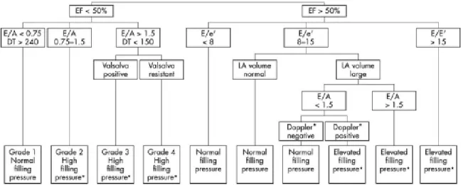
Doppler echocardiography became a fundamental tool in the evaluation of the diastolic function and of ventricular filling pressure [3]. The physiology of the diastole and diastolic dysfunction are seen in respect to different aspects. These are the cellular aspect (metabolic including relaxing), mechanical aspect (cavitary represented by the distensiblity or complacency) and clinical aspect (expressed by the signs and symptoms) can be studied and interpreted from the excellent synoptic state (Figure 1), proposed by OMMEN & NISHIMURA [7].
The patients are initially divided into two large subgroups in respect to the systolic function. As
mentioned, patients who already presented with systolic dysfunction have differing degrees of diastolic dysfunction characterized by Doppler echocardiography of the transmitral flow using the E/ A waves ratio and the E wave deceleration time. Thus, Grade 1 corresponds to alterations in the pattern of distensiblity and < A and Grade 2 to the pseudo-normal pattern which can already present signs of high filling pressures. Finally, Grades 3 and 4 correspond to a restrictive pattern which can be separated into reversible (Grade 3) and irreversible (Grade 4) by the Valsalva maneuver or reduction of the pre-load with significant prognostic implications [4,5]. On the other hand, patients with normal systolic function require a more detailed study, involving the E wave amplitude of the transmitral flow measured by Doppler echocardiography compared with the amplitude of the tissue e’ wave on the Doppler curve at the mitral ring. The left atrial volume separates the subgroup of patients with normal systolic function indexes (FE>50%) and intermediate indexes of diastolic dysfunction (E/e’= 8-15) and together with the E/A ratio <1.5 or >1.5 identifies patients with high ventricular filling pressures giving recognized clinical, prognostic and therapeutic relevance. The behavior of the left atrial volume post acute myocardial infarction in rats, studied by GELAPE et al. and published in this edition of the BJCVS on page 63, proved to be a sensitive indicator of the diastolic dysfunction.
P
BIBLIOGRAPHIC REFERENCES
1- Senni M, Tribouillay CM, Rodeheffer RJ, et al. Congestive heart failure in the community: a study of all incident cases in Olmsted County, Minnesota, in 1991. Circulation 1998; 98:2282-9.
2- Redfield MM, Jacobsen SJ, Burnett JC, et al. Burden of systolic and diastolic ventricular dysfunction in the community: Appreciating the scope of the heart failure epidemic. JAMA 2003; 289: 194-202.
3- Nishimura RA, Tajik AJ. Evaluation of diastolic filling of left ventricule in health and disease: Doppler echocardiography is the clinician’s Rosetta Stone. J Am Coll Cardiology 1997; 30: 8-18.
4- Xie GY, Berk MR, Smith MD, et al. Prognostic value of Doppler transmitral flow patterns in patients with congestive heart failure. J Am Coll Cardiology 1994; 24: 132-9.
5- Temporelli PL, Corra U, Imparato A, et al. Reversible restrictive left ventricular diastolic filling with optimized oral therapy predicts a more favorable prognosis in patients with chronic heart failure. J Am Coll Cardiology 1998; 31: 591-7.
6- Yamamoto K, Nishimura RA, Chaliki HP, et al. Determination of left ventricular filling pressure by Doppler echocardiography in patients with coronary artery disease: critical role of left ventricular systolic function. J Am Coll Cardiology 1997; 30: 1819-26. 7- Ommen SR, Nishimura RA. A clinical aproach to the
assessment of left ventricular diastolic function by Doppler echocardiography: Update 2003. Heart 2003; 89: 18-23.
Fig. 1- systematized evaluation of the diastolic function and filling pressures. Variables obtained from the bidimensional image and the Doppler echocardiography are utilized to immediately classify the diastolic function. In general, high filling pressures must be confirmed by several criteria such as: E/ e’, E/ Vp, difference dur-A, Valsalva control, TR speed, etc. [7].