Arq Neuropsiquiatr 2002;60(2-A):295-298
LUMBAR DISC HERNIATION ASSOCIATED WITH
SCOLIOSIS IN A 15-YEAR-OLD GIRL
Case report
Fernando Campos Gomes Pint o
1, Arthur W. Poet scher
2,
Faust o Ricardo Erba Quinhones
1, M ário Pena
2, M ário August o Taricco
2ABSTRACT - Intervertebral disc herniation is a rare condition in childhood and adolescence, although some cases have already been reported in the literature. We present the case of a 15 year-old-girl with low back pain and scoliosis. She had no previous history of trauma or collagen diseases. MRI showed L4-L5 and L5-S1 disc herniations and no further bone and structural changes. After two level discectomy, pain ceased and scoliosis improved, without further treatment. Based on her evolution and on what has already been reported in literature, we consider that scoliosis associated with disc herniation in young patients is most likely to be only an anthalgic position, not indicative of further structural changes.
KEY WORDS: lumbar disc herniation, low back pain, scoliosis, adolescence.
Hérnia de disco lombar associada a escoliose em uma jovem de 15 anos: relato de caso
RESUMO - Hérnia de disco intervertebral é condição rara em crianças e adolescentes. Alguns relatos isolados e algumas séries foram publicadas. Descrevemos o caso de uma paciente de 15 anos, sem antecedentes relevantes, que apresentou hérnia de disco intervertebral em dois níveis lombares (L4-L5 e L5-S1) associada a escoliose não estrutural que melhorou após a cirurgia, sem necessidade de órtese. Baseados neste caso e no que encontramos na literatura, acreditamos que escoliose associada a hérnia de disco em jovens é resultante apenas de posição antiálgica, não representando necessariamente alteração estrutural.
PALAVRAS-CHAVE: hérnia de disco lombar, escoliose, adolescente.
Division of Neurosurgery, Hospital das Clinicas, Faculdade de Medicina, Universidade de São Paulo, São Paulo SP, Brazil: 1Resident, 2Faculty.
Received 17 May 2001, received in final form 20 November 2001. Accepted 29 November 2001.
Dr. Fernando Campos Gomes Pint o - Rua Dr. Brasílio M achado 267/ 173 - 01230-010 São Paulo SP - Brazil. FAX 11 276 8367. E-mail: acapuano@nut ecnet .com.br
Intervertebral disc herniation is a rare condition in children and adolescents. Wahren (1946) was the first to report a case of a disc herniation surgery in a 12 years old boy. Since then, some single cases and series have been reported1-6. Less than 1% of
lum-bar disc herniation surgery occur in patients between 10 and 20 years old and only 0.5% of these discecto-mies are performed in patients under 16 years. Most of these patients (40% to 70%) have a history of traumatic injuries to the lumbar spine, usually rela-ted to sport activity, falls (higher than 1m) and mo-tor vehicle accidents. In patients under sixteen, about 2% of the cases occur at the L3-L4 level and 98% are distributed equally between L4-L5 and L5-S1 levels7.
Pain relieve posture is described in nearly 20% of pediatrics patients with lumbar disc herniation, whe-reby scoliosis is usually present, with the convexity turned to the affected side4.
We report the case of a 15-year-old girl, with no previous history of spinal disease, who presented with a non-traumatic intervertebral disc herniation in two lumbar levels (L4-L5 and L5-S1), associated with a non structural scoliosis, that improved after surgery, without need of further treatment.
CASE
296 Arq Neuropsiquiatr 2002;60(2-A)
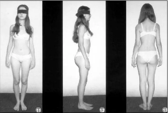
pain sensation in left L4, L5 and S1 dermatomes were re-duced. Lasègue sign (30º) and a crossed Lasègue sign (45º) were present. Plain X-rays showed a thoraco-lumbar scolio-sis with left convexity (Fig 1). On spine CT-scan and MRI films, a central L4-L5 and a paramedian right L5-S1 disc herniation were depicted (Fig 2).
As the patient did not improve with clinical manage-ment and physiotherapy, we decided for a two level dis-cectomy. After general anesthesia, we could already no-tice a significant reduction of the scoliosis. She was submit-ted to a left posterior approach to the L4-L5 and L5-S1 spa-ces. Discectomy was performed in both levels, when it became evident that there was an important anterior com-pression on L5 and S1 left nerve roots. Disc samples sho-wed cartilaginous tissue fragments with mixoid degen-eration.
Soon after surgery we could already observe an im-portant relieve of pain, scoliosis and weakness. One month after, the neurologic examination was normal, with a sig-nificant improvement of the scoliosis, also seen in plain X-ray films (Fig 1 and 3).
DISCUSSION
In childhood and adolescence, the signs and symptoms of root compression suggest the diagno-sis of tumors (osteoma and osteoblastoma), infec-tion or spondylolisthesis as the most likely. Due to its low incidence during the first and second decades of life, disc herniation should only be considered when the diseases above were ruled out. CT scan and MRI are also the best imaging methods to both show the disc herniation and exclude other
possi-Fig 1. (1), (2) On t his pre-op spinal plain X-rays w e can see a thoraco-lumbar scoliosis w ith the convexity on the left and ab-sence of lumbar spinal lordosis; (3), (4) The post-op spinal X-rays show s the improvement after surgery (1 month).
Arq Neuropsiquiatr 2002;60(2-A) 297
bilities. Disc herniation occurs when tears in the an-nulus fibrosus are frequent and significant enough to allow the nucleus pulposus to herniate. This tears are usually caused by aging and repeated uneven pressure distribution on axial loading.
Although during the first and second decades of life the nucleus pulposus is more expansible, disc herniation does not usually occur, due to the lack of tears in the annulus fibrosus at this age. However, in traumatic events, with heavy axial loading, or in pa-tients with collagen tissue diseases, the annulus fi-brosus may be damaged and disc herniation may follow. This explains why, in pediatric and adoles-cent patients, disc herniation is mostly related to traumatic events, collagen tissue diseases, congeni-tal weakness of the annulus fibrosus or spine mal-formations that cause inadequate pressure distribu-tion of axial loading. Our patient had no evidence of any of the conditions mentioned above.
Difficulty in walking, scoliosis and inability to perform anterior flexion of the trunk are frequent complaints of children and adolescents with lumbar disc herniation. Lumbar pain is the most common symptom; limitation of lumbar motility and Lasègue are the most common signs.
In seven patients described by Rugtveit4 , all of
them presented with difficulty to walk, scoliotic pos-ture, and inability to flex the trunk. It is important to notice that three of the seven patients harbored con-genital malformations. Two of them had six lumbar vertebrae and one, an asymmetric sacralization of the fifth lumbar vertebra. The unsatisfactory outcome he observed, was probably due to the long period of a symptoms before surgery.
Shillito5, during 37 years, operated on 20 patients
under fifteen years with disc herniation. Sixty-five percent of them were between 13 and 15 years old. Thirty-five percent related a growth spurt one year before the surgery and 60% were pubescent, like our patient, who also had a growth spurt recently.
Bradford and Garcia8 found microscopic
eviden-ces of degenerative changes in 8 of 25 patients youn-ger than 18 years submitted to discectomy. This could be the expression of a congenital defect of the an-nulus fibrosus leading to the nucleus pulposus her-niation. Shillito5 found ossification of the posterior
longitudinal ligament or of the annulus fibrosus in 8 of 20 patients under fifteen years. The microscopic study of our patient’s disc showed myxoid degen-eration.
The association between scoliosis and lumbar disc herniation is observed sometimes, although its
298 Arq Neuropsiquiatr 2002;60(2-A)
cal significance and pathophysiology are not well known. It is well established that children’s spines have better adaptive capacity, which helps to pro-tect the nervous tissue. An example of this could be scoliosis in patients with root compression, when he bends to the side contrary to the compression, causing an enlargement of the affected foramen and root release. Matsui et al.3 observed that 80% of the
patients with disc herniation and scoliosis had the convexity on the side of the root compression. Five of his patients were studied with MR, and it was evident that the scoliosis widened the foramen.
Grass et al.2 reported the youngest case, a
10-years-old girl with intervertebral disc herniation, re-lated to a fall from a low height, with unusual pre-sentation of a progressive structural scoliosis. She improved after surgery, with no need of orthesis. Our patient was 15 years older, pubescent, had no history of traumatic injury, but had a satisfactory evolution after surgery as well.
In the opinion of Rugtveit4, surgical treatment has
the same indications in children, adolescents and adults. Scoliosis, if present in patients in growth spurt, may worsen, if the intervertebral disc hernia-tion is not treated. Surgery results were excellent in all related cases, between 88% and 97%5,7,9-12.
The patient we describe in this report presented no one of the established predisposing factors but the relation with a growth spurt. A fast growing of the spine, muscles and ligaments, as well as a quick increase in height and weight, may lead to a tran-sient weakness of the disc or even an overload with inadequate load distribution. This could explain the disc herniation in pubescents in whom no other pre-disposing factor is found.
In conclusion, lumbar disc herniation in adoles-cents and children is not common and can be the cause of scoliosis and lumbar pain with or without sciatica. The scoliosis is not predictive of the level of disc herniation, but is suggestive of the affected side3.
A growth spurt may be the only predisposing factor for disc herniation in pubescents.An appropriated diagnostic investigation must be carried out, with spine MRI, in order to rule out other diseases. The presence of a disc herniation and the persistence of signs and symptoms are indications for surgical treat-ment.
Lumbar disc herniation can be alone the cause of scoliosis in children and adolescents. In this case the treatment of choice is discectomy.
REFERENCES
1. Basile R Jr. Hérnia de disco lombar em adolescentes: estudo crítico de cinco casos operados. Dissertação de Mestrado, Faculdade de Medicina da Universidade de São Paulo. São Paulo, 1979.
2. Grass JP, Dockendorff IB, Soto VA, Araya PH, Henriquez CM. Pro-gressive scoliosis with vertebral rotation after lumbar intervertebral disc herniation in a 10-year-old girl. Spine 1993;18:336-338. 3. Matsui H, Ohmori K, Kanamori M, Ishihara H, Tsuji H. Significance of
sciatic scoliotic list in operated patients with lumbar disc herniation. Spine 1998;23:338-342.
4. Rugtveit A. Juvenile lumbar disc herniations. Acta Orthop Scand 1966;37:348-356.
5. Shillito J Jr. Pediatric lumbar disc surgery: 20 patients under 15 years of age. Surg Neurol 1996;46:14-188.
6. Wahren H. Herniated nucleus pulposus in a child of twelve years. Acta Orthop Scand 1946;16:40-42.
7. Mason DE. Back pain in children. Pediatr Ann 1999;28:727-738. 8. Bradford DS, Garcia A. Herniations of the lumbar intervertebral disc
in children and adolescents. JAMA 1969;210:2045-2051.
9. Borgesen SE, Vang PS. Herniation of the lumbar intervertebral disc in children and adolescent. Acta Orthop Scand 1974;45:540-549. 10. Ebersold MJ, Quast LM, Bianco AJ. Results of lumbar discectomy in
the pediatric patient. J Neurosurg 1987;67:643-647.
11. Epstein AJ, Epstein NE, Marc J. Lumbar intervertebral disk herniation in teenage children, recognition and management of associated anoma-lies. Spine 1984;9:427-432.