INTRODUCTION
The sublingual glands are the smallest glands among the major salivary glands of the rat15,21. These glands are classified as mixed tubuloacinar glands which are predominantly of the mucous type, i.e., most of their terminal secretory units consist of typical mucous cells and are surrounded by relatively few peripheral serous cells forming
the serous demilunes4,6,8,9,17,19,25,26,30. The terminal secretory structures are connected to a highly branched duct system which starts with the inter-calated ducts, followed by the increasingly larger striated and excretory ducts, and terminating in a single wide-caliber duct which drains into the oral cavity, i.e., the main excretory duct21.
Morphometric characterization of sexual differences in the rat
sublingual gland
Caracterização morfométrica das diferenças sexuais na
glândula sublingual de rato
Marta da Cunha Lima* Dagoberto Sottovia-Filho** Tania Mary Cestari*** Rumio Taga****
* DDS; **PhD, Full Professor, Department of Biological Sciences; ***Graduate Student in Biological Science; Department of Biological Sciences; ****PhD, Chairman, Laboratory of Histology, Department of Biological Sciences – School of Dentistry of Bauru, University of São Paulo.
ABSTRACT: The presence of morphological differences in the sublingual gland of male and female adult rats was determined by morphometry. Absolute and relative glandular mass was 21% lower and 31% higher, respectively, in females than in males. The fractions of glandular volume occupied by the mixed acini, intercalated ducts and striated ducts did not differ significantly between genders; however, their absolute volume was respectively 29, 42 and 58% higher in males. Despite the differences in the volume of these morphological compartments, the number of cells did not differ significantly between genders, except for the excretory duct compartment, for which a larger number was observed in males. With respect to cell volume, 13, 33 and 47% higher volumes were observed in males for mucous acinar cells and striated and excretory duct cells, respectively, while a 38% higher volume of se-rous demilune cells was observed for females. The surface-to-volume ratio of acini and striated ducts was respec-tively 16 and 35% higher in females. Based on these results, we conclude that the sublingual gland of female rats possesses smaller acini, and shorter ducts whose caliber is narrower, smaller mucous acinar and larger serous cells than the ones found in the male gland, indicating the presence of sexual dimorphism as well as suggesting sexual differences in the quality of the secreted product.
DESCRIPTORS: Sublingual gland; Rats; Sex characteristics.
RESUMO: A ocorrência de diferenças morfológicas entre sexos na glândula sublingual de ratos adultos foi veri-ficada pela morfometria. As massas glandular absoluta e relativa das fêmeas foi, respectivamente, 21% menor e 31% maior que as dos machos. As frações de volume glandular ocupadas pelos ácinos mistos, ductos intercalares e ductos estriados não mostraram diferenças significantes entre sexos, no entanto, os seus volumes absolutos foram, respectivamente, 29%, 42% e 58% maiores nos machos. Apesar dessas diferenças nos volumes comparti-mentais, os seus conteúdos em número de células não apresentaram diferenças significantes entre sexos, exceto o compartimento dos ductos excretores, que mostrou maior número nos machos. Quanto ao volume celular, as células acinosas mucosas, as dos ductos estriados e as dos ductos excretores mostraram volumes, respectiva-mente, 13%, 33% e 47% maiores nos machos, e o das células das semiluas serosas foi 38% maior nas fêmeas. A relação superfície-volume dos ácinos e dos ductos estriados foi, respectivamente, 16% e 35% maior nas fêmeas. Baseados nos resultados obtidos, concluímos que as glândulas sublinguais das fêmeas exibem ácinos menores e ductos mais curtos e menos calibrosos e células acinosas mucosas menores e serosas maiores do que nos machos, indicando a ocorrência de dimorfismo sexual, e sugerindo que possa haver também diferenças na qualidade do produto secretado.
The clear predominance of mucous cells in this gland, which depend on a nervous stimulus for secretion of their content and respond to secre-tagogue-induced stimulation, makes this organ a useful model for the study of the biology of mucus secretion6.
Although our group has investigated various aspects of the postnatal development of the rat sublingual gland4,8,9,25,26, a study on its quantitative morphology in male and female adult animals is lacking, study which would permit the determi-nation of sexual dimorphism in this gland and the establishment of comparable morphometric parameters for future experimental research.
MATERIAL AND METHODS
Forty-four 120-day-old Wistar adult rats, ob-tained from the Central Animal House of the School of Dentistry of Bauru of the University of São Paulo, were used. The rats were divided into three groups of 12 (6 males and 6 females), 6 (3 males and 3 females) and 6 (3 males and 3 females), used for determination of morphometric dimensions, gland density and shrinkage caused by the histological procedures, respectively. The glands were always collected between 10:00 and 12:00 a.m. to avoid circadian variations. Before anesthesia, each rat was injected intraperitoneally with 1.25 atrophin solution (4 mg/kg body weight, Farmagricola S.A. Importação e Exportação, Mairiporã, SP, Brazil) to prevent salivary secretion. After five minutes, the rat was killed by excessive administration of chlorhydrate of ketamine and xylazine (Agribrands do Brasil Ltda., Paulínea, Brazil). The body mass of each animal was determined and sublingual glands were carefully removed and immediately weighed on an analytical scale. The glands were fixed in phosphate-buffered 10% formalin solution (Merck KGaA, Darmstadt, Germany) for one week at room temperature, rinsed overnight in running water, dehydrated in ethanol (Merck KGaA, Darm-stadt, Germany), cleared in xylene (Merck KGaA, Darmstadt, Germany) and embedded in Histosec (Merck KGaA, Darmstadt, Germany) (paraffin plus plastic resin) melted at 58°C.
Determination of the processed gland volume The volume of the processed gland (Vp) was calculated using the formula Vp = (m/δ) • Sf, where m = gland mass, δ = gland density and Sf = the correction factor for the shrinkage caused by his-tological processing. The gland density (δ) was de-termined by measuring 6 glands of the 2nd group
of rats using Mettler Toledo AR 261 Delta Rang scale (Mettler-Toledo GmbH, Greifensee, Switzer-land) containing accessories for determination of density, and the shrinkage caused by histological procedures was evaluated in 6 glands of the 3rd group of rats using the method proposed by Taga, Sesso24.
Stereologic measurements of volume density (Vvi), total volume (Vti), surface density (Svi), total surface (Sti), surface-to-volume ratio (s/vi) and absolute cell number (Ni) of each gland structure (i) and of nuclear density (ρni) of each cell type were taken.
These morphometric measurements were obtained using a Zeiss 8 X eyepiece containing a Zeiss II integration graticule (Carl Zeiss Jena GmbH, Jena, Germany) with 10 parallel lines and 100 points in quadrangular area, and a 100 X oil-immersion objective in an Olympus light micro-scope (Olympus America Inc., New York, USA). In 50 histological fields per animal, selected by sys-tematic sampling29, we counted: a) the number of points (Pi) over the images of each structure (i) and over the entire gland (P); b) the number of points over the nuclei (Pni) and the cytoplasm (Pcyti) of cells in each structure (i); c) the number of nu-clei (ni) of each gland structure; d) the number of intersections (Ii) of the contours of the structure with the lines of the graticule; and e) the number of intersections (c) of the nuclei with lines of the graticule. Given the processed gland volume (Vp), the distance between the lines of the graticules (d); the total area examined (A); the total length of the lines of the graticule used (L) and the thick-ness of the section (t), we calculated with the data obtained in the counts1,29: Vvi = Pi/P, Vti = Vvi Vp, Svi = 2Ii/L, Sti = Svi •Vp, s/vi = Svi/Vvi, Ni = (2ni •Vp)/A[(c/ni) • d + 2t], and ρni = Pni/Pni + Pcyti.
Determination of the volume of nucleus and cytoplasm of each cell type
thick-ness. The corrected volume densities of the nucle-us (ρni corr) and cytoplasm (ρcyti corr) are ρni corr = ρni/ Ko and ρcyti corr = 1 - ρni corr. Given the mean volume of the nucleus (Vni), the cytopasmic volume (Vcyti) was calculated by the relationship Vcyti = (Vni .
ρcyti corr)/ρni corr.
Statistical analysis
The results obtained for male rats were com-pared to those obtained for females by the Stu-dent’s t-test using the Sigma Stat – JadelTM Sci-entific software for Windows (Jadel Corporation, Chicago, USA) and the level of significance was set at 0.05. The volume densities were analyzed after arc sin transformation of the original data.
RESULTS
Body mass, absolute glandular mass, relative glandular mass and gland density data are shown in Table 1. Body mass and absolute glandular mass were respectively 40 and 21% lower in female rats compared to male animals, while relative glandu-lar mass was 31% higher in female rats. On the other hand, gland density did not differ between genders, and a mean density of 1.0545 mg/mm³ was observed for the two groups.
The morphometric dimensions of the different sublingual glandular structures obtained for male and female rats are shown in Table 2. A significant difference in volume density, i.e., the fraction of glandular volume occupied by each type of glandu-lar structure, was only observed between genders for the excretory ducts and stroma. This parameter is 1.9 times higher in males and 1.3 times higher in females, respectively. In absolute terms, the compartmental volume of acini, intercalated ducts, striated ducts and excretory ducts was respectively 29, 42, 58 and 155% higher in male rats. Despite these differences in compartmental volume, the absolute number of cells did not differ between groups for any of the epithelial compartments,
except for the excretory ducts where the number of cells was 193% larger in males compared to fe-males. However, the mean cell volume of mucous acinar cells and striated and excretory duct cells was significantly higher in male rats (13, 33, and 47%, respectively), while the mean cell volume of serous demilune cells was 38% higher in females. A significant difference in the total external surface of the various compartments was only observed for the excretory ducts, which was 74% greater in males. With respect to the surface-to-volume ratio, a 16 and 35% greater ratio was observed in females for the acini and striated ducts, respectively, indi-cating that these structures have smaller diameter in females.
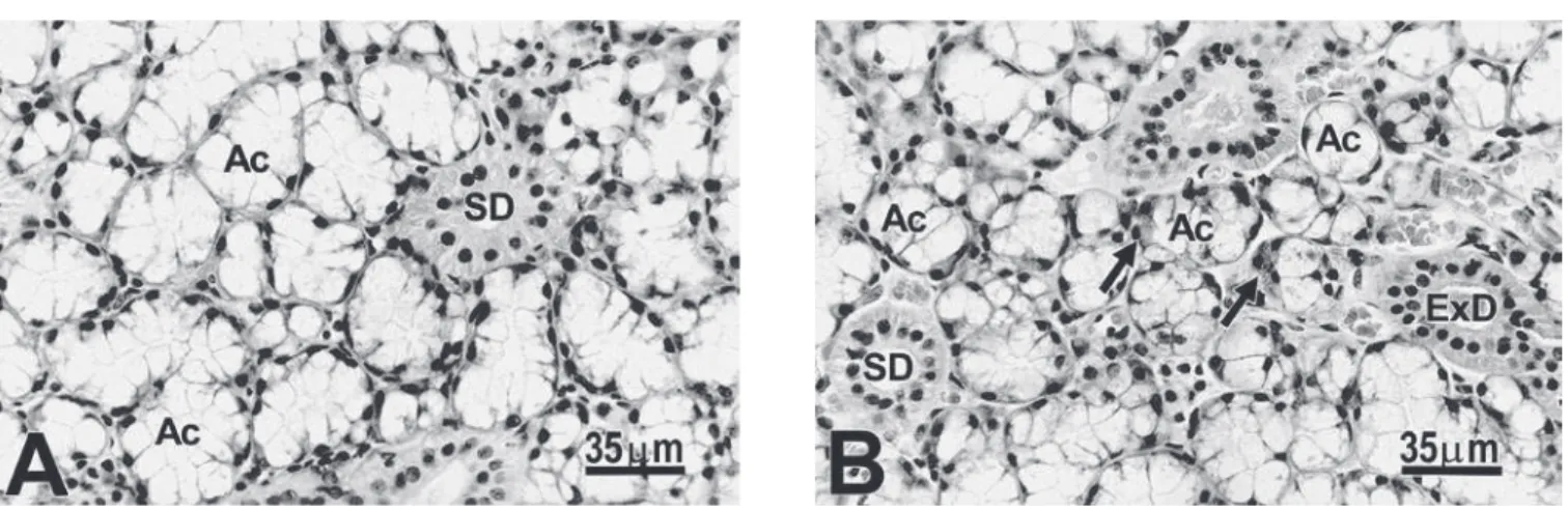
The differences in the mean volume of mu-cous, serous demilune and striated ducts cells and in the diameter of the acini and striated ducts between genders, could be morphologically con-firmed comparing Figures 1A and B.
DISCUSSION
Since the discovery by Lacassagne in 194011 that the submandibular gland of the laboratory mouse shows a marked sexual dimorphism char-acterized by the presence of a larger number of developed convoluted granular tubules in males than in females, this gland has been used as a model for the study of various aspects of second-ary sexual differences5,15,17,18.
Within this line of research, many other stud-ies have been carried out to determine the pres-ence of sexual differpres-ences in other major salivary glands of the mouse and, mainly, of other animal species (for a review and references therein, see Pinkstaff17). Based on these investigations, it be-came clear that sexual dimorphism in other mouse glands or in the salivary glands of other animals, if present, was not as conspicuous as in the mouse submandibular gland to be characterized by sim-ple morphological analysis of histological sections,
TABLE 1 - Body mass, absolute glandular mass, relative glandular mass and gland density obtained for male and female rats.
Parameter Male Female Statistical probability
Body mass (g) 362.33 ± 9.645* 217.31 ± 6.0287 p < 0.01 Absolute gland mass (mg) 78.49 ± 1.7297 61.64 ± 2.366 p < 0.01 Relative gland mass** 0.22 ± 0.007 0.28 ± 0.017 p < 0.01 Gland density (mg/mm³) 1.056 ± 0.002 1.053 ± 0.001 p > 0.05
but required other more sensitive methods for the characterization of these differences10,14,16,22. Among these methods, stereology has attracted attention due to its sensitivity and precision, permitting the determination of volumetric, superficial, linear and numerical dimensions of different structures and/ or cells present in an organ1.
In this respect, previous studies carried out in
our laboratory using this methodology have per-mitted, for example, the demonstration that sexual dimorphism in the mouse submandibular gland is not restricted to the convoluted granular tubules but is also present in the acini, intercalated ducts, striated ducts, excretory ducts and stroma15; that no sexual dimorphism is present in the granular or striated ducts of the rat submandibular gland23, TABLE 2 - Morphometric dimensions of the different sublingual gland structures obtained for male and female
rats.
Parameter Male Female Statistical probability
Volume density (%)
Mixed acini 82.51 ± 0.897 81.65 ± 0.861 p > 0.05
Intercalated duct 0.56 ± 0.059 0.49 ± 0.082 p > 0.05
Striated duct 3.61 ± 0.587 2.87 ± 0.293 p > 0.05
Excretory duct 2.88 ± 0.480 1.49 ± 0.341 p < 0.05
Stroma 10.44 ± 0.541 13.37 ± 1.104 p < 0.05
Total volume (mm³)
Mixed acini 51.59 ± 1.510 40.01 ± 1.432 p < 0.01
Intercalated duct 0.34 ± 0.035 0.24 ± 0.032 p < 0.05
Striated duct 2.23 ± 0.338 1.41 ± 0.164 p < 0.05
Excretory duct 1.81 ± 0.318 0.71 ± 0.137 p < 0.05
Stroma 6.40 ± 0.277 6.67 ± 0.693 p > 0.05
Cell number (× 105)
Mucous cells 112.87 ± 15.127 113.95 ± 3.353 p > 0.05
Serous demilune cells 56.62 ± 2.564 50.15 ± 3.806 p > 0.05 Intercalated duct cells 3.51 ± 0.571 2.32 ± 0.334 p > 0.05
Striated duct cells 10.31 ± 1.381 9.12 ± 0.905 p > 0.05
Excretory duct cells 8.02 ± 1.149 2.74 ± 0.293 p < 0.05
Cell volume (µm3)
Mucous cells 1,870.78 ± 68.970 1,650.21 ± 64.802 p < 0.05 Serous demilune cells 635.27 ± 16.726 876.41 ± 76.767 p < 0.05 Intercalated duct cells 285.77 ± 38.547 215.37 ± 45.187 p > 0.05 Striated duct cells 1,053.53 ± 56.567 792.115 ± 49.965 p < 0.05 Excretory duct cells 982.11 ± 60.65 667.59 ± 54.368 p < 0.05
Total surface (cm²)
Mixed acini 33.80 ± 1.367 30.77 ± 1.586 p > 0.05
Intercalated duct 0.52 ± 0.124 0.38 ± 0.090 p > 0.05
Striated duct 1.34 ± 0.171 1.22 ± 0.172 p > 0.05
Excretory duct 0.75 ± 0.087 0.43 ± 0.0515 p < 0.01
Surface-to-volume ratio (cm²/cm³)
Mixed acini 659.30 ± 38.38 766.66 ± 24.397 p < 0.05
Intercalated duct 1,836.37 ± 197.930 1,810.56 ± 17.962 p > 0.05
Striated duct 620.07 ± 52.25 835.84 ± 65.089 p < 0.01
Excretory duct 457.85 ± 62.700 561.44 ± 72.596 p > 0.05
while sexual differences exist in the acini, interca-lated ducts, excretory ducts and stroma4; and that in the mouse parotid gland the acini and interca-lated ducts are more developed in males20.
With respect to the sublingual gland of ro-dents, evidence for the occurrence of sexual di-morphism detected solely by histochemical meth-ods has only been provided for the mouse22 and hamster10, while no study is available regarding the presence of sexual differences in the rat sub-lingual gland.
With respect to its structure, the rat sublin-gual gland consists predominantly of mucous cells arranged in tubuloacinar structures which are surrounded by few serous cells arranged in demi-lunes6,17. The mucous acinar cells produce and secrete sublingual mucins or mucous glycopro-teins13,28, while the serous demilune cells secrete common salivary protein 1 (CSP-1), and neonatal submandibular gland secretory proteins B (SMGB) and D (SMGD)2,3,7,12. The innervation of these cells is mainly parasympathetic27 and they rapidly re-spond to stimulation with secretagogues6.
In order to exclude alterations in the results due to seasonal influences, all rats used in the present study came from a lot born on the same day and thus were of the same age. Mean body mass of the female rats used here was 40% lower than that of male animals, while absolute glandular mass showed a significant difference of only 21%. In this respect, relative glandular mass was 31% higher in females, i.e., females had larger glands than males in relation to their body mass.
Relative volumetric analysis showed no sig-nificant differences in volume density between genders for the acini, intercalated ducts or
stri-ated ducts, indicating a similar volumetric ratio for these structures in males and females. A sig-nificant difference in volume density was only ob-served for the excretory duct and stroma compart-ments, which were higher in males and females, respectively.
The absolute volume of all morphological com-partments, except for the stroma, was higher in male rats. These differences are due to the fact that glandular mass was higher in males, and that the fractions of glandular volume occupied by the acini and intercalated and striated ducts were the same in males and females.
It should be noted that the absolute volume of each compartment depends on the number of cells and on their volume. An interesting aspect is the fact that the number of cells of the acini, intercalated ducts or striated ducts did not differ between genders, suggesting that the differences in the absolute volume of these compartments were related to variations in the individual vol-ume of these cells. This hypothesis was confirmed for mucous acinar cells and striated duct cells, which presented 13 and 33% higher cell volumes in males, respectively. With respect to the serous demilune cells, a 38% higher cell volume was ob-served for females.
The surface-to-volume ratio, a relative mor-phometric parameter related to the shape and size of the structure, was found to be 16 and 35% higher in females for the acini and striated ducts, respectively. Since the shape of these structures is similar in the two genders, the differences observed indicate that the female acini and striated ducts have a smaller diameter than the same structures in males.
CONCLUSION
The present results led us to conclude that the sublingual gland of female rats possesses smaller mixed acini and shorter and narrower-caliber stri-ated ducts, smaller mucous acinar cells and larger serous demilune cells than the male gland, indicat-ing the presence of sexual dimorphism as well as suggesting sexual differences in the quality of the
REFERENCES
1. Aherne W, Dunnil MS. Morphometry. London: Edward Arnold; 1982.
2. Ball WD, Hand AR, Johnson AO. Secretory proteins as markers for cellular phenotypes in rat salivary glands. Dev Biol 1988;125:265-79.
3. Ball WD, Hand AR, Moreira JE. A neonatal secretory protein associated with secretion granule membranes in developing rat salivary glands. J Histochem Cytochem 1991;39:1693-706.
4. Barbosa DB, Hassunuma RM, Taga R. Estudo morfo-métrico e bioquímico do crescimento das glândulas sub-linguais do rato durante a vida pós-natal. Rev Ciênc Bi-omed 1997;18:83-94.
5. Chretien M. Action of testosterone on the differentiation and secretory activity of a target organ: the submaxillary gland of the mouse. Int Rev Cytol 1977;50:333-96. 6. Culp DJ, Graham LA, Latchney LR, Hand AR.Rat
sub-lingual gland as a model to study glandular mucous cell secretion. Am J Physiol 1991;260:1233-44.
7. Girard LR, Castle AM, Hand AR, Castle JD, Mirels L. Characterization of common salivary protein 1, a product of rat submandibular, sublingual, and parotid glands. J Biol Chem 1993;268:26592-601.
8. Hassunuma RM, Taga R. Allometric study of the postnatal development of the rat sublingual glands. Okajimas Folia Anat Jpn 1996;73:265-71.
9. Hernandes R, Bassi WE, Stipp ACM, Taga R. Estudo es-tereológico dos ácinos de glândulas sublinguais de ratos jovens e adultos. Rev Ciên Biomed 1995;15:31-9. 10. Kronman JH. Hamster salivary gland sexual dimorphism.
I. Protein histochemical study. J Dent Res 1963;42:123-7.
11. Lacassagne A. Dimorphisme sexuel de la glande sous-maxillaire chez la souris. C R Soc Biol 1940;133:180-1. 12. Mirels L, Miranda JA, Ball WD. Characterization of the
rat salivary gland B1-immunoreactive proteins. Biochem J 1998;330:437-44.
13. Moschera J, Pigman W. The isolation and characteriza-tion of rat sublingual mucus-glycoprotein. Carbohydr Res 1975;40:53-67.
14. Mudd BD, White SC. Sexual dimorphism in the rat sub-mandibular gland. J Dent Res 1975;54:193.
15. Pardini LC, Taga R. Stereological study of the sexual di-morphism in mouse submandibular glands. Okajimas Folia Anat Jpn 1996;73:119-24.
16. Pinkstaff CA. Sexual dimorphism of the miniature pig
submandibular glands. Am J Anat 1972;135:371-9. 17. Pinkstaff CA. The cytology of salivary glands. Int Rev Cytol
1980;63:141-261.
18. Raynaud J. Controle hormonal de la glande sous-maxil-laire de la souris. Bull Biol Fr Belg 1960,94:399-523. 19. Redman RS, Ball WD. Cytodifferentiation of secretory cells
in the sublingual gland of the prenatal rat: a histologi-cal, histochemical and ultrastructural study. Am J Anat 1978;153:367-89.
20. Ribeiro TT, Cestari TM, Taga R. Morphometric dimensions of the mouse parotid glands of both sexes. Ital J Anat Embryol 2001;106:27-34.
21. Riva A, Tandler B, Testa Riva F. Ultrastructural ob-servations on human sublingual gland. Am J Anat 1988;181:385-92.
22. Spicer SS. The use of various cationic reagents in histo-chemical differentiation of mucopolysaccharides. Am J Clin Pathol 1961;36:393-407.
23. Taga R, Achôa AS, Pardini LC. Estudo estereológico dos ductos granulosos e estriados de glândulas submandibu-lares de ratos machos e fêmeas. Rev FOB 1994;2:22-7. 24. Taga R, Sesso A. Avaliação do número de células de órgãos
pela dosagem bioquímica de DNA em homogeneizados e por contagem direta através de métodos morfométricos. Ciênc Cult 1978;30:1232-6.
25. Taga R, Sesso A. Postnatal development of the rat sublin-gual glands. A morphometric and radioautographic study. Arch Histol Cytol 1998;61:417-26.
26. Taga R, Sesso A. Ultrastructure of the rat sublingual gland during period of high proliferative activity in postnatal development. Braz J Morphol Sci 2002;19:55-62. 27. Templeton D, Thulin A. Secretory, motor and vascular
effects in the sublingual gland of the rat caused by auto-nomic nerve stimulation. Q J Exp Physiol Cogn Med Sci 1978;63:59-66.
28. Watson GE, Latchney LR, Luo W, Hand AR, Culp DJ. Biochemical and immunological studies and assay of rat sublingual mucins. Arch Oral Biol 1997;42:161-72. 29. Weibel ER. Stereological principles of morphometry in
electron microscopic cytology. Int Rev Cytol 1969;26:235-302.
30. Wolff MS, Mirels L, Lagner J, Hand AR. Development of the rat sublingual gland: a light and electron microscopic immunocytochemical study. Anat Rec 2002;266:30-42.
Received for publication on Feb 10, 2003 Sent for alterations on Nov 18, 2003 Accepted for publication on Dec 05, 2003 saliva secreted by the gland.