Antigen-Specific Interferon-Gamma
Responses and Innate Cytokine Balance in
TB-IRIS
Odin Goovaerts1,2*, Wim Jennes1, Marguerite Massinga-Loembe´1,3,4, Ann Ceulemans1, William Worodria5,6,7, Harriet Mayanja-Kizza5,6, Robert Colebunders8,9, Luc Kestens1,2, for the TB-IRIS Study Group
1.Department of Biomedical Sciences, Institute of Tropical Medicine, Antwerp, Belgium,2.Department of Biomedical Sciences, University of Antwerp, Antwerp, Belgium,3.Centre de Recherches Me´dicales de Lambare´ne´ (CERMEL), Albert Schweitzer Hospital, Lambare´ne´, Gabon,4.Institut fu¨r Tropenmedizin, Universita¨t Tu¨bingen, Tu¨bingen, Baden-Wu¨rttemberg, Germany,5.Makerere University College of Health Sciences, Kampala, Uganda,6.Infectious Diseases Institute, Makerere University College of Health Sciences, Kampala, Uganda,7.Infectious Diseases Network for Treatment and Research in Africa (INTERACT), Kampala, Uganda,8.Department of Clinical Sciences, Institute of Tropical Medicine, Antwerp, Belgium,9.Epidemiology and Social Medicine, University of Antwerp, Antwerp, Belgium
Abstract
Background:Tuberculosis-associated immune reconstitution inflammatory syndrome (TB-IRIS) remains a poorly understood complication in HIV-TB patients receiving antiretroviral therapy (ART). TB-IRIS could be associated with an
exaggerated immune response to TB-antigens. We compared the recovery of IFNc
responses to recall and TB-antigens and explored in vitro innate cytokine production in TB-IRIS patients.
Methods:In a prospective cohort study of HIV-TB co-infected patients treated for TB before ART initiation, we compared 18 patients who developed TB-IRIS with 18
non-IRIS controls matched for age, sex and CD4 count. We analyzed IFNcELISpot
responses to CMV, influenza, TB and LPS before ART and during TB-IRIS. CMV and LPS stimulated ELISpot supernatants were subsequently evaluated for
production of IL-12p70, IL-6, TNFaand IL-10 by Luminex.
Results: Before ART, all responses were similar between TB-IRIS patients and
non-IRIS controls. During TB-IRIS, IFNcresponses to TB and influenza antigens
were comparable between TB-IRIS patients and non-IRIS controls, but responses to CMV and LPS remained significantly lower in TB-IRIS patients. Production of innate cytokines was similar between TB-IRIS patients and non-IRIS controls. However, upon LPS stimulation, IL-6/IL-10 and TNFa/IL-10 ratios were increased in TB-IRIS patients compared to non-IRIS controls.
OPEN ACCESS
Citation:Goovaerts O, Jennes W, Massinga-Loembe´ M, Ceulemans A, Worodria W, et al. (2014) Antigen-Specific Interferon-Gamma Responses and Innate Cytokine Balance in TB-IRIS. PLoS ONE 9(11): e113101. doi:10.1371/ journal.pone.0113101
Editor:Yoshihiko Hoshino, National Institute of Infectious Diseases, Japan
Received:June 6, 2014
Accepted:October 19, 2014
Published:November 21, 2014
Copyright:ß2014 Goovaerts et al. This is an open-access article distributed under the terms of theCreative Commons Attribution License, which permits unrestricted use, distribution, and repro-duction in any medium, provided the original author and source are credited.
Data Availability:The authors confirm that all data underlying the findings are fully available without restriction. All relevant data are within the paper and its Supporting Information files.
Funding:The TB-IRIS project was funded by an EC FP6 Specific Targeted Research Project (STREP) grant LSHP-CT-2007-037659-TBIRIS. This work was done in association with the Infectious Diseases Network for Treatment and Research in Africa (INTERACT). The funders had no role in study design, data collection and analysis, decision to publish, or preparation of the manuscript.
Conclusion:TB-IRIS patients did not display excessive IFNcresponses to
TB-antigens. In contrast, the reconstitution of CMV and LPS responses was delayed in the TB-IRIS group. For LPS, this was linked with a pro-inflammatory shift in the innate cytokine balance. These data are in support of a prominent role of the innate immune system in TB-IRIS.
Introduction
Together with the HIV pandemic there has been a global increase in the number of tuberculosis (TB) infections [1]. An estimated 14 million individuals are dually
infected with HIV and TB worldwide [2]. Despite recent WHO recommendations
for early antiretroviral therapy (ART) [3], treatment is started at late stages of HIV infection in many developing countries [4]. This puts patients at increased risk of developing tuberculosis-associated immune reconstitution inflammatory
syn-drome (TB-IRIS) during ART [5]. TB-IRIS presents in up to 25% of HIV-TB
patients as worsening symptoms of TB during ART, despite a favourable response to TB-treatment (hence the name ‘‘paradoxical TB-IRIS’’) [6]. This complication typically occurs within the first 2 months after starting ART, with the majority occurring within the first few weeks [7]. TB-IRIS poses a significant diagnostic challenge to physicians and reliable laboratory markers to help detect this syndrome are urgently needed [8].
Known risk factors of TB-IRIS include a low CD4 count, high TB-antigen burden and short interval between initiation of TB treatment and ART [9,10]. The pathogenesis of TB-IRIS remains largely unclear, although there are clear signs of tissue-destructive inflammation during immune reconstitution (reviewed in [11,12]). This process could involve an amplified immune response to TB bacilli or their residual antigens [11,13]. Early research suggested that TB-IRIS development was linked to elevated T-helper type 1 (Th1) responses to TB-antigens. Indeed, studies in TB-IRIS patients reported elevated IFNcresponses to
several TB-associated antigenic compounds such as purified protein derivative (PPD), 6 kDa early secretory antigenic target (ESAT-6) and 10 kDa culture filtrate antigen (CFP-10) [14–18]. However, elevated IFNcresponses to TB-antigens are
often also seen in HIV-TB patients who do not develop TB-IRIS [19–21], casting doubt on the causal role of Th1 cells in TB-IRIS pathogenesis. It has been suggested that disturbances in the innate immune system [22] or in the interplay
between the innate and adaptive immune system [11] could drive TB-IRIS
pathogenesis. This is supported by repeated findings of elevated levels of IL-6 and TNFa, among other innate cytokines, during TB-IRIS [14,16,23–26].
in the innate and the adaptive arm of the immune system. However, the roles of
antigen-specific IFNcproduction and TLR stimulation in TB-IRIS remain to be
completely elucidated.
In this study, we aimed to assess the ART related recovery of IFNcresponses in
TB-IRIS patients. To this end, we determined specific responses to a number of TB and recall antigens in a well matched selection of TB-IRIS patients and controls from a large prospective cohort [28]. In addition, we explored the possible innate component of TB-IRIS by studying cytokine production upon TLR4 stimulation with lipopolysaccharide (LPS). We report a disturbed
reconstitution of the IFNcrecall response, without an excessive IFNcresponse to
TB-antigens. In addition, we observed a pro-inflammatory shift in the innate cytokine balance upon LPS stimulation during TB-IRIS, providing evidence of the involvement of the innate immune system in TB-IRIS.
Materials and Methods
Study population
Patients were recruited in a prospective observational study on paradoxical TB-IRIS at Mulago Hospital, Kampala, Uganda, between January 2008 and July 2010 as described previously [9,28,29]. The present study is based on HIV-TB co-infected adults who were being treated for active TB infection and put on ART within 2 months after starting TB treatment (median 6 weeks prior to ART). All HIV-patients received non-nucleoside reverse transcriptase inhibitor-based ART according to Ugandan national guidelines and were monitored for paradoxical TB-IRIS development during at least 3 months. Blood samples were taken before ART initiation (pre-ART) and when patients were diagnosed for TB-IRIS (IRIS event). Patients without IRIS-related symptoms were used as non-IRIS controls and had samples taken pre-ART, at 2 weeks and 1 month on ART. To compare with the expected immunocompetent antigen responses, two additional groups of HIV-uninfected subjects were recruited. One group was receiving treatment for active TB for less than 4 months (HIV2TB+ controls) while the other had no
clinical signs of active TB (HIV2TB2 controls). HIV-uninfected subjects had
samples taken only once.
Patient selection and matching
A large majority of patients from our cohort developed TB-IRIS within 1 month after ART initiation. To limit heterogeneity among TB-IRIS patients, we included only patients who developed TB-IRIS within one month on ART. This stricter selection of patients reduces potential bias due to differences in kinetics and immunopathology between early- and late-onset TB-IRIS. Selected TB-IRIS patients with PBMCs available pre-ART and at occurrence of IRIS were matched 1 by 1 with non-IRIS controls for sex, baseline CD4 count (+/215 CD4 cells/mm3)
control time point for their paired IRIS event, were selected at either 2 weeks or 1 month on ART. This selective pairing allowed for a matched time on ART between TB-IRIS patients and non-IRIS controls.
Definitions
Mycobacterium tuberculosis infection was diagnosed according to the TB/HIV WHO guidelines [30]. The diagnostic evaluation for TB included: clinical examination, chest X-rays and abdominal ultrasounds, sputum smear microscopy for acid-fast bacilli and mycobacterial culture of sputum, aspirate or effusion if available. TB-IRIS cases were classified by a committee of two co-authors (RC and WW) after reviewing all suspected TB-IRIS cases evaluated according to the International Network for the Study of HIV-associated IRIS (INSHI) clinical case-definition for resource limited settings [5]. This evaluation included: a symptom questionnaire, detailed physical examination to confirm TB-IRIS and exclude alternative causes and comparison of a second chest X-ray and abdominal ultrasound scan to the patient’s baseline examination. Biological TB-IRIS samples collected when patients developed new or worsening symptoms following initiation of ART. These symptoms included at least 1 major criterion, such as enlarged lymph nodes, or 2 minor criteria, such as fever and cough.
ELISpot assays
PBMCs were collected from TB-IRIS patients and cryopreserved in liquid nitrogen. Samples were consequently thawed and IFNcresponses to antigens were
measured by ELISpot (Diaclone SAS, Besanc¸on Cedex, France) according to the manufacturer’s instructions. In brief, PBMCs were cultured overnight in an antibody-coated ELISpot filter plate at 200,000 cells/200 ml in RMPI+2.5% human
serum in the presence of either 10 mg/ml cytomegalovirus (CMV) lysate (Institut
Virion\Serion GmbH, Wu¨rzburg, Germany), 5 mg/ml whole influenza virus
antigen (H3N2 A/Sydney/5/97, National Institute for Biological Standards and Control, Hertfordshire, Great Britain), 10 mg/ml PPD (Statens Serum Institute,
Copenhagen, Denmark), 10 mg/ml recombinant ESAT-6 (Statens Serum Institute,
Copenhagen, Denmark), and 10 mg/ml recombinant CFP-10 (a kind gift from
Lionex Diagnostics and Therapeutics, Braunschweig, Germany). In addition,
100 ng/ml LPS (E. coli O55:B5, Sigma-Aldrich BVBA, Diegem, Belgium) was
included separately as a strong inducer of innate cytokines. Medium only and
5 mg/ml staphylococcal enterotoxin B (SEB, Sigma-Aldrich BVBA, Diegem,
Belgium) were used as negative and positive controls, respectively. The number of
IFNcspot-forming cells (SFC) per 106PBMCs was determined using the ELISpot
Cytokine multiplex assay
ELISpot supernatants from PBMCs stimulated with CMV and LPS were stored at 280
˚
C until further use. Samples were thawed within 1 hour prior to analysing cytokine levels by using the Bio-Plex human cytokine assay kits (Bio-Rad Laboratories NV-SA, Nazareth, Belgium) according to the manufacturer’s instructions. We measured in vitro levels of IL-6, TNFaand IL-10 as a representation of innate pro- and anti-inflammatory cytokine production. In addition, IL-12p70 was measured as a link between changes in IFNcresponses andmonocyte function. Samples were diluted 26 for TNFaand IL-10 and 106 for
IL-6 to allow optimal detection of both high and low cytokine concentrations.
Ethical considerations
The study was approved by the Research Committee of the Infectious Diseases Institute (IDI), the ethical review board of Makerere University School of Medicine, the Uganda National Council of Science and Technology and by the institutional review board of the Institute of Tropical Medicine of Antwerp and the Ethics Committees of the Faculties of Medicine of the University of Antwerp. Written informed consent was obtained from all study participants.
Statistical analysis
Differences between paired patients and changes in concentration over time for each patient were analysed using the Wilcoxon signed-rank test for paired data. Differences between HIV-infected and HIV-uninfected patients were analysed using the Mann-Whitney U test. Correlations were calculated using the Spearman’s rank correlation. Statistics were performed using SPSS software (version 17.0) or GraphPad Prism (version 5) with significance level set at p,0.05. Because of the hypothesis driven nature of this study, no correction for multiple testing was applied [31,32].
Results
Study population
A total of 18 TB-IRIS patients were paired with 18 non-IRIS controls (Table 1). TB-IRIS patients and non-IRIS controls did not differ regarding baseline viral load, TB treatment duration prior to ART, or ART duration prior to IRIS event or corresponding control time point. Twenty two HIV uninfected controls were
included in the study, of whom 9 were being treated for TB (HIV-TB+controls)
and 13 did not have symptoms of TB (HIV-TB- controls). Neither HIV-TB+ nor
Antigen-specific IFN
c
ELISpot responses in HIV-TB patients
developing TB-IRIS
We examined IFNcresponses to TB-antigens, CMV, Influenza and LPS (a
well-known TLR4 antagonist) in order to assess the antigen-specific and non-specific
immunity in TB-IRIS patients compared to non-IRIS, HIV-TB+ and
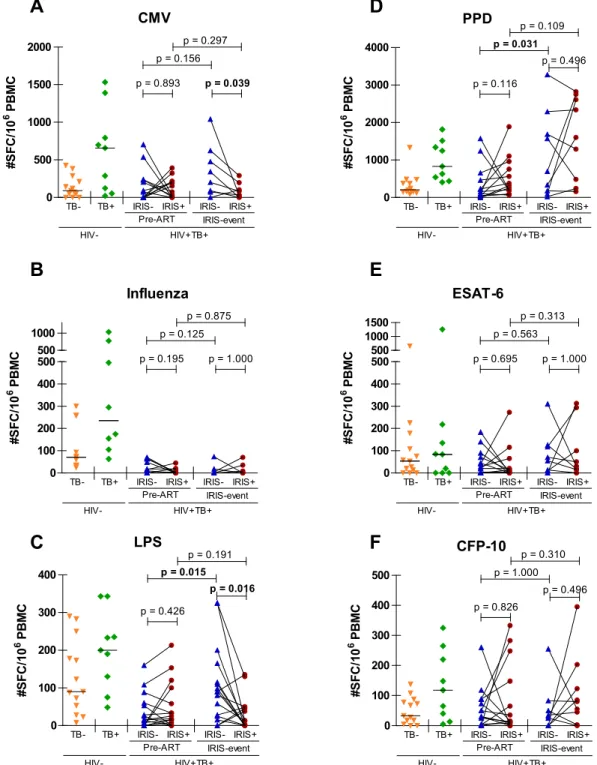
HIV-TB-controls (Figure 1). At pre-ART, we observed no difference in IFNcresponses
between TB-IRIS patients and non-IRIS controls for any of the antigens tested.
Both groups showed a diminished IFNc response to PPD, CMV, Influenza and
LPS when compared to HIV-TB+ controls (p#0.027) and to LPS and influenza
when compared to HIV-TB- controls (p#0.015).
IFNcresponses to the different TB-antigens were also similar between TB-IRIS
patients and non-IRIS controls at IRIS event (Figure 1). Both groups showed signs of higher PPD responses compared to before ART. This increase was statistically significant for non-IRIS controls (p50.031), while TB-IRIS patients
showed a trend (p50.109). Both groups also showed signs of higher PPD
responses compared HIV-TB- controls, yielding significant results for TB-IRIS patients only (p50.007). In contrast, IFNcresponses to CMV and LPS were
significantly lower in TB-IRIS patients compared to non-IRIS controls (p50.039
and p50.016, respectively). TB-IRIS patients also showed lower CMV responses
compared to HIV-TB+controls (p50.027), and lower LPS responses compared to
HIV-TB+ and HIV-TB- controls (p,0.001 and p50.001, respectively). In
contrast to TB-IRIS patients, non-IRIS controls showed a significantly recovered response to LPS compared to pre-ART (p50.015), which was still lower compared to HIV-TB+ controls (p50.034). Responses to influenza were not significantly different between TB-IRIS patients and non-IRIS controls. Responses to influenza did not recover within the first weeks of ART for either TB-IRIS patients or for Table 1.Characteristics of TB-IRIS patients and matched controls.
Characteristics TB IRIS (n518) non-IRIS controls (n518) pa
Prior to ART
Male sex, n (%) 8 (44) 8 (44) 0.815b
Age (years) 34 (33–43) 40 (31–44) 0.760
CD4 (cell/mm3) 19 (11–119) 23 (8–93) 0.477
TB treatment duration prior to ART (days) 36 (23–56) 46 (23–58) 0.663
Viral load (log copies/ml)c 5.4 (5.3–5.8) 5.53 (5.1–5.6) 0.646
During ART
Days between start of ART and TB-IRIS/control event 14 (12–22) 16 (14–28) 0.297
Values are shown as median values with interquartile range. The level of significance was set to p,0.05 for all tests. TB-IRIS patients were matched to non-IRIS controls for baseline CD4 count, age and sex.
a
Wilcoxon signed-rank test.
b
Mc Nemar test for binominal data.
c
n510.
Figure 1. Antigen-specific IFNcresponses in TB-IRIS patients and controls.Dots on these graphs represent IFNcspot-forming cells per 106PBMCs in
TB-IRIS patients (IRIS+) and non-IRIS controls (IRIS-) after stimulation with CMV lysate (A), influenza antigen A (B), LPS (C), PPD (D), ESAT-6 (E) and CFP-10 (F). Dots connected with full lines represent matched patient pairs. Horizontal full lines represent median values for HIV-TB+controls and HIV-TB-controls. Horizontal capped lines represent statistical comparisons between matched patients or between time points. The level of significance was set to p,0.05. A Wilcoxon signed-rank test was used to calculate p values between matched HIV patients and time points. Due to limited availability of viable PBMCs and pairwise exclusion, the number of patients across antigens and time points differed. Number of patients pre-ART were; 13 (A), 10 (B), 16 (C), 14 (D), 14 (E) and 14 (F). Number of patients during IRIS event were; 8 (A), 6 (B), 16 (C), 9 (D), 9 (E) and 9 (F).
non-IRIS controls compared to those in HIV-TB+ (p#0.003) and
HIV-TB-controls (p#0.015) at every time point.
Innate cytokine production upon stimulation with CMV, PPD and
LPS
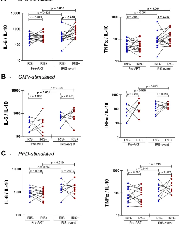
We next explored whether production of innate cytokines could provide an explanation for the delayed reconstitution of CMV and LPS responses in TB-IRIS patients. To this end, we analysed levels of the pro-inflammatory cytokines IL-12,
TNFa and IL-6 and of the anti-inflammatory cytokine IL-10 in ELISpot
supernatants after stimulation with CMV, PPD and LPS, but found no differences between TB-IRIS patients and non-IRIS controls (Table 2). Systemic inflamma-tion can be marked by increases in pro-inflammatory cytokines as well as by decreases in anti-inflammatory cytokines and their net balance has been shown to determine the clinical outcome of inflammation [33–37]. Accordingly, we
explored IL-6/IL-10 and TNFa/IL-10 ratios in TB-IRIS patients and non-IRIS
controls (Figure 2). After LPS stimulation, we observed significantly higher IL-6/ IL-10 (p50.025) and TNFa/IL-10 (p50.047) ratios in TB-IRIS patients compared to non-IRIS controls (Figure 2A). These differences corresponded to significant increases of IL-6/IL-10 (p50.005) and TNFa/IL-10 (p50.004) ratios from pre-ART to IRIS event in TB-IRIS patients but not in non-IRIS controls. Interestingly, IL-6/IL-10 and TNFa/IL-10 ratios correlated inversely with IFNcELISpot responses to LPS in the total HIV+/TB+ patient population (Figure 3).
Stimulation with CMV or PPD did not yield significant differences in cytokine ratios between patient groups (Figure 2B&C) nor resulted in significant
correlations between IFNcELISpot responses and cytokine ratios (Figure 3B&C).
Discussion
TB-IRIS could involve an amplified immune response to TB bacilli or their residual antigens [11,13]. Here, we aimed to study the recovery of antigen-specific responses of HIV-TB patients who developed TB-IRIS during ART. To that end,
we compared IFNcresponses of PBMCs to a panel of TB-associated antigens and
recall antigens between TB-IRIS patients and non-IRIS controls, matched for CD4 count, age and sex. In addition, we explored innate cytokine responses to CMV,
PPD and LPS. We report a disturbed reconstitution of the IFNcresponse to CMV
and LPS during TB-IRIS, without an excessive IFNcresponse to TB-antigens. In
addition, we observed a pro-inflammatory shift in the innate cytokine balance upon LPS stimulation during TB-IRIS.
TB-IRIS patients showed lower IFNcresponses to the recall antigen CMV and
to LPS during TB-IRIS, compared to non-IRIS controls. Unlike CMV and LPS, however, we did not observe significantly increased or decreased IFNcresponses
not in agreement with previous reports of elevated PPD-responses during TB-IRIS [14–18]. The magnitude of IFNc ELISPOT responses to TB-antigens has been
associated to the antigen-load, duration of TB-treatment and the extent of immune-suppression in HIV patients [38–40]. All three of these factors have been identified as risk-factors for TB-IRIS which could potentially influence
immunological measurements, consequently leading to discrepancies across studies. In the present study, we minimize this potential bias by directly comparing (time-)matched patients under very similar clinical conditions, possibly explaining the discrepancy with studies that observed elevations in TB-associated responses. Together, our results suggest that the conditions TB-associated with TB-IRIS, such as a high TB-antigen load and persistent inflammation [41–
44], could disturb the reconstitution of recall responses (to CMV in this case), rather than cause an excessive TB-specific IFNcresponse. In addition, the lowered
response to LPS could point towards a role of the innate immune system. Since we previously reported a significant rise in IL-6 plasma levels from pre-ART to TB-IRIS event, which lead to higher IL-6 levels compared to non-IRIS controls [26], we next hypothesised that TB-IRIS might result from an aberrant innate immune response. In contrast to our previous in vivo measurements, we found no significant differences in the in vitro production of pro- and anti-inflammatory cytokines between matched TB-IRIS patients and non-IRIS controls after exposure to CMV, PPD and LPS. In addition to absolute levels, however, the Table 2.In vitro cytokine production in response to CMV, PPD or LPS stimulation in TB-IRIS patients and non-IRIS controls.
Pre-ART IRIS event Change over time (pa)
TB-IRIS Control pa TB-IRIS Control pa TB-IRIS Control
CMV stimulationb
IL-12p70 (pg/ml) 1.3 (0.6–2.4) 0.9 (0.6–2.4) 0.866 2.6 (1.1–3.1) 2.2 (1.1–3.1) 0.725 0.225 0.107 IL-6 (pg/ml) 207.9 (64.1–1633.8) 103.3 (32.2–523.6) 0.735 1491.5 (118.9–2538.4) 933.7 (295.1–3126.7) 0.311 0.237 0.028 TNFa(pg/ml) 135.4 (37.9–338.8) 49.2 (2.1–145.3) 0.753 279.5 (52.4–354.0) 159.8 (100.65–380.05) 0.462 0.091 0.091
IL-10 (pg/ml) 0.6 (0.5–1.0) 0.2 (0.2–1.2) 0.237 1.4 (0.4–1.9) 1.6 (0.35–4.15) 1.000 0.753 0.043
PPD stimulationc
IL-6 (ng/ml) 16.3 (9.0–35.9) 15.9 (8.9–19.1) 0.345 54.2 (11.1–71.1) 33.1 (27.7–40.6) 0.594 0.128 0.116 TNFa(ng/ml) 1.3 (0.7–2.0) 1.0 (0.6–2.5) 0.221 3.8 (1.2–9.1) 3.4 (1.0–6.2) 0.953 0.043 0.463
IL-10 (pg/ml) 16.8 (10.5–47.0) 17.3 (8.5–30.8) 0.152 16.3 (7.8–47.5) 23.5 (8.8–30.1) 0.859 0.735 0.463
LPS stimulationd
IL-12p70 (pg/ml) 4.8 (2.3-7.6) 3.9 (3.0–5.9) 0.453 4.7 (3.1–9.5) 5.4 (3.0–7.5) 0.277 0.315 0.900 IL-6 (ng/ml) 25.7 (10.9–58.6) 22.2 (10.2–36.6) 0.215 22.9 (12.2–106.8) 25.6 (13.7–42.6) 0.352 0.156 0.245 TNFa(ng/ml) 4.9 (1.8–9.3) 4.2 (2.0–7.6) 0.352 5.0 (2.0–25) 7.5 (3.0–13.4) 0.469 0.307 0.363
IL-10 (pg/ml) 72.6 (31.4–304.6) 60.0 (23.2–215.2) 0.408 47.1 (18.9–121.6) 97.8 (41.2–207.7) 0.215 0.334 0.683
Values are shown as median values with interquartile range. The level of significance was set to p,0.05 for all tests.
a
Wilcoxon signed-rank test. Due to limited availability of PBMCs, the number of patients differed;
b
7 at pre-ART and 8 during IRIS event,
c
13 at pre-ART and 9 during IRIS event,
d
16 at pre-ART and 16 during IRIS event.
Figure 2. Pro- to anti-inflammatory ratios of innate cytokine production in TB-IRIS.Dots on these graphs represent cytokine ratios in PBMC supernatants after stimulation with LPS (A), CMV (B) and PPD (C). Dots connected with full lines represent matched pairs of TB-IRIS patients (IRIS+) with non-IRIS controls (IRIS-). Horizontal capped lines represent statistical comparisons between matched patients or between time points. A, pre-ART n516, IRIS event n516; B, pre-ART n57, IRIS event n58; C, pre-ART n513, IRIS event n59. A Wilcoxon signed-rank test was used to calculate p values between HIV patients. The level of significance was set to p,0.05.
balance between pro-inflammatory and anti-inflammatory cytokine levels has been shown to drive systemic inflammation [33–37]. Accordingly, we found that TB-IRIS was associated with a pro-inflammatory shift in the IL-6/IL-10 and TNFa/IL-10 ratios after stimulation with LPS, but not CMV or PPD. An increase in the IL-6/IL-10 ratio, caused by a decrease in IL-10, has previously been Figure 3. Correlation of cytokine ratios to IFNcresponses.Graphs represent the correlation between IFNcresponses and IL-6/IL-10 or TNFa/IL-10 ratios
after stimulation with LPS (A), CMV (B) or PPD (C). Dots represent both TB-IRIS patients and non-IRIS controls during TB-IRIS or corresponding control time point. The level of significance was set to p,0.05.
associated to the severity of systemic inflammatory response syndrome in patients with sepsis [34]. Of note, the IL-10 levels upon LPS stimulation in the current study were also somewhat lower during TB-IRIS. Although this difference did not reach statistical significance, it could have shifted the cytokine balance towards the pro-inflammatory side. In line with our findings, TB-IRIS patients from our cohort have previously been shown to have a pro-inflammatory monocyte-gene expression profile that is also perturbed in pattern recognition receptor pathways [45]. Another study previously reported elevated TNFaproduction during IRIS upon TLR2 stimulation with lipomannan, without an equivalent rise in IL-10 [16]. In the present study, we report a similar cytokine imbalance in the TLR4 branch of innate cytokine production. One could therefore argue that a disturbed equilibrium between pro- and anti-inflammatory cytokine-production upon TLR stimulation is implicated in the high degree of inflammation seen in TB-IRIS. This preferential involvement of TLRs in TB-IRIS could also explain why no cytokine shifts were observed after CMV- or PPD-stimulation, since these antigens preferentially induce an adaptive response via the major histocompatibility complex class II/T cell receptor pathway.
Intriguingly, the unbalanced cytokine ratios were inversely correlated to the LPS-induced IFNcresponses. This finding is somewhat contradictory, given the
fact that IL-6, TNFaand IFNcare all pro-inflammatory cytokines. However, IL-6 and TNFaare directly produced by monocytes after LPS stimulation, while IFNc is not. Rather, LPS-induced IFNcoriginates from T cells and NK cells in response
to monocyte derived cytokines [46,47]. We hypothesise that the cytokine ratio shifts result from aberrant monocyte behaviour in these patients, given the association of monocyte dysfunction with chronic HIV infection [48,49]. In fact, aberrant monocyte behaviour has previously been suggested to play a role in TB-IRIS [50] and is in line with our hypothesis on the role of TLRs in TB-IRIS. Since the balance between monocyte-derived cytokines seems to be disturbed upon TLR stimulation, we speculate that this negatively affected the subsequent induction of IFNc.
One limitation of our study was that patients and controls were not tested for CMV and influenza infection status, potentially complicating the interpretation of the CMV and influenza ELISPOT responses that we measured. This may not have been a major problem for CMV, which reaches a high seroprevalence in sub Saharan Africa [51,52]. While influenza is clearly present in sub Saharan Africa [53,54], limited availability of epidemiological data make it difficult to assess the expected recall response of our patients to this antigen.
Taken together, our data provide no evidence of an excessive IFNcresponse to
TB-associated antigens or other common recall antigens during TB-IRIS. In fact, TB-IRIS was associated with a disturbed reconstitution of the IFNcresponses to
Acknowledgments
The authors thank the study participants and the study team: D. Mazakpwe, K. Luzinda, P. Lwanga, M. Nakuya, C.O Namujju, C. Ahimbisibwe, J. Namaganda, A. Andama, E. Bazze and H. Kisembo. We thank N. Pakker and the data staff of the Infectious Diseases Network for Treatment and Research in Africa
(INTERACT) for assistance with data monitoring and management.
Lead author of the TB-IRIS study group: Luc Kestens ([email protected]), Institute of Tropical Medicine, Antwerp, Belgium. Other members of the TB-IRIS study group are as follows: Institute of Tropical Medicine, Antwerp, Belgium: Robert Colebunders, Marguerite Massinga Loembe´; Infectious Disease Institute, Kampala, Uganda: Harriet Mayanja, William Worodria; Joint Clinical Research Centre: Harriet Mayanja; Universite´ Libre de Bruxelles, Belgium: Francoise Mascart; VIB, Brussels, Belgium and Vrije Universiteit Brussel, Brussels, Belgium: Rafael van den Bergh; Institut Pasteur de Lille, France: Camille Locht; Academic Medical Centre, Department of Global Health and Amsterdam Institute for Global Health and Development, Amsterdam, The Netherlands: Peter Reiss, Frank Cobelens, Pascale Ondoa, Nadine Pakker; INTERACT, Kampala, Uganda: Roy Mugerwa, Harriet Mayanja, Nadine Pakker, William Worodria.
Author Contributions
Conceived and designed the experiments: OG LK. Performed the experiments: OG AC. Analyzed the data: OG WJ. Wrote the paper: OG WJ LK. Project management: MML WW HMK RC LK. Study physicians: WW RC.
References
1. Sharma SK, Mohan A, Kadhiravan T (2005) HIV-TB co-infection: epidemiology, diagnosis & management. Indian J Med Res 121: 550–567.
2. Getahun H, Gunneberg C, Granich R, Nunn P (2010) HIV infection-associated tuberculosis: the epidemiology and the response. Clin Infect Dis 50 Suppl 3:: S201–S207. 10.1086/651492 [doi].
3. WHO(2013) Consolidated guidelines on the Use of Antiretroviral Drugs for Treating and Preventing HIV Infection.
4. Kigozi IM, Dobkin LM, Martin JN, Geng EH, Muyindike W, et al. (2009) Late-disease stage at presentation to an HIV clinic in the era of free antiretroviral therapy in Sub-Saharan Africa. J Acquir Immune Defic Syndr 52: 280–289. 10.1097/QAI.0b013e3181ab6eab [doi].
5. Meintjes G, Lawn SD, Scano F, Maartens G, French MA, et al. (2008) Tuberculosis-associated immune reconstitution inflammatory syndrome: case definitions for use in resource-limited settings. Lancet Infect Dis 8: 516–523.
6. Muller M, Wandel S, Colebunders R, Attia S, Furrer H, et al. (2010) Immune reconstitution inflammatory syndrome in patients starting antiretroviral therapy for HIV infection: a systematic review and meta-analysis. Lancet Infect Dis 10: 251–261. S1473-3099(10)70026-8 [pii]; 10.1016/S1473-3099(10)70026-8 [doi].
8. Worodria W, Conesa-Botella A, Kisembo H, McAdam KP, Colebunders R(2009) Coping with TB immune reconstitution inflammatory syndrome. Expert Review of Respiratory Medicine 3: 147–152.
9. Conesa-Botella A, Loembe MM, Manabe YC, Worodria W, Mazakpwe D, et al. (2011) Urinary lipoarabinomannan as predictor for the tuberculosis immune reconstitution inflammatory syndrome. J Acquir Immune Defic Syndr. 10.1097/QAI.0b013e31823801de [doi].
10. Worodria W, Massinga-Loembe M, Mazakpwe D, Luzinda K, Menten J, et al.(2011) Incidence and predictors of mortality and the effect of tuberculosis immune reconstitution inflammatory syndrome in a cohort of TB/HIV patients commencing antiretroviral therapy. J Acquir Immune Defic Syndr 58: 32–37. 10.1097/QAI.0b013e3182255dc2 [doi].
11. Barber DL, Andrade BB, Sereti I, Sher A(2012) Immune reconstitution inflammatory syndrome: the trouble with immunity when you had none. Nat Rev Microbiol 10: 150–156. nrmicro2712 [pii]; 10.1038/ nrmicro2712 [doi].
12. Wilson EM, Sereti I (2013) Immune restoration after antiretroviral therapy: the pitfalls of hasty or incomplete repairs. Immunol Rev 254: 343–354. 10.1111/imr.12064 [doi].
13. French MA (2007) Disorders of immune reconstitution in patients with HIV infection responding to antiretroviral therapy. Curr HIV/AIDS Rep 4: 16–21.
14. Bourgarit A, Carcelain G, Martinez V, Lascoux C, Delcey V, et al.(2006) Explosion of tuberculin-specific Th1-responses induces immune restoration syndrome in tuberculosis and HIV co-infected patients. AIDS 20: F1–F7. 10.1097/01.aids.0000202648.18526.bf [doi]; 00002030-200601090-00001 [pii].
15. Tan DB, Yong YK, Tan HY, Kamarulzaman A, Tan LH, et al.(2008) Immunological profiles of immune restoration disease presenting as mycobacterial lymphadenitis and cryptococcal meningitis. HIV Med 9: 307–316.
16. Tan DB, Lim A, Yong YK, Ponnampalavanar S, Omar S, et al. (2011) TLR2-induced cytokine responses may characterize HIV-infected patients experiencing mycobacterial immune restoration disease. AIDS 25: 1455–1460. 10.1097/QAD.0b013e328348fb18 [doi].
17. Narita M, Ashkin D, Hollender ES, Pitchenik AE (1998) Paradoxical worsening of tuberculosis following antiretroviral therapy in patients with AIDS. Am J Respir Crit Care Med 158: 157–161.
18. Vignesh R, Kumarasamy N, Lim A, Solomon S, Murugavel KG, et al.(2013) TB-IRIS After Initiation of Antiretroviral Therapy Is Associated With Expansion of Preexistent Th1 Responses Against
Mycobacterium tuberculosis Antigens. J Acquir Immune Defic Syndr 64: 241–248. 10.1097/ QAI.0b013e31829f6df2 [doi].
19. Meintjes G, Wilkinson KA, Rangaka MX, Skolimowska K, van Veen K, et al.(2008) Type 1 helper T cells and FoxP3-positive T cells in HIV-tuberculosis-associated immune reconstitution inflammatory syndrome. Am J Respir Crit Care Med 178: 1083–1089.
20. Elliott JH, Vohith K, Saramony S, Savuth C, Dara C, et al.(2009) Immunopathogenesis and diagnosis of tuberculosis and tuberculosis-associated immune reconstitution inflammatory syndrome during early antiretroviral therapy. J Infect Dis 200: 1736–1745. 10.1086/644784 [doi].
21. Tieu HV, Ananworanich J, Avihingsanon A, Apateerapong W, Sirivichayakul S, et al. (2009) Immunologic markers as predictors of tuberculosis-associated immune reconstitution inflammatory syndrome in HIV and tuberculosis coinfected persons in Thailand. AIDS Res Hum Retroviruses 25: 1083–1089. 10.1089/aid.2009.0055 [doi].
22. Oliver BG, Elliott JH, Price P, Phillips M, Saphonn V, et al.(2010) Mediators of innate and adaptive immune responses differentially affect immune restoration disease associated with Mycobacterium tuberculosis in HIV patients beginning antiretroviral therapy. J Infect Dis 202: 1728–1737. 10.1086/ 657082 [doi].
23. Grant PM, Komarow L, Lederman MM, Pahwa S, Zolopa AR, et al.(2012) Elevated Interleukin 8 and T-Helper 1 and T-Helper 17 Cytokine Levels Prior to Antiretroviral Therapy in Participants Who Developed Immune Reconstitution Inflammatory Syndrome During ACTG A5164. J Infect Dis. jis604 [pii]; 10.1093/infdis/jis604 [doi].
25. Tadokera R, Meintjes G, Skolimowska KH, Wilkinson KA, Matthews K, et al. (2011) Hypercytokinaemia accompanies HIV-tuberculosis immune reconstitution inflammatory syndrome. Eur Respir J 37: 1248–1259. 09031936.00091010 [pii]; 10.1183/09031936.00091010 [doi].
26. Goovaerts O, Jennes W, Massinga-Loembe M, Ceulemans A, Worodria W, et al.(2013) LPS-binding protein and IL-6 mark paradoxical tuberculosis immune reconstitution inflammatory syndrome in HIV patients. PLoS One 8: e81856. 10.1371/journal.pone.0081856 [doi]; PONE-D-13-33780 [pii].
27. Fukuda T, Matsumura T, Ato M, Hamasaki M, Nishiuchi Y, et al.(2013) Critical roles for lipomannan and lipoarabinomannan in cell wall integrity of mycobacteria and pathogenesis of tuberculosis. MBio 4: e00472–12. mBio.00472-12 [pii]; 10.1128/mBio.00472-12 [doi].
28. Worodria W, Massinga-Loembe M, Mayanja-Kizza H, Namaganda J, Kambugu A, et al. (2011) Antiretroviral treatment-associated tuberculosis in a prospective cohort of HIV-infected patients starting ART. Clin Dev Immunol 2011: 758350. 10.1155/2011/758350 [doi].
29. Worodria W, Menten J, Massinga-Loembe M, Mazakpwe D, Bagenda D, et al. (2012) Clinical spectrum, risk factors and outcome of immune reconstitution inflammatory syndrome in patients with tuberculosis-HIV coinfection. Antivir Ther. 10.3851/IMP2108 [doi].
30. World Health Organisation (WHO)(2012) Improving the diagnosis and treatment of smear-negative pulmonary and extrapulmonary tuberculosis among adults and adolescents. Recommendations for HIV-prevalent and resource-contstrained settings.
31. Streiner DL, Norman GR(2011) Correction for multiple testing: is there a resolution? Chest 140: 16–18. 140/1/16 [pii]; 10.1378/chest.11-0523 [doi].
32. Perneger TV(1998) What’s wrong with Bonferroni adjustments. BMJ 316: 1236–1238.
33. Antoniades CG, Berry PA, Wendon JA, Vergani D(2008) The importance of immune dysfunction in determining outcome in acute liver failure. J Hepatol 49: 845–861. S0168-8278(08)00506-0 [pii]; 10.1016/j.jhep.2008.08.009 [doi].
34. Taniguchi T, Koido Y, Aiboshi J, Yamashita T, Suzaki S, et al. (1999) Change in the ratio of interleukin-6 to interleukin-10 predicts a poor outcome in patients with systemic inflammatory response syndrome. Crit Care Med 27: 1262–1264.
35. van Dissel JT, van Langevelde P, Westendorp RG, Kwappenberg K, Frolich M (1998) Anti-inflammatory cytokine profile and mortality in febrile patients. Lancet 351: 950–953.
S0140-6736(05)60606-X [pii]; 10.1016/S0140-S0140-6736(05)60606-X [doi].
36. Gogos CA, Drosou E, Bassaris HP, Skoutelis A(2000) Pro- versus anti-inflammatory cytokine profile in patients with severe sepsis: a marker for prognosis and future therapeutic options. J Infect Dis 181: 176–180. JID990857 [pii]; 10.1086/315214 [doi].
37. Walley KR, Lukacs NW, Standiford TJ, Strieter RM, Kunkel SL (1996) Balance of inflammatory cytokines related to severity and mortality of murine sepsis. Infect Immun 64: 4733–4738.
38. Millington KA, Innes JA, Hackforth S, Hinks TS, Deeks JJ, et al. (2007) Dynamic relationship between IFN-gamma and IL-2 profile of Mycobacterium tuberculosis-specific T cells and antigen load. J Immunol 178: 5217–5226. 178/8/5217 [pii].
39. Aiken AM, Hill PC, Fox A, McAdam KP, Jackson-Sillah D, et al.(2006) Reversion of the ELISPOT test after treatment in Gambian tuberculosis cases. BMC Infect Dis 6: 66. 1471-2334-6-66 [pii]; 10.1186/ 1471-2334-6-66 [doi].
40. Karam F, Mbow F, Fletcher H, Senghor CS, Coulibaly KD, et al.(2008) Sensitivity of IFN-gamma release assay to detect latent tuberculosis infection is retained in HIV-infected patients but dependent on HIV/AIDS progression. PLoS One 3: e1441. 10.1371/journal.pone.0001441 [doi].
41. Antonelli LR, Mahnke Y, Hodge JN, Porter BO, Barber DL, et al.(2010) Elevated frequencies of highly activated CD4+T cells in HIV+patients developing immune reconstitution inflammatory syndrome. Blood 116: 3818–3827. blood-2010-05-285080 [pii]; 10.1182/blood-2010-05-285080 [doi].
42. Lawn SD, Bekker LG, Miller RF(2005) Immune reconstitution disease associated with mycobacterial infections in HIV-infected individuals receiving antiretrovirals. Lancet Infect Dis 5: 361–373.
44. Beishuizen SJ, Geerlings SE (2009) Immune reconstitution inflammatory syndrome: immunopathogenesis, risk factors, diagnosis, treatment and prevention. Neth J Med 67: 327–331.
45. Tran HT, Van Den Bergh R, Vu TN, Laukens K, Worodria W, et al.(2014) The role of monocytes in the development of Tuberculosis-associated Immune Reconstitution Inflammatory Syndrome.
Immunobiology 219: 37–44. S0171-2985(13)00139-3 [pii]; 10.1016/j.imbio.2013.07.004 [doi].
46. Le J, Lin JX, Henriksen-DeStefano D, Vilcek J(1986) Bacterial lipopolysaccharide-induced interferon-gamma production: roles of interleukin 1 and interleukin 2. J Immunol 136: 4525–4530.
47. Raices RM, Kannan Y, Sarkar A, Bellamkonda-Athmaram V, Wewers MD(2008) A synergistic role for IL-1beta and TNFalpha in monocyte-derived IFNgamma inducing activity. Cytokine 44: 234–241. S1043-4666(08)00687-X [pii]; 10.1016/j.cyto.2008.08.004 [doi].
48. Pulliam L, Sun B, Rempel H(2004) Invasive chronic inflammatory monocyte phenotype in subjects with high HIV-1 viral load. J Neuroimmunol 157: 93–98. S0165-5728(04)00338-8 [pii]; 10.1016/
j.jneuroim.2004.08.039 [doi].
49. Dudhane A, Conti B, Orlikowsky T, Wang ZQ, Mangla N, et al.(1996) Monocytes in HIV type 1-infected individuals lose expression of costimulatory B7 molecules and acquire cytotoxic activity. AIDS Res Hum Retroviruses 12: 885–892.
50. Van Den Bergh R, Vanham G, Raes G, De Baetselier P., Colebunders R(2006) Mycobacterium-associated immune reconstitution disease: macrophages running wild? Lancet Infect Dis 6: 2–3.
51. Gompels UA, Larke N, Sanz-Ramos M, Bates M, Musonda K, et al.(2012) Human cytomegalovirus infant infection adversely affects growth and development in maternally HIV-exposed and unexposed infants in Zambia. Clin Infect Dis 54: 434–442. cir837 [pii]; 10.1093/cid/cir837 [doi].
52. Njeru DG, Mwanda WO, Kitonyi GW, Njagi EC(2009) Prevalence of cytomegalovirus antibodies in blood donors at the National Blood Transfusion Centre, Nairobi. East Afr Med J 86: S58–S61.
53. World Health Organisation (WHO)(2011) Influenza update.