ORIGINAL
ARTICLE
553
Incidence and risk factors of immune reconstitution
inflammatory syndrome in HIV-TB coinfected patients
Authors Dibyendu De1 Rathindra Nath Sarkar2 Sibaji Phaujdar1 Kuntal Bhattacharyya1 Hare Krishna Pal1
1MBBS; Postgraduate Trainees, Department of Medicine, Medical College, Kolkata, India
2MSc in Medicine; Professor, Department of Medicine, Medical College, Kolkata, India
Submitted on: 05/03/2011 Approved on: 07/17/2011
Correpondence to: Dibyendu De Tentulberia, Garia, Kolkata
West Bengal India, 700084 [email protected]
We declare no conflict of interest.
©2011 Elsevier Editora Ltda. All rights reserved. ABSTRACT
Tuberculosis is one of the leading causes of development of Immune reconstitution inflamma-tory syndrome (IRIS) in HIV patients receiving antiretroviral therapy (ART). Objective: To determine the incidence of IRIS in HIV-TB coinfected patients, and to find out the possible risk factors associated with IRIS. Materials and Methods: Study commenced with 96 patients adhered to standard antitubercular therapy (ATT) and ART without defaultering, and followed up for six months. Result: The mean (± SD) CD4 count and CD4 percentage at baseline was 59.16 (± 24.63) per mm3 and 4.59% (± 1.73) respectively. Only 18.75% developed IRIS after 57.05 (± 14.12) days of initiation of ART. Extrapulmonary tuberculosis was the most significant factor associated with IRIS (83.33%) than those without IRIS (44.87%) (p = 0.0032). Specifi-cally, tubercular lymphadenitis (38.88%, p = 0.0364) and disseminated tuberculosis (33.33%, p = 0.0217) were significantly associated with IRIS. The other risk factors associated with ap-pearance of IRIS were higher CD4 count (p = 0.0212) at three months after initiation of ART and increment of CD4 count (p = 0.0063) and CD4 percentage (p = 0.0016) during this pe-riod. The major manifestations of IRIS were fever (40%), followed by lymphadenitis (38%). The mortality rate in IRIS was not higher than those without IRIS. Conclusion: Patients with extrapulmonary tuberculosis, especially tubercular lymphadenitis, were more likely to develop IRIS and fever was associated in most of them. Higher increment of CD4 count may indicate development of IRIS in presence of new or worsening tuberculosis lesion.
Keywords: HIV; tuberculosis; immune reconstitution inflammatory syndrome; CD4 lympho-cyte count.
INTRODUCTION
Human immunodeficiency virus (HIV) in-fection today is one of the major causes of mortality and morbidity globally. Association of tuberculosis (TB) coinfection with HIV poses greater therapeutic challenge. In devel-oping nations, patients often present late with advanced HIV infection along with different opportunistic infections, most commonly tuberculosis.1 Tuberculosis in HIV-infected
patients often presents with diagnostic dif-ficulties, with increased chance of negative sputum smear results, atypical chest X-ray findings, negative tuberculin skin test, lack of classical granuloma formation in late stage and also presence of more fre-quent extrapulmonary tuberculosis. With the advent of highly active antiretrovi-ral therapy (HAART), incidence rate of im-mune reconstitution inflammatory syndrome
(IRIS) also raised significantly. Classically IRIS is defined as “occurrence or manifestation of new or existing opportunistic infection with-in six weeks to six months after with-initiation of antiretroviral therapy (ART), with associated increase in CD4 count”.2 Any
opportunis-tic infection may present with IRIS though tuberculosis (TB) is the major cause. Oth-er important manifestations include CMV retinitis, Pneumocystis jiroveci pneumonia, herpes simplex infection, varicella zoster virus infection, cryptococcal meningitis, hep-atitis B, hephep-atitis C infection and even some auto-immune diseases.3 The main features
554
space occupying lesions etc.4,5 It is hypothesized that
pro-inflammatory cytokines produced excessively in response to systemic bacterial lipopolysacharide, non-specifically act on latent mycobacterial antigens leading to clinical deterioration and paradoxical worsening of inflammatory responses.6 In most of the cases, IRIS
re-gress spontaneously without any significant mortality but sometimes it may cause worsening clinical scenario with respiratory failure and tracheal compression by enlarged lymph nodes resulting in death. It should be differentiated from exacerbation of tuberculosis due to drug resistance or non-compliance, appearance of new tubercular lesion or other opportunistic infection due to advancement of HIV. Till date, there is lack of clinical data about oc-currence of IRIS in HIV and TB coinfected patients in Indian population. Therefore, we conducted a cross-sectional study in a tertiary care set-up to find out prevalence and risk factors of IRIS in HIV-TB coinfected patients.
MATERIAL AND METHODS
Recently diagnosed HAART naive HIV-infected pa-tients, who had clinical, radiological or microbiological evidence of active TB, were selected for our study. Inclu-sion criteria were as follows:
Recently diagnosed HIV positive by ELISA method. (According to NACO guideline, a symptomatic sus-pected HIV-infected person is diagnosed HIV positive when the serum sample is reactive with at least two out of three different ELISA kit).2
Patient having active tuberculosis diagnosed by clinical features, radiological appearances, presence of granuloma in aspiration material, or microscopy and/or culture positive acid-fast bacilli (AFB).
Receiving antitubercular therapy (ATT) from Revised National Tuberculosis Control Programme (RNTCP).
Started ART after 15 days of ATT as per protocol fol-lowed in the institution.
All the patients were followed up for the period of six months. The patients with non-compliance with either ATT or ART during the follow-up period were excluded from the study. The diagnosis of IRIS was done using the pre-set criteria as follows:
The patient had to be on ART with rising CD4 count. Prior to starting ART, the patient must have docu-mented radiological, histological or microbiologically confirmed tuberculosis.
Prior to starting ART, patient must have improve-ment of tuberculosis by antitubercular therapy.
Following initiation of ART, the patients must have appearance of new symptoms or re-appearance of previ-ous symptoms.
The patients must have spontaneous resolution of symptoms with continuation of same treatment.
Our primary objective was to determine the incidence of IRIS in HIV-TB coinfected patients. Secondarily we tried to find out the possible risk factors for emergence of IRIS. The patients were divided into two groups according to develop-ment of IRIS or not. The demographic, clinical features, site of tuberculosis, presence of other opportunistic infection, base-line and follow-up CD4 count were measured and compared between the two groups. The statistical analysis was done, using chi-square test and Student’s t test using SPSS ver-16 to determine the significance of the variables. A p-value < 0.05 was taken to be statistically significant. A total of 96 patients, who were attending Medicine outdoor and indoor of Medical College Kolkata from May 2009 to April 2010, who met the eligibility criteria, were included in this study. The study was approved by the institutional ethical committee. The neces-sary informed consent was taken from all the patients.
RESULTS
Out of 96 patients suffering from both HIV and tubercu-losis, 84 (87.5%) of them male, only 18 (18.75%) patients developed IRIS (Table 1). The initial mean (± SD) CD4 count of these patients was 59.16 (± 24.63) per mm3 and initial mean
CD4 percentage was 4.59% (± 1.73). The baseline parameters of the patients developing IRIS and those who did not develop IRIS were compared (Table 1). The male:female ratios of both groups were comparable. There was no significant difference between the two groups in respect of mode of transmission of HIV. But the distribution of tuberculosis is strikingly differ-ent between the two groups. Those, who developed IRIS had significantly higher incidence of extrapulmonary tuberculo-sis (83.33%) than those who did not develop IRIS (44.87%) (p = 0.0032). Of the various forms of extrapulmonary tu-berculosis in IRIS positive patients, most had tubercular lymphadenitis (38.88%). None of the IRIS positive pa-tient had CNS tuberculosis. The initial CD4 count in both groups were comparable, but the patients with IRIS experi-enced significantly higher increments of mean CD4 count (45.22 ± 18.17 per mm3) than those without IRIS
(31.37 ± 14.88 per mm3), and this was statistically significant
(p = 0.0063) (Table 1). Also after three months, the mean CD4 count in patients with IRIS (105.56 ± 23.76 per mm3) were
significantly higher than those without IRIS (90.26 ± 24.23 per mm3) (p = 0.0212). The symptoms during development of
IRIS are described in Table 2 and Table 3. Among the various systemic symptoms developed with IRIS, fever was predomi-nant involving 40% patients of IRIS. The second most common symptom was generalized lymphadenopathy (38%). The incidence of flare up of pulmonary tuberculosis was 18%, while that of abdominal tuberculosis manifested by tubercular ascites and enlargement of abdominal lymph nodes was 27 percent. Only five patients required hospital admission for the severe manifestation of IRIS and intra-venous hydrocortisone was administered as a life-saving measure. Two of them died
555 Braz J Infect Dis 2011; 15(6):553-559
De, Sarkar, Phaujdar, et al.
Table 1. Baseline characteristics of all HIV-TB coinfected patients
IRIs pos Iris neg p value Total
(n = 18) (n = 78) (n = 96)
Sex
Male 16 (88.88%) 68 (87.18%) 0.8433 84 (87.5%)
Female 2 (11.12%) 10 (12.82%) 12 (12.5%)
Mode of infection
Sexual 16 (88.88%) 72 (92.31%) 0.6362 88 (91.67%)
Blood transfusion 2 (11.12%) 6 (7.69%) 8 (8.33%)
Site of tuberculosis
Pulmonary TB 3 (16.66%) 43 (55.13%) 0.0032 46 (47.92 %)
Extrapulmonary TB 15 (83.33%) 35 (44.87%) 0.0032 50 (52.08 %)
TB lymphadenitis 7 (38.88%) 13 (16.67%) 0.0364 20 (20.83%)
Abdominal TB 2 (11.12%) 8 (10.26%) 0.5374 10 (10.42%)
CNS TB 0 (0%) 5 (6.41%) 0.8926 5 (5.21%)
Disseminated TB 6 (33.33%) 9 (11.54%) 0.0217 15 (15.625%)
CD4 counts
CD4 baseline 60.33 ± 24.13 58.88 ± 24.89 0.8213 59.16 ± 24.63
CD4 % baseline 4.59 ± 1.64 4.59 ± 1.76 0.9938 4.59 ± 1.73
CD4 after 3 months 105.56 ± 23.76 90.26 ± 24.23 0.0212 93.13 ± 24.76
CD4 % after 3 months 8.67 ± 2.28 7.01 ± 1.99 0.0091 7.33 ± 2.14
CD4 increments 45.22 ± 18.17 31.37 ± 14.88 0.0063 33.97 ± 16.37
CD4 % increments 4.08 ± 1.85 2.42 ± 1.20 0.0016 2.73 ± 1.49
Death rate 2 (11.11%) 7 (8.97%) 0.3896 9 (9.375%)
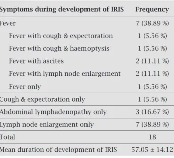
Table 2. Distribution of symptoms in the patients of TB-IRIS
symptoms during development of IRIs Frequency
Fever 7 (38.89 %)
Fever with cough & expectoration 1 (5.56 %)
Fever with cough & haemoptysis 1 (5.56 %)
Fever with ascites 2 (11.11 %)
Fever with lymph node enlargement 2 (11.11 %)
Fever only 1 (5.56 %)
Cough & expectoration only 1 (5.56 %)
Abdominal lymphadenopathy only 3 (16.67 %)
Lymph node enlargement only 7 (38.89 %)
Total 18
Mean duration of development of IRIS 57.05 ± 14.12
556
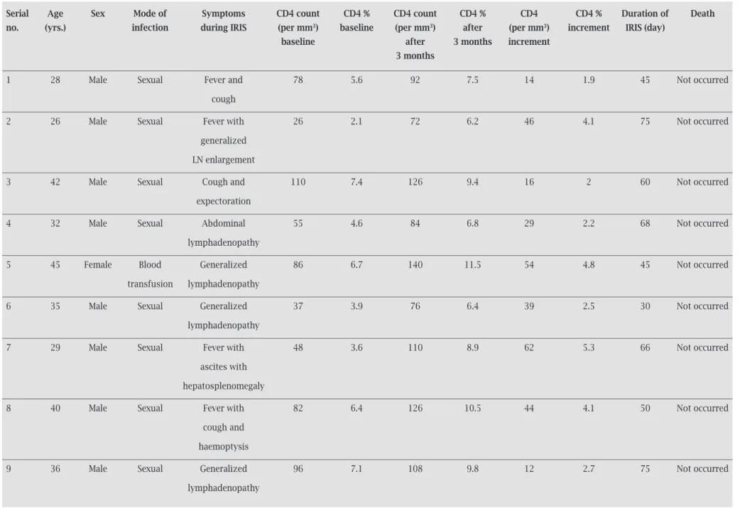
Table 3. Patient profiles of HIV-TB coinfected patients who developed IRIS
serial Age sex Mode of symptoms CD4 count CD4 % CD4 count CD4 % CD4 CD4 % Duration of Death
no. (yrs.) infection during IRIs (per mm3) baseline (per mm3) after (per mm3) increment IRIs (day)
baseline after 3 months increment
3 months
1 28 Male Sexual Fever and 78 5.6 92 7.5 14 1.9 45 Not occurred
cough
2 26 Male Sexual Fever with 26 2.1 72 6.2 46 4.1 75 Not occurred
generalized
LN enlargement
3 42 Male Sexual Cough and 110 7.4 126 9.4 16 2 60 Not occurred
expectoration
4 32 Male Sexual Abdominal 55 4.6 84 6.8 29 2.2 68 Not occurred
lymphadenopathy
5 45 Female Blood Generalized 86 6.7 140 11.5 54 4.8 45 Not occurred
transfusion lymphadenopathy
6 35 Male Sexual Generalized 37 3.9 76 6.4 39 2.5 30 Not occurred
lymphadenopathy
7 29 Male Sexual Fever with 48 3.6 110 8.9 62 5.3 66 Not occurred
ascites with
hepatosplenomegaly
8 40 Male Sexual Fever with 82 6.4 126 10.5 44 4.1 50 Not occurred
cough and
haemoptysis
9 36 Male Sexual Generalized 96 7.1 108 9.8 12 2.7 75 Not occurred
lymphadenopathy
cont.
In
ciden
ce a
n
d r
is
k fac
to
rs o
f imm
un
e r
eco
n
st
itu
tio
n infl
amm
at
o
ry sy
n
dr
o
m
e in HIV
-TB co
inf
ec
te
d p
at
ien
557
B
raz J I
n
fe
ct D
is 2011; 15(6):553-559
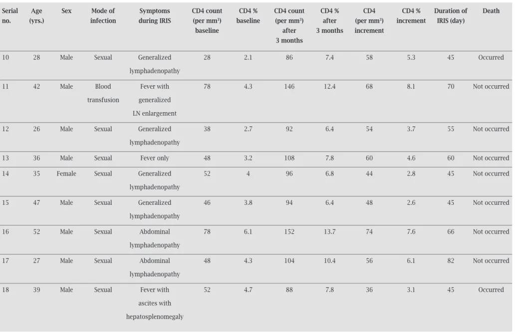
Table 3. Patient profiles of HIV-TB coinfected patients who developed IRIS (Cont.)
serial Age sex Mode of symptoms CD4 count CD4 % CD4 count CD4 % CD4 CD4 % Duration of Death
no. (yrs.) infection during IRIs (per mm3) baseline (per mm3) after (per mm3) increment IRIs (day)
baseline after 3 months increment
3 months
10 28 Male Sexual Generalized 28 2.1 86 7.4 58 5.3 45 Occurred
lymphadenopathy
11 42 Male Blood Fever with 78 4.3 146 12.4 68 8.1 70 Not occurred
transfusion generalized
LN enlargement
12 26 Male Sexual Generalized 38 2.7 92 6.4 54 3.7 55 Not occurred
lymphadenopathy
13 36 Male Sexual Fever only 48 3.2 108 7.8 60 4.6 60 Not occurred
14 35 Female Sexual Generalized 52 4 96 6.8 44 2.8 45 Not occurred
lymphadenopathy
15 47 Male Sexual Generalized 46 3.8 94 6.4 48 2.6 45 Not occurred
lymphadenopathy
16 52 Male Sexual Abdominal 78 6.1 152 13.7 74 7.6 66 Not occurred
lymphadenopathy
17 27 Male Sexual Abdominal 48 4.3 104 10.4 56 6.1 82 Not occurred
lymphadenopathy
18 39 Male Sexual Fever with 52 4.7 88 7.8 36 3.1 45 Occurred
ascites with
hepatosplenomegaly
LN, lymph node.
D
e, Sa
rka
r, P
h
au
jd
ar
, et a
558
DISCUSSION
The incidence of IRIS varies throughout the world. In pre-HAART era, incidence of IRIS in developed countries was reported to be about 36%.7 In developing countries, due to
higher prevalence of tuberculosis, the incidence of IRIS is also higher, though remains largely under reported. The in-cidence in developing countries ranges from 11% to 43%, whereas in India it was reported as 8%.6,8 In our study,
incidence of TB-IRIS was 18.75%. As HAART is now widely available in India, the incidence of IRIS is like-ly to be increasing. The IRIS appears mostlike-ly within first three months of initiating ART. In our study the mean duration of appearance of IRIS was 57.05 days. The early appearance of IRIS may be due to early initiation of ART, after 15 days of ATT, in all patients. Other studies from developing countries like Brazil also documented simi-lar result.9 Shelburne et al.10 showed that IRIS is more
likely to develop earlier if HAART is initiated earlier. Most of the IRIS patients experienced an exacerbation of extrapulmonary tuberculosis, tubercular lymphadenitis being most common. Significantly 27% of the patients pre-sented with intra-abdominal flaring up of tuberculosis. Various previous studies also showed similar result though occurrence of intra- abdominal tuberculosis was not high.11-13
This highlights that one must look out for the enlargement of abdominal lymph nodes as a marker of IRIS. Interestingly, in our study, none had CNS tubercular manifestation. This may be because CNS involvement in TB-IRIS usually oc-curs late with median interval being 5-10 months.14 The risk
factor emerged from the study was presence of extrapulmo-nary tuberculosis, of which tubercular lymphadenitis and disseminated tuberculosis predominates. These findings corroborates with the findings of previous western studies, though they did not show disseminated tuberculosis as a risk factor.15 This may be due to the fact, that exposure to high
load of mycobacterium and their interaction with the im-mune system in disseminated form of tuberculosis increases the risk of IRIS.16 The baseline CD4 count was not a risk
factor for emergence of IRIS. Although previous studies, including those from different developing countries with high prevalence of HIV, postulated that CD4 counts less than 100 per mm3 are associated with increased incidence
of IRIS, this was not present in our study.9,17 Rather, the
in-crease in absolute as well as percentage of CD4 count was significantly higher in IRIS group of patients than the other. Similar result was shown by Breton G et al.12 This may be due
to the inherent pathophysiology of IRIS development. With the initiation of ART, there is partial restoration of immune response, which causes increase in CD4 count. As IRIS occurs due to excessive effect of various inflammatory me-diators liberated from activated lymphocytes, so increment of CD4 count is related with occurrence of IRIS and it may act as a surrogate marker of development of IRIS, though
more detailed studies need to be done to know the actual predictive value. The major limitation of the study was exclusion of subclinical tuberculosis patients who may develop IRIS after initiation of ART. Also all the patients were given standard ATT and ART, so effect of variation of therapy with emergence of IRIS was not evaluated. In addition, because ART was started after 15 days of ATT in all patients, any effect of delay of ART with appearance of IRIS could not be studied. All the patients recruited in the study were in advanced HIV. Further study is needed to know the variation of incidence of IRIS at different stages of HIV. Although most of our patients recovered spontaneously without requiring any deviation from their treatment protocol, large prospective study will guide us about proper management of IRIS.
CONCLUSION
In conclusion, IRIS in HIV-TB coinfected patients is an important entity, 12% of the patients developed IRIS in our study within two months of starting ART. Patients with extrapulmonary tuberculosis, especially tubercular lymphadenitis are more prone to develop IRIS. Fever is associated in most of these patients. Higher increment of CD4 count may give warning for development of IRIS in presence of new or worsening tuberculosis lesion.
REFERENCES
1. John M, French MA. Exacerbation of the inflammatory response to Mycobacterium tuberculosis after antiretroviral therapy. Med J Aust. 1998; 169(9):473-4.
2. Antiretroviral Therapy Guidelines for HIV-infected Adults and adolescents including post-exposure prophylaxis by NACO 2007. 3. Murdoch DM, Venter WD, Van Rie A, Feldman C. Immune re-constitution inflammatory syndrome (IRIS): review of common infectious manifestations and treatment options. AIDS Res Ther. 2007; 4:9.
4. Lawn SD, Bekker LG, Miller RF. Immune reconstitution disease associated with mycobacterial infections in HIV-infected individ-uals receiving antiretrovirals. Lancet Infect Dis. 2005; 5(6):361-73. 5. Sungkanuparph S, Vibhagool A, Mootsikapun P, Chetchotisakd
P, Tansuphaswaswadikul S, Bowonwatanuwong C. Opportunistic infections after the initiation of highly active antiretroviral thera-py in advanced AIDS patients in an area with a high prevalence of tuberculosis. AIDS. 2003; 17(14):2129-31.
6. Shankar EM, Vignesh R, Murugavel KG, et al. Immune recon-stitution inflammatory syndrome in association with HIV/AIDS and tuberculosis: views over hidden possibilities. AIDS Res Ther. 2007; 4:29-42.
7. Narita M, Ashkin D, Hollender ES, Pitchenik AE. Paradoxi-cal worsening of tuberculosis following antiretroviral therapy in patients with AIDS. Am J Respir Crit Care Med. 1998; 158(1):157-61.
8. Gazzard B; BHIVA Writing Committee. British HIV Asso-ciation (BHIVA) guidelines for the treatment of HIV-infected adults with antiretroviral therapy (2005). HIV Med. 2005; 6 Suppl 2:1-61.
559 Braz J Infect Dis 2011; 15(6):553-559
9. Serra FC, Hadad D, Orofino RL, et al. Immune reconstitution syndrome in patients treated for HIV and tuberculosis in Rio de Janeiro. Braz J Infect Dis. 2007; 11(5): 462-65.
10. Shelburne SA, Visnegarwala F, Darcourt J, et al. Incidence and risk factors for immune reconstitution inflammatory syn-drome during highly active antiretroviral therapy. AIDS. 2005; 19(4):399-406.
11. Furrer H, Malinverni R. Systemic inflammatory reaction after starting highly active antiretroviral therapy in AIDS patients treated for extrapulmonary tuberculosis. Am J Med. 1999; 106(3):371-2.
12. Breton G, Duval X, Estellat C, et al. Determinants of immune reconstitution inflammatory syndrome in HIV type 1-infected patients with tuberculosis after initiation of antiretroviral ther-apy. Clin Infect Dis. 2004; 39(11):1709-12.
13. Manosuthi W, Kiertiburanakul S, Phoorisri T, Sungkanuparph S. Immune reconstitution inflammatory syndrome of tubercu-losis among HIV-infected patients receiving antituberculous and antiretroviral therapy. J Infect. 2006; 53 (6):357-63.
14. Crump JA, Tyrer MJ, Lloyd-Owen SJ, Han LY, Lipman MC, Johnson MA. Military tuberculosis with paradoxical expan-sion of intracranial tuberculomas complicating human immu-nodeficiency virus infection in a patient receiving highly active antiretroviral therapy. Clin Infect Dis. 1998; 26(4):1008-9. 15. Wendel KA, Alwood KS, Gachuhi R, Chaisson RE, Bishai WR,
Sterling TR. Paradoxical worsening of tuberculosis in HIV-infected persons. Chest. 2001; 120(1):193-7.
16. Jon F. Tuberculosis. In: William P, editor. Infectious Diseases. Vol. 1. Spain: Mosby; 2004. p. 401-18.
17. Michailidis C, Pozniak AL, Mandalia S, Basnayake S, Nelson MR, Gazzard BG. Clinical characteristics of IRIS syndrome in patients with HIV and tuberculosis. Antivir Ther. 2005; 10(3):417-22.