Clinical Study
Serum Oxidative Stress Markers and Genotoxic Profile Induced
by Chemotherapy in Patients with Breast Cancer: A Pilot Study
Antonio Luiz Gomes Júnior,
1Marcia Fernanda Correia Jardim Paz,
1Laís Iasmin Soares da Silva,
1Simone da Costa e Silva Carvalho,
2André Luiz Pinho Sobral,
1Kátia da Conceição Machado,
1Paulo Michel Pinheiro Ferreira,
3Prabodh Satyal,
4Rivelilson Mendes de Freitas,
5and Ana Amélia de Carvalho Melo Cavalcante
11Laboratory of Genetic Toxicity, Postgraduate Program in Pharmaceutical Sciences, Federal University of Piau´ı,
64049-550 Teresina, PI, Brazil
2Postgraduate Program in Genetics, State University of S˜ao Paulo, 14049-900 Ribeir˜ao Preto, SP, Brazil 3Laboratory of Experimental Cancerology, Department of Biophysics and Physiology,
Postgraduate Program in Pharmaceutical Sciences, Federal University of Piau´ı, 64049-550 Teresina, PI, Brazil
4Chemistry Department, University of Alabama in Huntsville, Huntsville, AL 35899, USA
5Laboratory of Research in Experimental Neurochemistry, Postgraduate Program in Pharmaceutical Sciences,
Federal University of Piau´ı, 64049-550 Teresina, PI, Brazil
Correspondence should be addressed to Rivelilson Mendes de Freitas; rmendesfreitas@hotmail.com
Received 29 November 2014; Revised 2 March 2015; Accepted 2 March 2015
Academic Editor: Amit Tyagi
Copyright © 2015 Antonio Luiz Gomes J´unior et al. his is an open access article distributed under the Creative Commons Attribution License, which permits unrestricted use, distribution, and reproduction in any medium, provided the original work is properly cited.
he aim of this study was to evaluate the oxidative parameters of erythrocytes and genotoxicity in leukocytes of patients with breast cancer. Oxidative parameters were detected by spectrophotometry and genotoxic damage by single cell gel electrophoresis. Twenty-eight women with breast cancer were monitored before chemotherapy and ater the second and fourth cycles of therapy with cyclophosphamide and doxorubicin. Ater the fourth cycle, increases (� < 0.05) in the reactive substances to thiobarbituric acid levels, nitrite content, and superoxide dismutase activity and high rates of DNA damage in leukocytes were observed when compared with healthy women group and baseline levels. Similarly, ater the second cycle, the same parameters were increased
(� < 0.05) when compared with baseline levels. Increase in catalase activity was detected only ater the fourth cycle and reduced
glutathione levels and glutathione peroxidase activity were decreased in all cycles when compared with healthy women, as well as ater the second and fourth chemotherapy cycles compared to baseline (� < 0.05). Patients with breast cancer presented an indicative of oxidative stress before, during, and ater chemotherapy, as well as increased genotoxic damage in all stages of treatment, demonstrating the clinical applicability of this investigation.
1. Introduction
he etiology of breast cancer possesses a multifactorial origin [1,2], showing as risk factors reproductive age, early menarche, late menopause, nulliparity, exogenous hormones, smoking, obesity, diet, alcohol consumption, physical inac-tivity, and genetic and environmental factors [1–5].
Most chemotherapeutic agents are not speciic against neoplastic cells, also afecting normal cells [6], which results in a wide range of adverse reactions in virtually all tissues of body such as bone marrow suppression, alopecia, fatigue, generalized rash, diarrhea, and dizziness [7, 8]. Cyclophos-phamide, one of the most used anticancer compounds, is a bifunctional alkylating member of the nitrogen mustard Volume 2015, Article ID 212964, 11 pages
family that induces various types of DNA damage, such as DNA adducts, gene mutations, and chromosomal aberrations [6, 9]. In clinical and trials protocols, cyclophosphamide is used in combination with doxorubicin, an anthracycline agent capable of intercalating into DNA [10]. heir mecha-nism of cytotoxicity includes intracellular production of free radicals, DNA intercalation, and subsequent inhibition of DNA topoisomerase II [6,9,10].
Reactive oxygen species (ROS) represent important factor in carcinogenesis and may play a role in initiation and progression of tumors. Free radicals stimulate oxidative DNA damage, contributing to mutagenesis, which is essential for the process of tumor initiation [11–13]. Unrepaired DNA damage has been associated with a variety of human dis-orders including cancer and neurodegenerative diseases. When DNA is properly repaired, the injuries are inactivated and the cells return to normal cell cycle operation. If this damage is not repaired, speciic cellular responses such as cell death, senescence, or uncontrolled proliferation could result. his damage may consist of small lesions in very speciic sites within the DNA molecule, as adducts, cross-links, abasic sites, and points of gross abnormalities [14,
15]. he extent of this damage caused by ROS can be maximized or minimized by enzymatic (catalase, superoxide dismutase, and glutathione peroxidase) or nonenzymatic (vitamins A, C, and E, selenium, and reduced glutathione (GSH)) [2,16–18] mechanisms of antioxidant defense. Based on this approach, the present study evaluated the antioxidant and genotoxic proile in blood cells of patients receiving a combined chemotherapy of adriamycin (doxorubicin) and cyclophosphamide (AC).
2. Materials and Methods
2.1. Study Population and Sample Collection. he subjects were patients diagnosed with ductal breast cancer under treatment at the Department of Oncology, S˜ao Marcos Hos-pital, Teresina, Piau´ı, Brazil, from August 2012 to February 2013. his clinical study was approved by the Research Ethics Committee of University Center UNINOVAFAPI (registration number 0406.0.043.00011). his study involved a total of 56 individuals including 28 patients exposed to chemotherapy by the AC protocol (adriamycin 60 mg/m2and cyclophosphamide 600 mg/m2) and 28 patients not exposed to the chemotherapy. he patients were exposed to four 21-day cycles with intravenous AC. he unexposed group con-sisted of individuals who had not been exposed to genotoxic agents (including radiation and chemicals) and who were free of any malignant neoplasm or clinical, biochemical, hematological, hepatic, cardiovascular, renal, or endocrine manifestations. Blood samples were collected with EDTA or heparin by venipuncture using vacutainers, maintained at 4∘C during transport to the laboratory, and immediately processed. hree collections of the peripheral blood were carried out during four cycles of chemotherapy: the irst collection was performed before the beginning of treatment (C0), 21 days ater the second cycle of chemotherapy (C2), and 21 days ater the fourth cycle (C4).
All individuals in this study were submitted to a ques-tionnaire from International Commission for Protection against Environmental Mutagens and Carcinogens [19], which included questions regarding standard demographic data (e.g., age and gender), medical issues (e.g., exposure to X-rays, vaccinations, and medications), lifestyle (e.g., smok-ing, cofee and alcohol consumption, diet, etc.) and occupa-tion, such as number of working hours per day and protective measures adopted (PPE). In all groups, individuals who smoked more than 20 cigarettes per day were considered smokers [20].
Ater the questionnaires, data were analyzed using SPSS 17.0. (Chicago: SPSS Inc.) and the demographic, medical and lifestyle were summarized inTable 1.
Details about clinical features, such as cancer site, clinical stage and HER-2/neu, ER (estrogen receptor), and PR (pro-gesterone receptor)status, were obtained and analyzed from medical records. he descriptive statistics for such variables are listed inTable 2.
2.2. Comet Assay. he alkaline comet (single cell gel elec-trophoresis (SCGE)) assay was performed as described by Singh et al. [21] with modiications suggested by Tice et al. [22]. Blood cells (5�L) were embedded in 95�L of 0.75% low-melting point agarose, which was immediately added to the surface of a precoated (1.5% agarose) microscope slide. When the agarose had solidiied, the slides were placed in lysis bufer (2.5 M NaCl, 100 mM EDTA, and 10 mM Tris; pH 10.0–10.5) containing freshly added 1% (v/v) Triton X-100 and 10% (v/v) dimethyl sulfoxide (DMSO) for a minimum of 1 day and a maximum of 7 days. Ater treatment with lysis bufer, the slides were incubated in freshly made alkaline bufer (300 mM NaOH and 1 mM EDTA; pH N 13) for 20 min and the DNA was electrophoresed for 20 min at 25 V (0.90 V/cm) and 300 mA ater which the bufer was neutralized with 0.4 M Tris (pH 7.5) and dried overnight. Gels were rehydrated for 5 min in distilled water and then stained for 15 min (37∘C) with a solution containing the following sequence: 34 mL of Solution B (0.2% w/v ammonium nitrate, 0.2% w/v silver nitrate, 0.5% w/v tungstosilicic acid, 0.15% v/v formaldehyde, and 5% w/v sodium carbonate) and 66 mL of Solution A (5% sodium carbonate). he staining was stopped with 1% acetic acid and the gels were air dried. Analyses (100 cells/patient) were carried out by light microscopy (Olympus CX40) at 100x magniication with immersion oil. Images of cells (50 cells/slide in two replicates) were evaluated for the following: (i) damage index (DI), in which each cell was classiied into classes (no damage = 0, maximum damage = 4) according to tail size and cell shape [23], with resulting values for each individual ranging from 0 (0×100) to 400 (4×100); (ii) damage frequency (DF), calculated as the percentage of injured cells. International guidelines and recommendations for the comet assay consider the visual scoring of comets to be a well-validated evaluation method. Although the DI parameter is oten subjective, it has high correlation with computer-based image analysis [22,24,25].
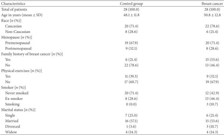
Table 1: Demographic, medical, and lifestyle data of the patients.
Characteristics Control group Breast cancer
Total of patients 28 (100.0) 28 (100.0)
Age in years (mean±SD) 48.1±11.8 50.8±12.8
Race [�(%)]
Caucasian 20 (71.4) 22 (78.6)
Non-Caucasian 8 (28.6) 6 (21.4)
Menopause [�(%)]
Premenopausal 19 (67.9) 20 (71.4)
Postmenopausal 9 (32.1) 8 (28.6)
Family history of breast cancer [�(%)]
Yes 6 (21.4) 15 (53.6)
No 22 (78.6) 13 (46.4)
Physical exercises [�(%)]
Yes 11 (39.3) 9 (32.1)
No 17 (60.7) 19 (67.9)
Smoker [�(%)]
Never smoked 20 (71.4) 12 (42.9)
Ex-smoker 8 (28.6) 13 (46.4)
Smoking 0 (0.0) 3 (10.7)
Marital status [�(%)]
Single 7 (25.0) 6 (21.4)
Married 16 (57.1) 15 (53.6)
Divorced 1 (3.6) 3 (10.7)
Widow 4 (14.3) 4 (14.3)
SD: standard deviation.
Table 2: Clinical characteristics of patients with breast ductal carcinoma (� = 28).
Characteristics Breast cancer Cancer sites [�(%)]
Let breast 11 (39.3)
Right mama 17 (60.7)
Clinical stage [�(%)]
Grade 1 4 (14.3)
Grade 2 10 (35.7)
Grade 3 14 (50.0)
Estrogen receptor [�(%)]
Negative 7 (25.0)
Positive 21 (75.0)
Progesterone receptor [�(%)]
Negative 7 (25.0)
Positive 21 (75.0)
HER2/neu [�(%)]
Score 0 9 (32.1)
Score +1 10 (35.7)
Score +2 1 (3.6)
Score +3 8 (28.3)
HER2/neu: human epidermal growth factor receptor 2.
reagent was added in white tube plus 500�L of distilled water (Blank) was added in white tube. In another test tube
500�L of Griess reagent and 500�L of the homogenate at 10% of the erythrocytes (sodium phosphate bufer 50 mM pH 7.4) (Test) were added. he spectrophotometric measurement was performed at 560 nm. Results were expressed in�M/mg protein.
2.4. hiobarbituric Acid Reactive Substances (TBARS) Levels. Blood samples were centrifuged at 3000 rpm at 4∘C during 5 minutes. Plasma was removed and a pellet of erythro-cytes was washed with a cold solution of NaCl 0.9% and centrifuged. An erythrocytes’ homogenate 10% diluted in phosphate bufer sodium 50 mM and pH 7.4 was stored at
−20∘C. Lipid peroxidation was measured by TBARS levels, a method previously described by Draper and Hadley (1990) [27,28]. 250�L of homogenate, 1 mL of trichloroacetic acid 10%, and 1 mL of thiobarbituric acid 0.67% were mixed and stirred. Subsequently, this mixture was maintained in a bath of boiling water for 15 min and freshened under running water. Ater cooling, 2 mL of n-butanol was added and centrifuged at 1.200 rpm/5 min and the butanol phase was read spectrophotometrically at 535 nm. Results were expressed as nmol/mL.
320�L of distilled water and 80�L of trichloroacetic acid 50%. Ater centrifugation at 3.000 rpm for 15 min, 400�L from the supernatant was collected and added to 800�L of Tris-HCl 0.4 M, pH 8.9, and 20�L of DTNB 0.01 M. One minute later, spectrophotometric measurement was performed at 412 nm. Concentration of GSH was expressed in mg/g of hemoglobin.
2.6. Glutathione Peroxidase (GPx) Activity. he glutathione peroxidase activity coupled assay was determined by Paglia and Valentine [30]. GPx catalyzes the reduction of hydrogen peroxide (H2O2), oxidizing reduced glutathione (GSH) to form oxidized glutathione (GSSG). GSSG is then reduced by glutathione reductase (GR) and�-nicotinamide adenine dinucleotide phosphate (NADPH) forming NADP+ (result-ing in decreased absorbance at 340 nm) and recycl(result-ing the GSH. Because GPx is limiting, the decrease in absorbance at 340 nm is directly proportional to the GPx concentration. 1 unit of GPx-1 = the amount of enzyme necessary to catalyze the oxidation (by H2O2) of 1.0�mole GSH to GSSG, per minute at 25∘C, pH 7.0. Results were expressed in U/g of hemoglobin.
2.7. Catalase (CAT) Activity. Erythrocytes’ homogenate in pH 7.4 was centrifuged (800 g, 20 min) and the supernatant was used to quantify catalase activity. he reaction medium was prepared with H2O2(18 mL), Tris HCl 1 M, EDTA pH 8.0 5 mM (1.0 mL), and H2O (0.8 mL). he reading was carried out in a quartz cuvette at 230 nm with 980�L of reaction medium plus 20�L erythrocytes’ homogenate prepared in sodium phosphate bufer 50 mM, pH 7.4 [31].
2.8. Superoxide Dismutase (SOD) Activity. Erythrocytes homogenate prepared in sodium phosphate bufer 50 mM, pH 7.4, was centrifuged (800 g, 20 min) and supernatants were used for testing superoxide dismutase (SOD) activity. Cytochrome�reduction rate was determined by superoxide radicals using the xanthine-xanthine oxidase system as a source of superoxide anion (O2−) [32]. Results were expressed as U/mg protein. One unit (U) of SOD activity corresponds to the inhibition of 50% of O2−in the presence of cytochrome�. 2.9. Statistical Analyses. In order to determine statistical diferences, data expressed as mean ± standard error of the mean (S.E.M.) were compared by one-way analysis of variance (ANOVA) followed by the Newman-Keuls test (� <
0.05) using the Graphpad program (Intuitive Sotware for Science, San Diego, CA) and SPSS (version 19, SPSS Inc.). Correlations among data obtained were calculated using Spearman’s correlation coeicient.
3. Results
3.1. Evaluation of Oxidative Stress. Evaluation of oxidative stress in patients with breast cancer in AC chemotherapy was performed by analyzing enzymatic and nonenzymatic parameters in erythrocytes by serum thiobarbituric acid reactive substances (TBARS) level, nitrite content, GSH concentration, and GPx, CAT, and SOD activities.
Results showed that the status of oxidative stress (� <
0.05) increased, as demonstrated by basal TBARS (1.42 ±
0.45nM/mg of protein) and nitrite (1.16 ± 0.62 �M/mg protein) contents in erythrocytes of patients with breast cancer when compared with the control group (0.37 ±
0.09nM/mg of protein and 0.16 ± 0.05 �M/mg of protein, resp.) (� < 0.05). When these same patients were submitted to chemotherapeutics (combination of cyclophosphamide and doxorubicin), such increases in both TBARS (4.76 ± 0.68 and 11.98 ± 0.65nM/mg of protein) and nitrite ion levels (1.81 ± 0.02 and 3.49 ± 0.07 �M/mg of protein) were also detected in C2 and C4, respectively (� < 0.05;Table 3).
Red blood cells of the patients revealed decrease in reduced glutathione concentration at 36.1% (24.94 ± 1.51U/g protein) in comparison with control group (36.13 ± 7.65U/g protein) (� < 0.05). With AC chemotherapy, there was decrease in GSH levels in C2 (46.6%) and C4 groups (50.9%) (� < 0.05). Similarly, baseline levels (C0) also presented diminution of 21.4% and 27.7% in C2 and C4 groups, respectively (Table 3).
GPx activity (Figure 1(a)) showed reduction (63.32, 78.31, and 81.0%) in erythrocytes of the patients with breast cancer activity in all groups analyzed (101.90±29.48,60.25±4.66, and 52,77±3.26U/g for C0, C2, and C4, resp.) when compared to the control group (277.8±15.88U/g), respectively (� < 0.05). In relation to the catalase levels, only treated patients (C4,
22.83 ± 1.17 �M/mg) exhibited a signiicant increase (44.3%) when compared to the control group (15.82 ± 1.21 �M/mg), C0, and C2 (18.44 ± 1.24 and 18.77 ± 0.96 �M/mg, resp.) (Figure 1(b)). Similarly, superoxide dismutase activity
(Figure 1(c)) also increased (33.1%) ater the second cycle
of chemotherapy (1.81 ± 0.63 �M/mg) when compared with control group. Ater chemotherapy (2.63 ± 0.65 �M/mg), its activity increased about 93.4 and 54.7% in relation to the control group (1.36 ± 0.62 �M/mg) and baseline (1.70 ±
0.43 �M/mg), respectively.
3.2. Index and Frequency of DNA Damage. DNA in the tail was organized into ive classes: (i) class 0: undamaged, with no tail; (ii) class 1: with tail shorter than the diameter of the head (nucleus); (iii) class 2: with tail length between one and two times the diameter of the head; (iv) class 3: with tail longer than two times the diameter of the head; and (v) class 4: comets with no heads [33] (Figure 2).
With the application of alkaline comet assay it was possible to observe an increase (� < 0.05) in the classes of DNA damage in lymphocytes of patients with breast cancer (C0) in the control group. his condition is increased (� <
0.05) in C2 and C4 (Figure 3).
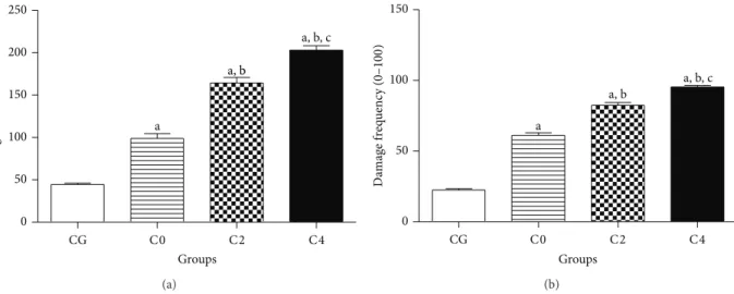
DNA damage in lymphocytes of patients with breast cancer increased by 122.6% (98.89 ± 5.56) compared to the control group (44.43 ± 1.67). Ater AC chemotherapy, there was an increase of 66.25 and 105.2% of damage index in C2 (164.4 ± 6.36) and C4 groups (202.9 ± 5.34) in comparison with C0 group (98.89 ± 5.56). In a similar way, an increase of 23.2% ater the fourth cycle was noted when compared to the C2 group (Figure 4(a)).
Table 3: Biomarkers levels of oxidative stress in antioxidant enzymatic system of patients with breast cancer before (C0), during (C2), and ater chemotherapy (C4) and control group.
Groups TBARS levels (nM/mg de protein)
NO2−content (�M/mg protein)
GSH concentration (U/g protein)
Control 0.37±0.09 0.16±0.05 36.13±7.65
C0 1.42±0.45a 1.16±0.62a 24.94±1.51a
C2 4.76±0.68a,b 1.81±0.02a,b 19.30±0.74a,b
C4 11.98±0.65a.b,c 3.49±0.07a,b,c 17.75±0.46a,b
TBARS: thiobarbituric acid reactive substances levels, NO2−: nitrite content, and GSH: reduced glutathione concentration. Values represent mean±S.E.M.
a� < 0.05when compared with control group (CG) by ANOVA followed by�-Student-Newman-Keuls.b� < 0.05when compared with C0 group (before
chemotherapy) andc� < 0.05when compared with C2 group (second cycle of chemotherapy).
0 100 200 300 400
a
a a
Groups
CG C0 C2 C4
GPx ac
ti
vi
ty (U/g)
(a)
0 10 20 30
a, b, c
Groups
CG C0 C2 C4
CA
T ac
ti
vi
ty (
𝜇
M/m
g)
(b)
0 1 2 3
a a
a, b, c
Groups
CG C0 C2 C4
SO
D ac
ti
vi
ty (
𝜇
M/m
g)
(c)
Figure 1: Antioxidant enzymes activity in erythrocytes of patients with breast cancer before (C0), during (C2), and ater (C4) AC chemotherapy. Control group (CG) is represented by healthy patients. Values represent mean±S.E.M.a� < 0.05when compared with control group (CG) by ANOVA followed by�-Student-Newman-Keuls.b� < 0.05when compared with C0 group (before chemotherapy) and
c� < 0.05when compared with C2 group (second cycle of chemotherapy).
when compared to the CG (22.50 ± 0.94) (Figure 4(b)). AC chemotherapy raised this frequency by 34.9 and 56.3% in C2 (82.32 ± 2.08) and C4 (95.36 ± 0.99) groups, respectively, in comparison with the base status (C0). Similarly, in C4 patients, an increase of 15.8% was observed in comparison with the frequency of C2.
0
0 0
0 0 0
0
0
(a)
0 2
1 2
1 0
(b)
0
0
1 1
1
3
3
0
0 0
(c)
4
4 1
3
1 3 2
2 2
(d)
Figure 2: Photomicrograph of the comet test indicative of the types of damage. (a) Control group (CG); (b) beginning of treatment (C0); (c) 21 days ater second cycle of chemotherapy (C2); (d) 21 days ater the fourth cycle (C4).
CG C0 C2 C4
0 20 40 60 80 100
Damage 0 Damage 1 Damage 2
Damage 3 Damage 4 a
a, b
a, b, c a
a a
a
a, b a, b
a, b
a, b, c a, b, c
a, b, c
Groups
N
u
m
b
er o
f cells (
0
–
100
)
Figure 3: Proile of DNA damage in lymphocytes evaluated by the alkaline comet assay (single cell gel electrophoresis) for each phase of chemotherapy. Control group (CG) is represented by healthy patients. Values represent mean±S.E.M.a� < 0.05when compared with control group (CG) by ANOVA followed by�-Student-Newman-Keuls.b� < 0.05when compared with C0 group (before chemotherapy) and
c� < 0.05when compared with C2 group (second cycle of chemotherapy).
correlation (correlation factor = 0.389 and� = 0.041) was observed between race and nitrite levels ater chemotherapy (Q4) and a negative correlation was observed with nitrite levels (correlation factor =−0.474,� = 0.011) in the diagnosis and activity of superoxide dismutase (correlation factor =
0 50 100 150 200 250
a
a, b
a, b, c
Groups
CG C0 C2 C4
D
amag
e index (
0
–
40
0
)
(a)
0 50 100 150
a
a, b
a, b, c
Groups
CG C0 C2 C4
D
amag
e f
req
uenc
y (
0
–
100
)
(b)
Figure 4: DNA damage investigation by the alkaline comet assay (single cell gel electrophoresis) carried out in lymphocytes of patients with breast cancer before (C0), during (C2), and ater AC (C4) chemotherapy. Control group (CG) is represented by healthy patients. Analyses were performed by light microscopy at 100x magniication with immersion oil. Values represent mean±S.E.M.a� < 0.05when compared
with control group (CG) by ANOVA followed by�-Student-Newman-Keuls.b� < 0.05when compared with C0 group (before chemotherapy)
andc� < 0.05when compared with C2 group (second cycle of chemotherapy).
with oxidative stress, except between HER2 and glutathione peroxidase in Q2 group with 0.412 correlation factor� =
0.29.
Regarding the genotoxicity and oxidative stress, positive correlations were observed for the contents of DNA damage assessed at diagnosis (QD) compared to those obtained dur-ing (Q2) and ater (Q4) chemotherapy, with 0.663 correlation factor 0.537 and� = 0.000and 0.003, respectively. A negative correlation was observed between levels of DNA damage during chemotherapy (Q2) and nitrite levels (Q4), as well as between frequency of damage (QD) and nitrite in group Q4 and Q2 catalase group.
4. Discussion
Breast cancer is the second most common cancer in women over the age of 50. It is oten irst detected as an abnormality on a mammogram before the patient or health care provider feels it. Early cases may be asymptomatic, and pain and discomfort are typically not present. Breast cancer can begin in diferent areas of the breast, such as ducts and lobules. Ductal carcinomain situ(DCIS) is the most common nonin-vasive or preinnonin-vasive type with chances of a recurrence under 30% within 5–10 years ater initial diagnosis. On the other hand, invasive ductal carcinoma (IDC), known as iniltrating ductal carcinoma, is the most common type of invasive breast cancer, representing around 80% of cases. About two-thirds of women are 55 or older when they are diagnosed with an IDC [34].
Many studies have reported that reactive oxygen species (ROS) and reactive nitrogen species (RNS) are involved in the etiology and progression of various cancers [35–
38]. hese reactive species have been associated with the development of carcinogenesis by activating diverse types of DNA damage, contributing to the emergence of mutations
and chromosomal aberrations in the inlammatory process and leading to intense tissue disorganization and injuries [39].
An alternative method of analyzing oxidative stress is achieved by quantiication of lipid peroxidation. he lipid radical is unstable and degrades very rapidly into secondary products. Most of them are electrophilic aldehydes, such as TBARS, which is the main marker of oxidative injury in the unsaturated lipids in cell membranes, leading to oxidation of fatty acids (LH) and formation of the lipid radical (L∙) [40]. herefore, TBARS is an important indicator of oxidative stress [16]. he present study demonstrated an elevation of TBARS levels in AC-treated breast cancer patients com-pared to controls, corroborating previous studies [1, 35] and suggesting severe lipid peroxidation. hese changes may be attributed to the production of hydroxyl radicals, which participate directly in the lipid peroxidation process, inducing a disturbance in membrane structure [41].
hypothesis that breast cancers are associated with increased nitric oxide levels whose changes are linked to inlamma-tory process [38]. Prior analyses in 14 patients with breast carcinomas showed no elevation of serum TBARS. However, increased NO concentrations were detected [42].
he extent of oxidative damage depends not only on ROS levels, but also on mechanisms of cellular antioxidant defenses. Low level of GSH, a molecule of critical importance in maintaining the stability of erythrocytes membranes, is related to cellular defense against xenobiotics and harmful compounds such as free radicals and hydroperoxides [43]. his drop in GSH was also observed in erythrocytes of the patients. An additional reduction in GSH levels was observed in healthy patients and those under chemotherapy. Glutathione acts as the irst line of defense against free radicals produced by antitumor molecules. Decreased GSH levels can be explained by a decrease in GSH synthesis and/or increased consumption to remove peroxides and xenobiotics [44].
Metabolites generated by CMF (cyclophosphamide, methotrexate, and 5-luorouracil) induced lipid peroxidation by inactivation of GSH levels and SOD, CAT, GPx, and GST activities in erythrocytes of patients with breast cancer, thereby rendering the system ineicient in management of the free radical attack. Acrolein and phosphoramide mustard are the metabolites of cyclophosphamide that are among the causative agents, which reduce the activity of SOD, CAT, GPX, glutathione-S-transferase, and glucose-6-phosphate dehydrogenase in erythrocytes of CMF treated breast cancer patients [45]. In the present study, GSH concentration and GPx activity were also observed just before AC chemotherapy. Our data demonstrate that GPx activity decreased, compared to the control group. However, this decrease was seen before the start of chemotherapy, suggesting no change in the activity of this enzyme for the therapeutic protocols used, since the reductions for during and ater chemotherapy evaluation were similar to those observed prior to chemotherapy. Furthermore, these results suggest that the establishment of the pathophysiology of breast cancer may be a compromise in the activity of this enzyme. Present results and outcomes of Singh et al. [46] and Prabasheela et al. [47] also revealed, during chemotherapy FAC (5-luorouracil, doxorubicin, and cyclophosphamide) or AC, a decrease in the nonenzymatic antioxidant GSH levels in patients with breast cancer before chemotherapy. On the other hand, additional studies did not ind decrease in GPx activity before or ater administration of chemotherapeutics [47,48].
In relation to the CAT levels, our indings did not show diferences before and during chemotherapy, presenting only increasing activity ater treatment. On the other hand, while some studies found increases only ater chemotherapy [48,
49], others observed decreases in CAT activity before and ater chemotherapy [5,40]. Since antioxidants can activate gene expression via the antioxidant response element [50], overexpression of enzymatic activities can explain these indings [40]. Similarly, SOD activity was elevated in patients with breast cancer before, during, and ater chemotherapy. Hasan et al. [51] also showed plasma SOD activity increasing
in patients with breast carcinoma compared to patients with benign tumors, suggesting that elevated total SOD might relect a response to oxidative stress and then may predict a state of excess reactive oxygen species in the carcinogenesis process. Analogous outcomes were described by Badid et al. [52] before chemotherapy in erythrocytes of 38 patients with ductal breast cancer. Nevertheless, Gupta et al. [53] found a decrease in SOD activity in serum from 30 women. Patients with breast cancer in chemotherapy with epirubicin (90 mg/m2) and cyclophosphamide (600 mg/m2) also showed reduced CAT, SOD, GSH, and GPx activity and increased TBARS levels [54]. Some of these parameters are contradictory when compared to the outcomes in the present study. hese diferences are probably explained by the fact that enzymatic activity of antioxidant defenses is more expressed at the cytoplasmic and mitochondrial cellular level, especially for SOD [46].
In this study, the genotoxic proile assay of the patients with breast cancer under treatment with AC was also investi-gated. his evaluation was carried out by alkaline comet assay, a well-established, simple, versatile, rapid, visual, and sensi-tive tool used to assess DNA damage and repair quantitasensi-tively as well as qualitatively in individual cell populations [55]. Some other forms of DNA damage such as DNA cross-links (e.g., thymidine dimers) and oxidative DNA damage may also be assessed using lesion-speciic antibodies or speciic DNA repair enzymes in the comet assay. his technique has gained wide acceptance as a valuable tool in fundamental DNA dam-age and repair studies, genotoxicity testing, and human bio-monitoring [56]. Relative to other genotoxicity tests, such as chromosomal aberrations, sister chromatid exchanges, alka-line elution, and micronucleus assay, the advantages of the comet assay include its demonstrated sensitivity for detecting low levels of DNA damage (one break per 1010Da of DNA) [57].
he pathological condition signiicantly raised the dam-age indices and frequencies in lymphocytes when compared with the normal control group, conirming previous investi-gations performed by S´anchez-Su´arez et al. [6] and Agnoletto et al. [4]. hese efects on DNA structure remained elevated up to 80 days ater the end of exposure to FEC (5-luorouracil, epirubicin, and cyclophosphamide) [6]. As seen in this work, Vaghef et al. [9] showed signiicant increase in DNA damage on lymphocytes of patients treated with cyclophosphamide.
5. Conclusion
Patients with breast cancer under chemotherapy presented antioxidant status indicative of oxidative stress before, dur-ing, and ater chemotherapy, as well as increasing genotoxic damage in all stages of the treatment. hese results highlight the importance of monitoring patients in chemotherapy, especially using cytogenetic and molecular markers in order to provide new prognostic indings to the treatment as a stra-tegy to reduce recurrences and to improve quality of life.
Ethical Approval
his study was previously approved by Committee in Ethical Research at UNINOVAFAPI (N. 0406.0.043.00011) and is in accordance with Brazilian research guidelines (Law 466/2012, National Council of Health, Brazil) and with Declaration of Helsinki.
Conflict of Interests
he authors declare no conlict of interests.
Acknowledgments
he authors are grateful to the Brazilian agencies Con-selho Nacional de Desenvolvimento Cient´ıico e Tecnol´ogico (CNPq), Coordenac¸˜ao de Aperfeic¸oamento de Pessoal de N´ıvel Superior (CAPES) and Fundac¸˜ao de Amparo e Pesquisa do Estado do Piau´ı(FAPEPI) for inancial support in the form of grants and fellowship awards.
References
[1] R. Kumaraguruparan, J. Kabalimoorthy, and S. Nagini, “Corre-lation of tissue lipid peroxidation and antioxidants with clinical stage and menopausal status in patients with adenocarcinoma of the breast,”Clinical Biochemistry, vol. 38, no. 2, pp. 154–158, 2005.
[2] S. M. Tsai, M. F. Hou, S. H. Wu et al., “Expression of manganese superoxide dismutase in patients with breast cancer,”Kaohsiung Journal of Medical Sciences, vol. 27, pp. 167–172, 2011.
[3] A. G¨onenc¸, D. Erten, S. Aslan, M. Akinci, B. S¸ims¸ek, and M. Torun, “Lipid peroxidation and antioxidant status in blood and tissue of malignant breast tumor and benign breast disease,”Cell Biology International, vol. 30, no. 4, pp. 376–380, 2006. [4] M. H. Agnoletto, T. N. Guecheva, F. Dond´e et al., “Association
of low repair eiciency with high hormone receptors expression and SOD activity in breast cancer patients,”Clinical Biochem-istry, vol. 40, no. 16-17, pp. 1252–1258, 2007.
[5] K. A. Amin, B. M. Mohamed, M. A. M. El-Wakil, and S. O. Ibrahem, “Impact of breast cancer and combination chemother-apy on oxidative stress, hepatic and cardiac markers,”Journal of Breast Cancer, vol. 15, no. 3, pp. 306–312, 2012.
[6] P. S´anchez-Su´arez, P. Ostrosky-Wegman, F. Gallegos-Hern´andez et al., “DNA damage in peripheral blood lympho-cytes in patients during combined chemotherapy for breast cancer,”Mutation Research—Fundamental and Molecular Mech-anisms of Mutagenesis, vol. 640, no. 1-2, pp. 8–15, 2008.
[7] E. P. M. de Almeida, M. G. R. de Guti´errez, and N. P. Adami, “Monitoramento e avaliac¸˜ao dos efeitos colaterais da quimio-terapia em pacientes com cˆancer de c´olon,” Revista Latino-Americana de Enfermagem, vol. 12, no. 5, pp. 760–766, 2004. [8] C. M. Walko and C. Grande, “Management of common adverse
events in patients treated with Sorafenib: nurse and pharmacist perspective,”Seminars in Oncology, vol. 41, supplement 2, pp. S17–S28, 2014.
[9] H. Vaghef, P. Nygren, C. Edling, J. Bergh, and B. Hellman, “Alka-line single-cell gel electrophoresis and human biomonitoring for genotoxicity: a pilot study on breast cancer patients under-going chemotherapy including cyclophosphamide,”Mutation Research—Genetic Toxicology and Environmental Mutagenesis, vol. 395, no. 2-3, pp. 127–138, 1997.
[10] S. Marsh and G. Liu, “Pharmacokinetics and pharmacoge-nomics in breast cancer chemotherapy,”Advanced Drug Deliv-ery Reviews, vol. 61, pp. 381–387, 2009.
[11] J. Sastre-Serra, A. Valle, M. M. Company, I. Garau, J. Oliver, and P. Roca, “Estrogen down-regulates uncoupling proteins and increases oxidative stress in breast cancer,”Free Radical Biology and Medicine, vol. 48, no. 4, pp. 506–512, 2010.
[12] L. Vera-Ramirez, P. Sanchez-Rovira, M. C. Ramirez-Tortosa et al., “Does chemotherapy-induced oxidative stress improve the survival rates of breast cancer patients?”Antioxidants and Redox Signaling, vol. 15, no. 4, pp. 903–909, 2011.
[13] L. R. Freeman and J. N. Keller, “Oxidative stress and cere-bral endothelial cells: regulation of the blood-brain-barrier and antioxidant based interventions,”Biochimica et Biophysica Acta—Molecular Basis of Disease, vol. 1822, no. 5, pp. 822–829, 2012.
[14] H. Rodriguez-Rocha, A. Garcia-Garcia, M. I. Panayiotidis, and R. Franco, “DNA damage and autophagy,”Mutation Research, vol. 711, no. 1-2, pp. 158–166, 2011.
[15] J. Vijg, “Somatic mutations and aging: a re-evaluation,” tion Research/Fundamental and Molecular Mechanisms of Muta-genesis, vol. 447, no. 1, pp. 117–135, 2000.
[16] N. Badjatia, A. Satyam, P. Singh, A. Seth, and A. Sharma, “Altered antioxidant status and lipid peroxidation in Indian patients with urothelial bladder carcinoma,”Urologic Oncolog, vol. 28, no. 4, pp. 360–367, 2010.
[17] C. Glorieux, N. Dejeans, B. Sid, R. Beck, P. B. Calderon, and J. Verrax, “Catalase overexpression in mammary cancer cells leads to a less aggressive phenotype and an altered response to chemotherapy,”Biochemical Pharmacology, vol. 82, no. 10, pp. 1384–1390, 2011.
[18] A. G¨onenc¸, A. Hacis¸evki, S. Aslan, M. Torun, and B. S¸ims¸ek, “Increased oxidative DNA damage and impaired antioxidant defense system in patients with gastrointestinal cancer,” Euro-pean Journal of Internal Medicine, vol. 23, no. 4, pp. 350–354, 2012.
[19] A. V. Carrano and A. T. Natarajan, “Considerations for popula-tion monitoring using cytogenetic techniques Internapopula-tional Comission for protection against Environmental Mutagens and Carcinogens (ICPEMC publication 14),”Mutation Research, vol. 204, no. 3, pp. 379–406, 1988.
[20] H. Hofmann, J. H¨ogel, and G. Speit, “he efect of smoking on DNA efects in the comet assay: a meta-analysis,”Mutagenesis, vol. 20, no. 6, pp. 455–466, 2005.
[22] R. R. Tice, E. Agurell, D. Anderson et al., “Single cell gel/comet assay: guidelines for in vitro and in vivo genetic toxicology test-ing,”Environmental and Molecular Mutagenesis, vol. 35, no. 3, pp. 206–221, 2000.
[23] O. Garc´ıa, T. Mandina, A. I. Lamadrid et al., “Sensitivity and variability of visual scoring in the comet assay: results of an inter-laboratory scoring exercise with the use of silver staining,”
Mutation Research, vol. 556, no. 1-2, pp. 25–34, 2004.
[24] A. R. Collins, “he comet assay for DNA damage and repair: principles, applications, and limitations,”Applied Biochemistry and Biotechnology, Part B Molecular Biotechnology, vol. 26, no. 3, pp. 249–261, 2004.
[25] M. Dusinska and A. R. Collins, “he comet assay in human biomonitoring: gene-environment interactions,”Mutagenesis, vol. 23, no. 3, pp. 191–205, 2008.
[26] L. C. Green, S. R. Tannenbaum, and P. Goldman, “Nitrate syn-thesis in the germfree and conventional rat,”Science, vol. 212, no. 4490, pp. 56–58, 1981.
[27] E. D. Wills, “Mechanisms of lipid peroxide formation in animal tissues,”Biochemical Journal, vol. 99, no. 3, pp. 667–676, 1966. [28] H. H. Draper and M. Hadley, “Malondialdehyde determination
as an index of lipid peroxidation,” inMethods in Enzymology, vol. 186, pp. 421–431, 1990.
[29] J. Sedlak and R. H. Lindsay, “Estimation of total, protein-bound, and nonprotein sulhydryl groups in tissue with Ellman's reagent,”Analytical Biochemistry, vol. 25, no. 1, pp. 192–205, 1968.
[30] D. E. Paglia and W. N. Valentine, “Studies on the quantitative and qualitative characterization of erythrocyte glutathione per-oxidase,”he Journal of Laboratory and Clinical Medicine, vol. 70, no. 1, pp. 158–169, 1967.
[31] B. Chance and A. C. Maehly, “Assay catalases and peroxidases,”
Methods in Enzymology, vol. 2, pp. 764–768, 1995.
[32] J. R. Arthur and R. Boyne, “Superoxide dismutase and glu-tathione peroxidase activities in neutrophils from selenium deicient and copper deicient cattle,”Life Sciences, vol. 36, no. 16, pp. 1569–1575, 1985.
[33] S. T. Miorelli, R. M. Rosa, D. J. Moura et al., “Antioxidant and anti-mutagenic efects of ebselen in yeast and in cultured mam-malian V79 cells,”Mutagenesis, vol. 23, no. 2, pp. 93–99, 2008. [34] E. A. Rakha, J. S. Reis-Filho, F. Baehner et al., “Breast cancer
prognostic classiication in the molecular era: the role of histological grade,”Breast Cancer Research, vol. 12, pp. 1–12, 2010.
[35] D. Pande, R. Negi, S. Khanna, R. Khanna, and H. D. Khanna, “Vascular endothelial growth factor levels in relation to oxida-tive damage and antioxidant status in patients with breast cancer,”Journal of Breast Cancer, vol. 14, no. 3, pp. 181–184, 2011. [36] S. J. Ralph, S. Rodr´ıguez-Enr´ıquez, J. Neuzil, E. Saavedra, and R. Moreno-S´anchez, “he causes of cancer revisited: ‘Mitochon-drial malignancy’ and ROS-induced oncogenic transforma-tion—why mitochondria are targets for cancer therapy,” Molec-ular Aspects of Medicine, vol. 31, no. 2, pp. 145–170, 2010. [37] G. Rockenbach, P. F. di Pietro, C. Ambrosi et al., “Dietary intake
and oxidative stress in breast cancer: before and ater treat-ments,”Nutricion Hospitalaria, vol. 26, no. 4, pp. 737–744, 2011. [38] C. G. Vallejo, A. Cruz-Berm´udez, P. Clemente, R. Hern´andez-Sierra, R. Garesse, and M. Quintanilla, “Evaluation of mito-chondrial function and metabolic reprogramming during tumor progression in a cell model of skin carcinogenesis,” Bio-chimie, vol. 95, no. 6, pp. 1171–1176, 2013.
[39] N. Gupta, B. Goswami, and P. Mittal, “Efect of standard anthracycline based neoadjuvant chemotherapy on circulating levels of serum IL-6 in patients of locally advanced carcinoma breast—a prospective study,”International Journal of Surgery, vol. 10, no. 10, pp. 638–640, 2012.
[40] T. T. Reed, “Lipid peroxidation and neurodegenerative disease,”
Free Radical Biology and Medicine, vol. 51, no. 7, pp. 1302–1319, 2011.
[41] C. Panis, A. C. S. A. Herrera, V. J. Victorino et al., “Oxidative stress and hematological proiles of advanced breast cancer patients subjected to paclitaxel or doxorubicin chemotherapy,”
Breast Cancer Research and Treatment, vol. 133, no. 1, pp. 89–97, 2012.
[42] H. Alag¨ol, E. Erdem, B. Sancak, G. Turkmen, M. Camlibel, and G. Bugdayci, “Nitric oxide biosynthesis and malondialdehyde levels in advanced breast cancer,”Australian and New Zealand Journal of Surgery, vol. 69, no. 9, pp. 647–650, 1999.
[43] A. Pastore, G. Federici, E. Bertini, and F. Piemonte, “Analysis of glutathione: implication in redox and detoxiication,”Clinica Chimica Acta, vol. 333, no. 1, pp. 19–39, 2003.
[44] S. Aggarwal, M. Subberwal, S. Kumar, and M. Sharma, “Brain tumor and role of�-carotene,�- tocopherol, superoxide dis-mutase and glutathione peroxidase,”Journal of Cancer Research and herapeutics, vol. 2, no. 1, pp. 24–27, 2006.
[45] S. Subramaniam, S. Subramaniam, and C. S. Shyamala Devi, “Erythrocyte antioxidant enzyme activity in CMF treated breast cancer patients,”Cancer Biochemistry Biophysics, vol. 14, no. 3, pp. 177–182, 1994.
[46] G. Singh, S. K. Maulik, A. Jaiswal, P. Kumar, and R. Parshad, “Efect on antioxidant levels in patients of breast carcinoma during neoadjuvant chemotherapy and mastectomy,”Malaysian Journal of Medical Sciences, vol. 17, no. 2, pp. 24–28, 2010. [47] A. B. Prabasheela, A. K. Singh, A. Fathima, K. Pragulbh, N. J.
Deka, and R. Kumar, “Association between antioxidant enzymes and breast cancer,”Recent Research in Science and Technology, vol. 3, no. 11, pp. 93–95, 2011.
[48] J. Kasapovi´c, S. Peji´c, V. Stojiljkovi´c et al., “Antioxidant status and lipid peroxidation in the blood of breast cancer patients of diferent ages ater chemotherapy with 5-luorouracil, doxoru-bicin and cyclophosphamide,”Clinical Biochemistry, vol. 43, no. 16-17, pp. 1287–1293, 2010.
[49] C. P. Rajneesh, A. Manimaran, K. R. Sasikala, and P. Adaikap-pan, “Lipid peroxidation and antioxidant status in patients with breast cancer,”Singapore Medical Journal, vol. 49, no. 8, pp. 640– 643, 2008.
[50] M. Gago-Dominguez, J. E. Castelao, M. C. Pike, A. Sevanian, and R. W. Haile, “Role of lipid peroxidation in the epidemiology and prevention of breast cancer,”Cancer Epidemiology Biomark-ers and Prevention, vol. 14, no. 12, pp. 2829–2839, 2005. [51] H. R. Hasan, T. H. Mathkor, and M. H. Al-Habal, “Superoxide
dismutase isoenzyme activities in plasma and tissues of Iraqi patients with breast cancer,”Asian Paciic Journal of Cancer Prevention, vol. 13, no. 6, pp. 2571–2576, 2012.
[52] N. Badid, F. Z. Baba Ahmed, H. Merzouk et al., “Oxidant/anti-oxidant status, lipids and hormonal proile in overweight women with breast cancer,”Pathology and Oncology Research, vol. 16, no. 2, pp. 159–167, 2010.
[54] M. J. Ram´ırez-Exp´osito, E. S´anchez-L´opez, C. Cueto-Ure˜na et al., “Circulating oxidative stress parameters in pre- and post-menopausal healthy women and in women sufering from breast cancer treated or not with neoadjuvant chemotherapy,”
Experimental Gerontology, vol. 58, pp. 34–42, 2014.
[55] P. L. Olive and J. P. Ban´ath, “he comet assay: a method to measure DNA damage in individual cells,”Nature Protocols, vol. 1, no. 1, pp. 23–29, 2006.
[56] P. Møller, “he alkaline Comet assay: towards validation in biomonitoring of DNA damaging exposures,”Basic & Clinical Pharmacology & Toxicology, vol. 98, no. 4, pp. 336–345, 2006. [57] C. M. Gedik, S. W. B. Ewen, and A. R. Collins, “Single-cell gel
electrophoresis applied to the analysis of UV-C damage and its repair in human cells,”International Journal of Radiation Biology, vol. 62, no. 3, pp. 313–320, 1992.
[58] A. N. C. Sortibr´an, M. G. O. T´ellez, and R. R. Rodr´ıguez-Arnaiz, “Genotoxic proile of inhibitors of topoisomerases I (camp-tothecin) and II (etoposide) in a mitotic recombination and sex-chromosome loss somatic eye assay of Drosophila mela-nogaster,”Mutation Research—Genetic Toxicology and Environ-mental Mutagenesis, vol. 604, no. 1-2, pp. 83–90, 2006. [59] G. M. Z´u˜niga-Gonz´alez, O. Torres-Bugar´ın, A. L. Zamora-Perez
et al., “Induction of micronucleated erythrocytes in mouse peripheral blood ater cutaneous application of 5-luorouracil,”
Archives of Medical Research, vol. 34, no. 2, pp. 141–144, 2003. [60] P. Noordhuis, U. Holwerda, C. L. van der Wilt et al.,
“5-Fluorouracil incorporation into RNA and DNA in relation to thymidylate synthase inhibition of human colorectal cancers,”
Annals of Oncology, vol. 15, no. 7, pp. 1025–1032, 2004. [61] L. P. Swit, A. Rephaeli, A. Nudelman, D. R. Phillips, and S.
M. Cutts, “Doxorubicin-DNA adducts induce a non-topoiso-merase II-mediated form of cell death,”Cancer Research, vol. 66, no. 9, pp. 4863–4871, 2006.
Submit your manuscripts at
http://www.hindawi.com
Stem Cells
International
Hindawi Publishing Corporation
http://www.hindawi.com Volume 2014
Hindawi Publishing Corporation
http://www.hindawi.com Volume 2014
INFLAMMATION
Hindawi Publishing Corporation
http://www.hindawi.com Volume 2014
Behavioural
Neurology
Endocrinology
International Journal ofHindawi Publishing Corporation
http://www.hindawi.com Volume 2014
Hindawi Publishing Corporation
http://www.hindawi.com Volume 2014
Disease Markers
Hindawi Publishing Corporation
http://www.hindawi.com Volume 2014
BioMed
Research International
Oncology
Journal ofHindawi Publishing Corporation
http://www.hindawi.com Volume 2014
Hindawi Publishing Corporation
http://www.hindawi.com Volume 2014 Oxidative Medicine and Cellular Longevity Hindawi Publishing Corporation
http://www.hindawi.com Volume 2014
PPAR Research
The Scientiic
World Journal
Hindawi Publishing Corporationhttp://www.hindawi.com Volume 2014
Immunology Research
Hindawi Publishing Corporation
http://www.hindawi.com Volume 2014
Journal of
Obesity
Journal ofHindawi Publishing Corporation
http://www.hindawi.com Volume 2014
Hindawi Publishing Corporation
http://www.hindawi.com Volume 2014 Computational and Mathematical Methods in Medicine
Ophthalmology
Journal of Hindawi Publishing Corporationhttp://www.hindawi.com Volume 2014
Diabetes Research
Journal ofHindawi Publishing Corporation
http://www.hindawi.com Volume 2014
Hindawi Publishing Corporation
http://www.hindawi.com Volume 2014 Research and Treatment
AIDS
Hindawi Publishing Corporation
http://www.hindawi.com Volume 2014
Gastroenterology Research and Practice
Hindawi Publishing Corporation
http://www.hindawi.com Volume 2014
Parkinson’s
Disease
Evidence-Based Complementary and Alternative Medicine
Volume 2014