Investigation of the antioxidant activity of chitooligosaccharides on mice
with high-fat diet
Daofeng Qu1, Jianzhong Han1
1 Zhejiang Gongshang University, School of Food Science and Biotechnology, Food Safety Key Laboratory of Zhejiang Province, Hangzhou,
China.
ABSTRACT - The objective of this study was to analyze the antioxidant activities of chitooligosaccharides (COS) both
in vitro and in high-fat diet (HFD)-mouse model. In antioxidant assays in HFD-mouse model, mice were administered with normal diet, HFD, or HFD with 0.5% COS for six weeks. The administration of HFD with 0.5% COS resulted in significant increase in the activity of superoxide dismutase, catalase, and glutathione peroxidase in stomach, liver, and serum of mice as compared with the HFD-fed group, which means that COS may have certain antioxidant activity and can restore the activity of the enzymes affected by the HFD. Through morphological measurements of the small intestinal mucosa, mice fed HFD showed decreased villus height compared with other groups. On the other hand, HFD with 0.5% COS group showed similar ratio of villus height to depth compared with control mice, indicating that intestinal integrity was improved when COS was added. Chitooligosaccharides have potent antioxidant activity that can protect mice from oxidative stress.
Key Words: COS, in vitro, HFG, mouse, radical scavenging ISSN 1806-9290
www.sbz.org.br R. Bras. Zootec., 45(11):661-666, 2016
Received January 30, 2016 and accepted July 16, 2016. Corresponding author: [email protected] http://dx.doi.org/10.1590/S1806-92902016001100004
Copyright © 2016 Sociedade Brasileira de Zootecnia. This is an Open Access article distributed under the terms of the Creative Commons Attribution License (http://creativecommons.org/licenses/by/4.0/), which permits unrestricted use, distribution, and reproduction in any medium, provided the original work is properly cited.
Introduction
Oxidative stress is believed to cause impairment to important cellular components; thus, the excess production of reactive oxygen species (ROS) in tissues can lead to death of cells. Many data indicate that ROS are correlated with age-related and various degenerative diseases, such as cardio-cerebrovascular diseases, cancers, and Parkinson’s and Alzheimer’s diseases, which have some correlation with oxidation of cellular components (Finkel and Holbrook, 2000; Ngo et al., 2012). Antioxidants are emerging as prophylactic and therapeutic agents, which scavenge free radicals and prevent the damage caused by them (Ou et al., 2007).
Chitooligosaccharides (COS) are homo- or hetero-oligomers of N-acetylglucosamine and D-glucosamine that can be produced using chitin or chitosan as starting material (Aam et al., 2010). More recently, COS has been shown to have anti-bacterial characteristics, immune-enhancing effects, and protection against pathogenic infections (Liu et al., 2008; Okamoto et al., 2002; Xu et al., 2012). Qiao et al. (2011) showed that mice can be protected
from the LPS challenge by the anti-inflammatory effects of chitosan oligosaccharides. Liu et al. (2008) demonstrated that supplementation of diet with 100 and 200 mg/kg COS enhanced growth performance by decreasing the incidence of diarrhea, increasing apparent digestibility, and improving small intestinal morphology in weaning pigs, while the anti-oxidation effect in vitro and in mice models has been little reported and needs to be researched further.
In this study, the scavenging effects of chitooligosaccharides on free radicals in vitro and in vivo
were investigated.
Material and Methods
Chitooligosaccharides, which are composed of five oligomers (chitobiose, chitotriose, chitotetrose, chitopentose, and chitohexose) with an average molecular weight of 1,500 Da, were provided by Jinan Beiyang Bioengineering Institute. All reagents used for analyzing oxidative indices were obtained from Sigma chemicals (St Louis, Mo, USA). Other reagents of analytical grade were purchased from normal commercial sources.
reaction was terminated by adding 1 mL of 8 mM HCl. Ascorbic acid (VC), which was obtained from Aladdin Reagent Co. (Shanghai, China), was used as the positive control. The absorbance was determined at 420 nm. The following formula was used to calculate the superoxide radical scavenging activity:
Scavenging activity (%) = [A (control) – A (sample) /A (control)] × 100,
in which A (sample) was the absorbance of the sample contained in the tested samples (0.25, 0.5, 1.0, 2.5, and 5 mg/mL of COS) and A (control) was the absorbance in the presence of all the reaction reagents, except the tested samples.
The hydroxyl radical-scavenging assay was done by the method of Qi with some modifications (Qi, 2010). The reaction mixture contained 1 mM H2O2,2.8 mM
2-deoxyribose, 104 µM EDTA, 100 mM ascorbate and FeCl 3,
and different concentrations of sample (0.25, 0.5, 1.0, 2.5, and 5 mg/mL of COS). The volume was made up to 1.0 mL with 20 mM potassium phosphate buffer (pH 7.4) and then was incubated at 37 ºC for 1 h. One percent thiobarbituric acid was added to the mixture and the absorbance of the supernatants was measured spectrophotometrically at 535 nm. The following formula was used to calculate the scavenging effect of hydroxyl radicals (%):
Scavenging activity (%) = [A (control) – A (sample) /A (control)] × 100,
in which A (sample) was the absorbance of the sample contained in the tested samples (0.25, 0.5, 1.0, 2.5, and 5 mg/mL of COS) and A (control) was the absorbance in the presence of all the reaction reagents except the tested samples.
The effect of antioxidants on 2,2-diphenyl-1-picrylhydrazyl (DPPH) is thought to be due to their hydrogen-donating ability. It was performed according to the method of Yeo with modifications (Yeo et al., 2010). Test samples, which were dissolved in methanol, were serially diluted into different concentrations (0.25, 0.5, 1.0, 2.5, and 5 mg/mL of COS) and taken in test tubes in triplicates. To each of the test tubes, 2 mL of 0.1 mM DPPH was added and then were shaken vigorously. After 30 min at room temperature, the absorbance was measured at 517 nm using UV-VIS spectrophotometer (Shimadzu). The following formula was used to calculate DPPH radical scavenging activity:
Scavenging activity (%) = [A (control) – A (sample) /A (control)] × 100,
in which A (control) was the absorbance of the control contained all the reaction reagents except the tested
samples (0.25, 0.5, 1.0, 2.5, and 5 mg/mL of COS) and A (sample) was the absorbance in the presence of the tested samples.
All of the experiments were carried out according to the guidelines for animal experiments at the National Institute of Animal Health. Thirty female ICR mice (24±2 g), obtained from Zhejiang Experimental Animal Center (Certificate No. 22-2001001, Hangzhou, China), were used for the study. They were kept in rat cages in well-ventilated house, 12 h natural light and 12 h darkness, and temperature of 27-30 ºC, with free access to dry rat pellet and tap water. Treatment of the mice was according to the Principles of Laboratory Animal Care. Mice were divided into three groups, each consisting of 10 mice and three replicates each. Group A, control group fed the normal diet (10.2% fat); Group B, fed high-fat diet (HFD)(44.8% fat); Group C, fed HFD with 0.5% COS. The experimental period was extended to six weeks.
The mice in all groups were monitored daily and the body weight was recorded pre-treatment and six weeks post-treatment.
Six weeks after the last HFD administration, blood samples were collected from the mice and centrifuged at 4,000 g at 4 ºC for 10 min to afford the serums. The stomachs and livers were rapidly removed, washed, and homogenized in ice-cold physiological saline to prepare homogenate (10%, w/v). The supernatant was collected for analysis after centrifugation (4,000 g at 4ºC for 10 min).
The biochemical assays were performed in accordance with the procedures of kits purchased from Hangzhou Jitai Bioengineering Institute. Briefly, glutathione peroxidase (GSH-Px) activity was determined through the reaction of five 50-dithiobis-(2-nitrobenzoic acid) and glutathione. Superoxide dismutase (SOD) activity was measured on the basis of the inhibition of hydroxylamine oxidation by the superoxide radicals generated in the xanthine–xanthine oxidase system. Catalase activity was measured through the absorbance of the yellow H2O2-ammonium molybdate
complex at 405 nm. All above activities were expressed as unit per milligram of protein (U/mg protein) in stomachs and livers or unit per milliliter (U/mL) in serums.
photographs were taken. Crypt depth and villus height were determined by Image J analysis software. The crypts were measured from the villus-crypt junction to the baseline; villus height was measured from the villus-crypt junction to the top of the villus. Thirty crypts and thirty vertically oriented villi were measured per sample, with a total of 120 readings per variable for each treatment.
The results are expressed as mean ± standard deviation. The results were subjected to Duncan’s multiple-range test, followed by Student’s t test to determine the significant differences among the samples. Difference was considered to be statistically significant if P<0.05.
Results
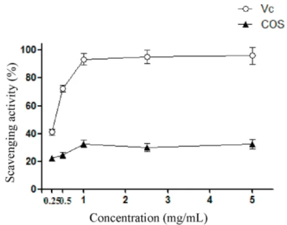
The scavenging activities of all samples increased with the increasing concentrations (Figure 1). The scavenging activity for COS and Vc was 32.5% and 96.1%, respectively, at the concentration of 5 mg/mL. In addition, Vc showed higher superoxide radical scavenging activity than COS (P<0.05).
The results indicate that the hydroxyl radical scavenging activities increased with the increasing concentration of COS, demonstrating a correlated increase in antioxidant activity (Figure 2). Specifically, the highest COS concentration of 5 mg/mL resulted in 41.9% hydroxyl radical scavenging.
The results revealed that the COS had the similar free radical scavenging activity when compared with standard control Vc (Figure 3). The scavenging activity for COS and Vc was 89.1% and 98.8%, respectively, at the concentration of 5 mg/mL. In addition, Vc showed higher DPPH radical scavenging activity than COS (P<0.05).
Compared with the control diet, the intake of HFD resulted in a significant increase in body weight at week six (P<0.05) (Table 1). There was no significant difference in body weight between the COS + HFD group and control group (P>0.05).
In this study, changes of the antioxidant enzyme activities in mice were determined. For all the samples tested, COS groups significantly increased the activities of antioxidant enzymes (SOD, GSH-Px, and CAT) in HFD with 0.5% COS group as compared with HFD group (P<0.05) (Tables 2, 3, and 4).
Mice fed HFD showed decreased villus height compared with other groups (P<0.05) (Table 5). On the other hand, HFD with 0.5% COS group showed similar ratio of villus height to depth compared with control mice, indicating that
Data are presented as means ± standard deviation of triplicates. COS - chitooligosaccharides; Vc - ascorbic acid.
Figure 1 - Scavenging activity on superoxide radical of COS and Vc.
Figure 2 - Scavenging activity on hydroxyl radical of COS and Vc.
Data are presented as means ± standard deviation of triplicates. COS - chitooligosaccharides; Vc - ascorbic acid.
Figure 3 - Scavenging activity on DPPH radical of COS and Vc.
Data are presented as means ± standard deviation of triplicates.
adding COS improved the intestinal integrity (Figure 4). Higher and lower villus:crypt ratios (P<0.05) in ileum were shown in HFD with 0.5% COS group compared with HFD group. There was no significant difference between 0.5% COS group and control group.
Discussion
Reactive oxygen species have been associated with the pathogenesis of various disorders such as diabetes, cancer, inflammation, hypertension, neurodegenerative, aging, and arthritis (Chai et al., 2012).
Reactive oxygen species, such as superoxide anion, hydroxyl radical, and so on, are by-products of biological metabolism that have been implicated in the pathogenesis of a variety of diseases (Ou et al., 2007). Superoxide radical, arising either from oxygen activation by physical irradiation or through metabolic process, is considered as the primary ROS. Superoxide radical can further interact with other molecules to generate secondary ROS (e.g., hydrogen peroxide, singlet oxygen, and hydroxyl radical), either directly or prevalently through enzyme or metal catalyzed processes (Liu et al., 2013). Hydroxyl radical, the most active and toxic free radical, can attack and damage almost every bio-macromolecule in living cells. Hydroxyl ions are formed through the redox reaction between metal ions and peroxide, whose biological damage is its capacity to stimulate lipid peroxidation, which occurs when hydroxyl radical is generated close to membranes and attacks the fatty acid side chains of the membrane phospholipids (Urban et al., 1995; Chandel et al., 2012). 2,2-diphenyl-1-picrylhydrazyl radicalis a stable, nitrogen-centered free radical that produces a violet color in ethanol solution. When
Table 3 - Effect of COS on the activities of CAT, SOD, and GSH-Px in livers of mice (U mg protein–1)
Group CAT GSH-Px SOD
Control 88.2±7.4a 44.8±2.1a 90.9±4.5a
HFD 62.5±5.2b 24.4±3.0c 61.8±3.8c
HFD+0.5%COS 79.3±6.1a 37.1±3.6b 80.5±10.7b
In the same row, values with different letters mean significant differences (P<0.05), while values followed by the same letter or by no letter mean no significant differences (P>0.05).
HFD - high-fat diet; COS - chitooligosaccharides; CAT - catalase; GSH-Px - glutathione peroxidase; SOD - superoxide dismutase.
Table 4 - Effect of COS on the activities of CAT, SOD, and GSH-Px in stomach of mice (U mg protein–1)
Group CAT GSH-Px SOD
Control 59.3±6.2a 16.0±0.9a 45.9±1.9a
HFD 45.8±4.5b 8.7±2.0c 32.0±3.3c
HFD+0.5%COS 53.9±3.9ab 14.9±0.6b 39.7±2.8b
In the same row, values with different letters mean significant differences (P<0.05), while values followed by the same letter or by no letter mean no significant differences (P>0.05).
HFD - high-fat diet; COS - chitooligosaccharides; CAT - catalase; GSH-Px - glutathione peroxidase; SOD - superoxide dismutase.
Table 5 - Villus height, crypt depth, and villus height:crypt depth of ileum of mice fed the normal diet, HFD, or HFD with 0.5% COS, respectively
Group Villus height (µm) Crypt depth (µm) Villus height:crypt depthof ileum (µm µm−1)
Control 226.3±11.7a 51.7±3.2a 4.4±0.8a
HFD 112.8±9.1c 53.3±3.1a 2.1±0.4b
HFD+0.5%COS 179.5±10.2b 49.1±2.7a 3.7±0.7a
In the same row, values with different letters mean significant difference (P<0.05), while values followed by the same letter or by no letter mean no significant differences (P>0.05).
HFD - high-fat diet; COS - chitooligosaccharides.
Table 1 - Body weight changes (g) of mice in each group
Group Initial weight 6-week weight
Control 26.45±0.9a 36.50±0.6a
HFD 26.37±0.7a 38.82±1.3b
HFD+0.5%COS 26.08±1.4a 36.86±1.2a
In the same row, values with different letters mean significant differences (P<0.05), while values followed by the same letter or by no letter mean no significant differences (P>0.05).
HFD - high-fat diet; COS - chitooligosaccharides.
Table 2 - Activities of SOD, GSH-Px, and CAT in serum of mice (U mL–1)
Group CAT GSH-Px SOD
Control 31.2±1.9a 206.8±14.9a 14.7±0.8a
HFD 24.9±0.8b 114.2±16.5b 11.3±0.6b
HFD+0.5%COS 28.5±1.3a 177.8±17.2ab 12.9±0.8ab
In the same row, values with different letters mean significant differences (P<0.05), while values followed by the same letter or by no letter mean no significant differences (P>0.05).
HFD - high-fat diet; COS - chitooligosaccharides; CAT - catalase; GSH-Px - glutathione peroxidase; SOD - superoxide dismutase.
Figure 4 - Morphology of ileum slices in each group of mice.
A - control group; B - HFD group; C - HFD+0.5% COS group.
DPPH encounters proton donors, such as antioxidants, it is reduced to a yellow product, diphenyl picryl hydrazine, and then absorbance decreases. The antioxidant activity of compounds was evaluated by measuring its scavenging capacity for the DPPH free radical (Islam et al., 2012). Therefore, DPPH assay, superoxide anion radical scavenging assay, and hydroxyl free radical scavenging assay were used as relevant indicator of antioxidant activity. Three
in vitro studies showed that COS hasa certain antioxidant
activity and definite radical scavenging activity, which is related with the concentration. When the concentration of COS was increased, the radical scavenging activity was improved. However, this increasing tendency with the higher concentration is limited.
Cells possess a variety of primary and secondary defenses against lipid peroxidation and other deleterious effect of oxidative damage. Primary defenses were mainly preventative, which depend on scavenging/inactivation of reactive oxygen species (ROS) or redox metal ions before lipid peroxidation takes place. They include glutathione peroxidase (GPx); superoxide dismutase (SOD), which scavenge superoxide anion radical and hydrogen peroxide at low concentration; catalase, which scavenges hydrogen peroxide at high concentration, whereas secondary defenses have protective role, which involve excision/repair of any lesion that develop(Girotti, 1990 et al.; Nadithe and Bae., 2010; Mohamed Sadek, 2012).
We evaluated the potential antioxidant activity of COS
in vivo. From the results, we observed that the activity of
SOD, CAT, and GPx decreased in mice fed HFD compared with those in mice fed control group (P<0.05), which is in agreement with another study (Feillet-Coudray et al., 2009). However, the administration of HFD with 0.5% COS resulted in significant increase in the activity of SOD, CAT, and GPx in stomach, liver, and serum of mice as compared with the HFD-fed group (P<0.05). This means that COS has certain antioxidant activity and can restore the activity of the enzymes affected by the HFD.
Conclusions
This study shows that chitooligosaccharides have a certain in vitro radical scavenging activity. Administration of high-fat diet with 0.5% chitooligosaccharides can significantly decrease the damage caused by the high-fat diet, in which the activities of antioxidant enzymes (superoxide dismutase, catalase, and glutathione peroxidase) in serums, livers, and stomachs of mice decreased. These results suggest that chitooligosaccharides have potent antioxidant activity that can protect mice from oxidative stress.
Acknowledgments
This work was supported by Food Science and Engineering, the most important discipline of Zhejiang province, and the National University Student Innovation Program (GJ201510002). The authors thank the Zhejiang Provincial Key Laboratory of Food Safety for providing the equipment and facilities.
References
Aam, B. B.; Heggset, E. B.; Norberg, A. L.; Sørlie, M.; Vårum, K. M. and Eijsink, V. G. H. 2010. Production of chitooligosaccharides and their potential applications in medicine. Marine Drugs 8:1482-1517. doi: 10.3390/md8051482
Chai, Y. Y.; Wang, F.; Li, Y. L.; Liu K. and Xu, H. 2012. Antioxidant activities of stilbenoids from Rheum emodi wall. Evid-based Complementary and Alternative Medicine 2012:603678. doi: 10.1155/2012/603678
Chandel, M.; Sharma, U.; Kumar, N.; Singh, B. and Kaur, S. 2012. Antioxidant activity and identification of bioactive compounds from leaves of Anthocephalus cadamba by ultra-performance liquid chromatography/electrospray ionization quadrupole time of flight mass spectrometry. Asian Pacific Journal of Tropical Medicine 5:977-985. doi: 10.1016/S1995-7645(12)60186-2 Feillet-Coudray, C.; Sutra, T.; Fouret, G.; Ramos, J.;
Wrutniak-Cabello, C.; Wrutniak-Cabello, G.; Cristol, J. P. and Coudray, C. 2009. Oxidative stress in rats fed a high-fat high-sucrose diet and preventive effect of polyphenols: Involvement of mitochondrial and NAD(P)H oxidase systems. Free Radical Biology and Medicine 46:624-632. doi: 10.1016/j.freeradbiomed.2008.11.020
Finkel, T. and Holbrook, N. J. 2000. Oxidants, oxidative stress and the biology of ageing. Nature 408:239-247. doi: 10.1038/35041687 Girotti, A. W. 1990. Photodynamic lipid peroxidation in biological
systems. Photochemistry and Photobiology 51:497-509.
Islam, M. R.; Parvin, M. S. and Islam, M. E. 2012. Antioxidant and hepatoprotective activity of an ethanol extract of Syzygium jambos
(L.) leaves. Drug Discoveries & Therapeutics 6:205-211. Kim, S. J.; Han, D.; Moon, K. D. and Rhee, J. S. 1995. Measurement
of superoxide dismutase-like activity of natural antioxidants. Bioscience, Biotechnology, and Biochemistry 59:822-826. doi: 10.1271/bbb.59.822
Liu, J.; Jia, L.; Kan, J. and Jin, C. H. 2013. In vitro and in vivo
antioxidant activity of ethanolic extract of white button mushroom (Agaricus bisporus). Food and Chemical Toxicology 51:310-316. doi: 10.1016/j.fct.2012.10.014
Liu, P.; Piao, X. S.; Kim, S.W.; Wang, L.; Shen, Y. B.; Lee, H. S. and Li, S. Y. 2008. Effects of chito-oligosaccharide supplementation on the growth performance, nutrient digestibility, intestinal morphology, and fecal shedding of Escherichia coli
and Lactobacillus in weaning pigs. Journal of Animal Science 86:2609-2618. doi: 10.2527/jas.2007-0668
Mohamed Sadek, K. 2012. Antioxidant and immunostimulant effect of carica papaya linn. Aqueous extract in acrylamide intoxicated rats. Acta Informatica Medica 20:180-185. doi: 10.5455/aim.2012.20.180-185
Ngo, D. N.; Kim, M. M. and Kim, S. K. 2012. Protective effects of aminoethyl-chitooligosaccharides against oxidative stress in mouse macrophage RAW 264.7 cells. International Journal of Biological Macromolecules 50:624-631. doi: 10.1016/j.ijbiomac.2012.01.036 Okamoto, Y.; Watanabe, M.; Miyatake, K.; Morimoto, M.; Shigemasa,
Y. and Minami, S. 2002. Effects of chitin/chitosan and their oligomers/monomers on migrations of fibroblasts and vascular endothelium. Biomaterials 23:1975-1979.
Ou, S. Y.; Jackson, G. M.; Jiao, X.; Chen, J.; Wu, J. Z. and Huang, X. S. 2007. Protection against oxidative stress in diabetic rats by wheat bran feruloyl oligosaccharides. Journal of Agricultural and Food Chemistry 55:3191-3195. doi: 10.1021/jf063310v
Qi, X. 2010. Reactive oxygen species scavenging activities and inhibition on DNA oxidative damage of dimeric compounds from the oxidation of (-)-epigallocatechin-3-O-gallate. Fitoterapia 81:205-209. doi: 10.1016/j.fitote.2009.09.004
Qiao, Y.; Bai, X. F. and Du, Y. G. 2011. Chitosan oligosaccharides protect mice from LPS challenge by attenuation of inflammation and oxidative stress. International Immunopharmacology 11:121-127. doi: 10.1016/j.intimp.2010.10.016
Urban, T.; Hurbain, I.; Urban, M.; Clément, A. and Housset, B. 1995. Oxidants and antioxidants. Biological effects and therapeutic perspectives. Annales de Chirurgie 49:427-434.
Xu, W.; Jiang, C.; Kong, X. and Liu, W. S. 2012. Chitooligosaccharides and N-acetyl-D-glucosamine stimulate peripheral blood mononuclear cell-mediated antitumor immune responses. Molecular Medicine Reports 6:385-390. doi: 10.3892/mmr.2012.918 Yeo, J. D.; Jeong, M. K.; Park, C. U. and Lee, J. H. 2010. Comparing