UNIVERSIDADE DE LISBOA Faculdade de Medicina de Lisboa
SOCIAL DISTRESS AND PAIN MODULATION: FINDINGS FROM HEALTHY AND CHRONIC PAIN PATIENTS
Rita Isabel Mangerico Canaipa
Advisor: Alexandre Lemos de Castro Caldas, MD, PhD Co-Advisor: Roi Treister, PhD
Thesis specially prepared for the degree of Doctor in Biomedical Sciences, Specialty Neuroscience
1 UNIVERSIDADE DE LISBOA
Faculdade de Medicina de Lisboa
SOCIAL DISTRESS AND PAIN MODULATION: FINDINGS FROM HEALTHY AND CHRONIC PAIN PATIENTS
Rita Isabel Mangerico Canaipa
Advisor: Alexandre Lemos de Castro Caldas, MD, PhD Co-Advisor: Roi Treister, PhD
Thesis specially prepared for the degree of Doctor in Biomedical Sciences, Specialty Neuroscience
Jury:
President: José Luís Bliebernicht Ducla Soares, MD, PhD Roi Treister, PhD
Jaime da Cunha Branco, MD, PhD
Fernando Manuel Pimentel dos Santos, MD, PhD Sónia Gomes da Costa Figueira Bernardes, PhD Alexandre Lemos de Castro Caldas, MD, PhD Daniel José Branco de Sampaio, MD, PhD
Maria Isabel Segurado Pavão Martins Catarino Petiz, MD, PhD
This thesis was supported by the PhD grant SFRH/BD/42709/2008 from Fundação para a Ciência e a Tecnologia.
2 The opinions expressed in this publication are the exclusive responsibility of the author.
3 The impression of this work was approved by the Scientific Council of the Faculdade de Medicina de Lisboa in the meeting of 17th November 2015.
4
To Margarida, the shiny little flower that blossomed during this work
5 ACKNOWLEDGMENTS
This dissertation was a challenging journey that reached its final goal thanks to the efforts of many. The list is long, as long as my PhD. First, I wish to express my gratitude for those who participated in the studies reported here. Some enthusiastically and some fearfully, all generously accepted the painful challenge and rejections feelings inflected in these studies. I also thank their smiles at their encouraging words at the end, when they understood the real aim of the study.
I want to thank Prof. Alexandre Castro Caldas for giving me the honor of accepting to supervise this dissertation when it only had an aim but no method, for accepting my ideas and proposals, for providing all the resources and support for developing this work and patiently recognize the difficulties we faced.
I gratefully acknowledge Prof. Roi Treister for giving me the honor of accepting this challenge, for teaching me about pain research and supervising all the details even with an ocean between us. Thanks for his unique way of motivating me, stimulating me to do better, and for helping me enjoy this journey.
I thank Prof. João Manuel Moreira, Prof. Jaime Branco and Prof. Fernando Pimentel Santos for all their help in this project, for open their institutions to this work and for collaborating in the studies.
For Myos, their members, particularly Carolina Lopes, Inês Afonso, Maria João Freire, Florinda Salgueiro and António Amaral, and for all the Fibromyalgia patients, my words can never be enough. They were the driving force for my scientific interrogations and for this PhD. They helped teach me how devastating pain can be, and committed to contribute to the fight against pain and suffering. A fight that helped me develop as a professional and a human being.
I also thank to Dr. Patricia Nero, Dr. Teresa Pedrosa, Nurse Maria José Martins and Ana Catarina Matias, Prof. Sónia Gonçalves, Prof. Fatima Serralha and Dr. Licinia Alfaiate for their help in the recruitment of participants. I thank Dr. Débora Oliveira for the kind efficiency, helping resolve all the troubles and fulfill my needs during this dissertation.
6
Thank to Eng. Tiago Araújo for its brilliant “SOS technical support” during data collection and to Salomé Fletcher e Ruben Fletcher for their friendship and help with the English editing of the second study.
I would like to express my gratitude to all my friends. I cannot say all their names, but should mention at least Sara Fernandes, Carla Abreu, Rute Pires, Carlos Botelho, Luísa Patrão and Jacinto Nunes. My special gratitude goes also to Inês Rodrigues that was going through similar challenges and was always trying to look on the bright side. I also want to thank her for the small piece of paper that changed everything.
Thanks to my family, my big and strong family that soon taught me the benefits of social inclusion, particularly to my precious sister Célia and “brother” Hugo, to my special cousin Mafalda, my grandmother (an inspiring woman!), to my parents-in-law and to my parents. For them specially, because they teach me that there are no limits to love and knowledge, for giving me the confidence and freedom to choose my steps and to learn from my mistakes. Thanks to my wonderful three little girls, my nieces Beatriz, Maria Leonor and to my daughter Margarida. For their unconditional love that help me realize during this work that I was, at least to some extent, going in the right direction.
My final and most special words are for Luis, for love and for being the most sacrificed person with this dissertation, for helping with the technical details and so many other things, for supporting all the bad moments and the difficulties raised by my sometime childish ideas and ideals.
This dissertation was supported by a Portuguese Foundation for Science and Technology PhD grant SFRH / BD / 42709 / 2008. Gratefully Acknowledged.
7 This pain
It is a glacier moving through you
And carving out deep valleys
And creating spectacular landscapes
And nourishing the ground
With precious minerals and other stuff
So, don't you become paralyzed with fear
When things seem particularly rough
John Grant in Glacier, Pale Green Ghosts, (The perfect soundtrack for this dissertation)
8 TABLE OF CONTENTS Page Abstract Resumo Chapters outline 1. Pain ……… 22 1.1. Generation of pain ……….. 22 1.1.1 Transduction ……… 22 1.1.2 Transmission ……… 23 1.1.3 Modulation ……….. 29
Descending Pain Modulatory System ……… 30
Opioids ……… 33 Serotonin ……….. 33 Noradrenaline ……… 34 Dopamine ………. 35 Other neurotransmitters ……… 35 Glial Cells ……… 36 1.1.4 Perception ……… 37 Cognitive Modulation ……… 37 Placebo Analgesia ……… 39 Emotional Modulation ………. 40 1.2 Pain assessment ………. 41
1.2.1 Pain assessment in clinic ……… 41
1.2.2 Pain assessment in laboratory settings ………. 42
2. Chronic Pain ……….……… 44
2.1 Changes in pain-related brain areas ………. 46
2.2 Fibromyalgia ………. 48
2.2.1 Studied mechanisms ……….. 50
2.2.2 Structural findings ……… 53
2.2.3 Functional findings ……….. 54
2.2.4 Neurochemical findings ……… 56
2.3 Differences between FM and other Rheumatic diseases……… 57
2.3.1 Emotional modulation of pain in FM and other Rheumatic diseases ………. 59
2.3.2 Social modulation of pain in FM and other Rheumatic diseases ……… 61
3. Social modulation of pain ……….. 63
3.1 Social pain/social distress theory ……… 63
3.2. Social distress and pain ……… 68
3.3 Induction of social distress in laboratory settings ………. 70
4. Studies’ aims and hypotheses ……… 72
9
5.1 Abstract ……… 74
5.2 Introduction ……….. 75
5.3 Material and methods ……… 76
5.3.1 Participants ……….. 76
5.3.2 Tools ……….... 77
5.3.2.1 Experimental apparatus ……… 77
5.3.2.2 Social distress manipulation and assessment … 77 5.3.2.3 Pain stimulation Pre-Cyberball ……… 78
Familiarization trial ………. 78
Calibration ………. 78
5.4.2.4 Pain stimulation Post-Cyberball ……….. 79
5.3.3 Questionnaires ……….. 79
5.3.4 Procedure ………. 80
5.3.5 Statistical analysis ……… 81
5.4 Results ……… 80
5.4.1 Subjects’ characteristics and manipulation check ………… 81
5.4.2 Baseline pain and unpleasantness thresholds ………. 82
5.4.3 Social distress ………. 82
5.4.4 Relations between baseline pain, unpleasantness thresholds and social distress ……….. 84
5.4.5 Post-Cyberball pain and unpleasantness ……… 84
5.5 Discussion ……… 85
6. Second Study – Fibromyalgia patients ……….. 89
6.1 Abstract ………..………. 89
6.2 Introduction ……….. 90
6.3 Methods ……….. 91
6.3.1 Participants ………. 91
6.3.2 Social distress manipulation ……… 92
6.3.3 Experimental pain assessment ……….. 92
6.3.3.1 Electrical Stimulation ……….. 92
Familiarization ……… 93
Stimulation before and during each study condition ……… 93
6.3.3.2 Cold Stimulation ………. 93
Familiarization ……… 93
Cold pressor test during each study condition …… 94
6.3.4 Clinical Assessment ……… 94
6.3.4.1 Clinical pain intensity ……….. 94
6.3.4.2 Brief Pain Inventory ………. 94
6.3.4.3 Fibromyalgia Impact Questionnaire ………. 94
6.3.4.4 Health Assessment Questionnaire ………. 95
6.3.4.5 Short-Form Health Survey SF-36 ………. 95
6.3.4.6 Hospital Depression and Anxiety Scale ………….. 95
6.3.4.7 Medical Consumption ………. 95
6.3.5 Procedure ………. 95
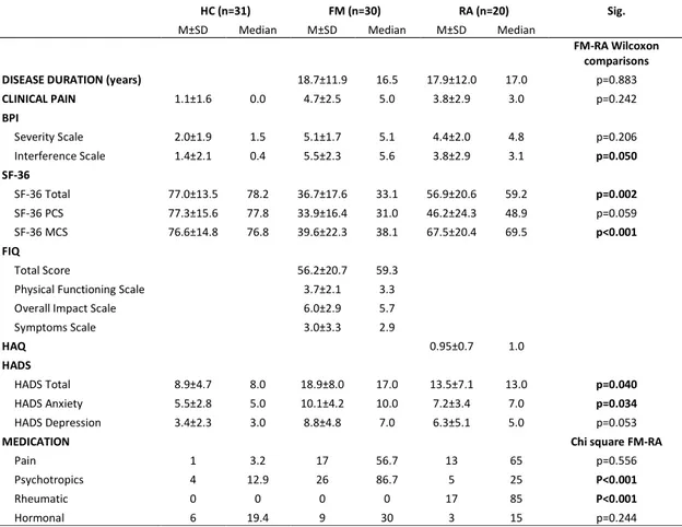
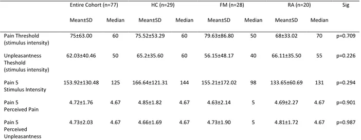
10 6.4 Results ……… 96 6.4.1 Subjects characterization ……….. 96 6.4.2 Manipulation check ……… 99 6.4.3 Baseline Pain ……….. 99 6.4.4 Social Distress ………. 100
6.4.5 The effect of social distress on pain ……… 101
6.4.5.1 Between Groups Comparison ……… 101
Electrical pain ………. 101
Cold pain ……… 101
6.4.5.2 Within Groups Comparison ………. 101
Electrical pain ………. 101
Cold pain ……… 103
6.4.6 Correlations between social distress and effects of Cyberball on pain ……….. 103
6.5 Discussion ……… 103
7. General conclusions ……….. 106
8. References ……… 110
11 LIST OF TABLES
Page Table 1: Sensory testing of peripheral and central mechanisms in
fibromyalgia, osteoarthritis and rheumatoid arthritis ……… 59
Table 2: Participants’ demographic characteristics ……….. Table 3: Participants’ scores in the Experiences in Close Relationships Questionnaire and Neuroticism Scale of Big Five Inventory ……… 82
Table 4: Participants’ demographic characteristics .………..…………. 96
Table 5: Patients’ characteristics in each study group ……..……… 98
Table 6: Baseline pain sensitivity ……….. 99
Table 7: Social distress scores ……….. 100
Table 8: Percentage of change in pain and unpleasantness during each Cyberball condition ……… 102
12 LIST OF FIGURES
Page Figure 1: Cortical and subcortical brain regions related to pain perception
connections ……… 27
Figure 2: The lateral pain system processing sensory components of pain
……….... 28
Figure 3: Development of pain modulation concept .………. 30 Figure 4: Schematic representation of the descending pain modulatory system
………. 32
Figure 5: Neurochemical control of descending pain modulatory system ………… 36 Figure 6: Representation of Cyberball Inclusion and Exclusion condition as the participant sees it in the computer ………... 65 Figure 7: Social distress after Cyberball in the 3 game conditions ………. 83 Figure 8: Pain and unpleasantness intensity after Cyberball in the 3 game
13 LIST OF ABREVIATIONS
ACC = anterior cingulate cortex AI = anterior insula
BDNF = brain-derived nerve growth factor CGRP = calcitonin gene-related peptide CPM = conditioned pain modulation CVLM = caudal ventrolateral medulla DLF = dorsolateral funiculus
DLPFC = dorsolateral prefrontal cortex DMN = default mode network
DNIC = diffuse noxious inhibitory control EAN = executive attention network FM = fibromyalgia
fMRI = functional magnetic resonance imaging GABA = gamma-aminobutyric acid
HC = healthy controls
HPA = hypothalamic-pituitary-adrenal
IASP = International Association for Study of Pain MVN = medial visual network
NA = nucleus accumbens NMDA = N-methyl-D-aspartate
14
NPS = numerical pain scale OA = osteoarthritis
PAG = periaqueductal gray PFC = prefrontal cortex RA = rheumatoid arthritis
RVM = rostral ventromedial medulla TRP = transient receptor potential VAS = verbal analogue scale VPL = ventral posterolateral
15 ABSTRACT
Pain is a complex experience that integrates sensory, emotional and cognitive dimensions. Understanding how these different dimensions integrate this experience and how each of these dimensions can modulate pain is thus a challenging task. Growing body of evidence showed in the last decades that the central nervous system can increase and decrease the noxious information via the descending pain modulatory system, a conjunction of pro-nociceptive and anti-nociceptive projections tracks that modulate pain. Deficiencies in this system have been proposed has a key element of some chronic pain conditions, mostly in those particularly known to involve central sensitization mechanisms, as Fibromyalgia syndrome.
Among several emotional dimensions that can modulate pain, it has been proposed that social distress threatens well being in a similar mode as pain does, and may share neurocognitive resources and mechanisms with physical pain. In this view it would be expected that social distress would significantly modulate pain experience, but this prediction has not been well established in healthy subjects. Furthermore, this was not, to the best of our knowledge, tested in chronic pain, which is a huge public health problem that, according to the International Association for the Study of Pain, is believed to affect more than 20% of the population worldwide. Based on these theoretical grounds, two studies were developed with the aim of investigating how social distress manipulations modulate pain experience in healthy and chronic pain patients.
In the first study, we aimed to understand the relationship between social distress and pain intensity and unpleasantness in healthy individuals. Sixty participants were enrolled to one condition of a well validated paradigm that induce social distress, the Cyberball game. Electrical stimulation protocol was induced before and after playing the game.
It was found that participants that had a lower electrical unpleasantness threshold were also more distressed by the Cyberball game (p=0.012) and that the manipulation itself affected pain intensity ratings (p=0.001). The relationship between social distress and physical pain was not related to attachment styles or neuroticism. Overall, this study
16
provided evidence that sensitivity to social distress is related to sensitivity to physical pain and that social distress modulates pain in healthy individuals.
In the second study, 90 participants were recruited to a study aimed to further investigate how social distress could modulate the pain experience in response to experimental pain models in healthy and two chronic pain conditions: Fibromyalgia, a condition that although recently recognized to have peripheral abnormalities is classically related to central sensitization mechanisms, and Rheumatoid Arthritis, a condition with a well described peripheral inflammation mechanism but less information regarding central sensitization mechanisms. Each participant played the Inclusion and Exclusion condition of Cyberball while pain was induced before and during each condition.
In line with the first study, healthy controls (pain=-13.71±45.28; unplesentness,=-20.78±28.7) and rheumatoid arthritis patients (pain=-7.50±34.54; unplesentness=-5.60±38.04) demonstrated a reduction in pain intensity ratings in response to the electrical induced pain in the Inclusion condition, suggesting the recruitment of the anti-nociceptive projections of the descending pain modulatory system, while in fibromyalgia patients, pain (7.50±26.04, p=0.019) and unplesentness (2.86±31.98, p=0.021) were significantly increased during the same condition. This suggests an impairment of the descending pain modulatory system in fibromyalgia.
These results are discussed in line with evidence of impaired anti-nociceptive projections and changes related to chronic pain that have been found to occur in brain areas as insula, anterior cingulate and midbrain projections, fundamental areas for social connection. Further studies are needed to collect additional information on the nature of the descending pain modulatory system deficits in fibromyalgia. We hope that the increased knowledge regarding the relationships between social events and pain modulation will provide relevant insights for new social and emotional therapeutic approaches in chronic pain conditions, and ultimately contribute to reducing suffering.
Key words:
17 RESUMO
A dor é uma experiência complexa que integra dimensões sensoriais, emocionais e cognitivas. Compreender de que forma estas diferentes dimensões se integram nesta experiência e como é que cada uma delas modula a dor tem-se revelado uma tarefa desafiadora do ponto de vista científico. O crescente desenvolvimento de investigação nas últimas décadas tem demonstrado que essa integração se relaciona com a capacidade do sistema nervoso central inibir ou potenciar o processamento da informação dolorosa através do sistema de modulação descendente da dor.
Este sistema integra áreas como o córtex préfrontal, o córtex do cíngulo anterior e o córtex da ínsula, áreas relacionadas com a componente emocional da dor, em ligação com diversos núcleos do tronco cerebral, sobretudo a substância periaqueductal cinzenta e os núcleos ventromediais rostrais do bolbo raquidiano. Estes núcleos comunicam com a espinhal medula através de projeções serotonergicas, noradrenergicas e dopaminergicas descendentes, aumentando ou diminuindo o processamento de informação. Deste modo, essas projeções tanto poderão ter um efeito inibitório no processamento da dor, isto é, antinociceptivo, como poderão ter um efeito excitatório no processamento da dor, isto é pronociceptivo. Tem sido proposto que deficiências neste sistema modulador descendente poderão ser um aspeto central de algumas síndromes de dor crónica, nomeadamente naquelas que parecem ter um maior envolvimento de mecanismos de sensibilização central, como a Fibromialgia. De facto, diversos estudos têm evidenciado a existência de deficiências no recrutamento de projeções antinociceptivas e um aumento no recrutamento de projeções pronociceptivas nesta síndrome, facto que poderá contribuir significativamente para a dor generalizada reportada por estes doentes.
Diversos investigadores da área das neurociências sociais acreditam que de entre as emoções que podem relacionar-se com a experiência da dor, o sofrimento social que decorre de situações de perda ou ameaça de relações sociais significativas, poderá ter um papel particularmente importante na sua modulação, partilhando com a experiência da dor diversos mecanismos comportamentais e neurocognitivos. De acordo com esta abordagem, as semelhanças entre estas duas experiências resultam do facto de os
18
humanos, tal como outros mamíferos, serem animais que se desenvolvem em grupos sociais, dependendo não apenas de uma boa condição física mas também de uma boa integração social. Isto poderia, na argumentação dos autores, ter implicado que este sistema social tivesse co-optado os recursos neurocognitivos da dor física, nomeadamente no que diz respeito ao recrutamento das áreas de processamento da componente emocional da dor, como o córtex do cíngulo anterior e a ínsula anterior.
Com base nesta perspetiva, seria de esperar que situações de sofrimento social alterassem significativamente a experiência da dor, mas esta predição tem sido difícil de verificar experimentalmente em indivíduos saudáveis. Acresce ainda que, tanto quanto é do nosso conhecimento, ela nunca foi testada em indivíduos com dor crónica, um importante problema de saúde pública que, de acordo com a International Association for the Study of Pain afeta cerca de 20% da população em todo o mundo.
Esta dissertação foi desenvolvida com o objetivo de integrar as duas áreas de conhecimento apresentadas, o estudo da dor e as neurociências sociais, investigando através de dois estudos, de que forma o sofrimento social poderá modular a experiência da dor, em indivíduos saudáveis e em indivíduos com dor crónica.
O primeiro estudo teve como objetivo compreender as relações entre o sofrimento social e, a desagradabilidade e intensidade da dor, em indivíduos saudáveis. Sessenta participantes foram recrutados e sujeitos a uma condição de um paradigma desenvolvido para induzir sofrimento social, o Cyberball. O Cyberball trata-se de um jogo de computador criado para estudar rejeição social, onde se pretende que o participante passe a bola a outros dois jogadores, que ele pensa serem jogadores “reais” ligados online. Na verdade, o participante está, sem saber, a jogar sozinho com o computador que determina até que ponto será excluído. Neste primeiro estudo, depois de preencherem um conjunto de questionários, os participantes jogaram o Cyberball, tendo-lhes sido aplicado um protocolo de estimulação elétrica antes e depois do jogo.
Os resultados mostraram que os indivíduos que apresentavam um limiar de desagradabilidade da dor mais baixo eram os que sentiam mais sofrimento social durante o jogo (p=0.012). Em segundo lugar, verificou-se que a manipulação induzida pelo jogo alterava a perceção da intensidade da dor aos estímulos elétricos aplicados depois do
19
jogo (p=0.001). Foi ainda possível verificar que a relação entre o sofrimento social e a dor física não se relacionava com o estilo de vinculação ou com o neuroticismo, duas dimensões que têm sido teoricamente relacionadas com a sensibilidade ao sofrimento social. Em resumo, este estudo forneceu evidências de que a sensibilidade ao sofrimento social está relacionada com a sensibilidade à dor física, sobretudo nas suas dimensões emocionais, e que o sofrimento social modula significativamente a dor física em indivíduos saudáveis.
No segundo estudo, noventa participantes foram recrutados com o objetivo de compreender de que forma o sofrimento social modula a dor em indivíduos com dor crónica. Nesse sentido, dois modelos experimentais de dor foram investigados em indivíduos saudáveis e em duas condições de dor crónica: na Fibromialgia, síndrome onde têm sido amplamente estudados os mecanismos de sensibilização central, mas onde só recentemente se reconheceu o envolvimento de mecanismos periféricos e na Artrite Reumatóide, onde pelo contrário, os mecanismos inflamatórios periféricos se encontram bem descritos, mas só recentemente se têm reconhecido evidências relacionadas com mecanismos de sensibilização central. Cada participante jogou duas condições do jogo, Inclusão e Exclusão, sendo-lhe induzidos estímulos dolorosos antes e durante cada condição.
Tal como no primeiro estudo, verificou-se que os indivíduos saudáveis (intensidade da dor=-20.78±28.7; desagradabilidade=-13.71±45.28) e com Artrite Reumatóide (intensidade da dor=-7.50±34.54; desagradabilidade=-5.60±38.04) evidenciavam uma redução na intensidade da dor resultante da estimulação elétrica quando participavam na condição de Inclusão do jogo, sugerindo o recrutamento das projeções antinociceptivas do sistema modelador descendente da dor. Pelo contrário, os indivíduos com Fibromialgia revelaram um aumento de dor durante a mesma condição, sugerindo a existência de deficiências no sistema modulador descendente da dor nesta síndrome, que poderão ser particularmente acentuadas em resposta a emoções ou situações sociais positivas (intensidade da dor=7.50±26.04, p=0.019 e desagradabilidade=2.86±31.98, p=0.021).
20
Estes resultados são discutidos tendo em conta os dados de outros estudos que reportam dificuldades no recrutamento de projeções antinociceptivas na Fibromialgia. Para além disso, estes resultados corroboram também os estudos de neuroimagem que descrevem alterações estruturais e funcionais na dor crónica em áreas como o córtex da ínsula, o córtex da cingulo anterior e as projeções do mesencefalo, áreas que são fundamentais para as motivações e ligações sociais. Alterações nestas áreas poderão ser também centrais nas reorganizações das redes neuronais que se verificam nos processos de transição da dor aguda para os estados de dor crónica.
Os resultados evidenciados pelos estudos aqui descritos destacam a necessidade de desenvolvimento da investigação direcionada à compreensão da natureza das deficiências no sistema modulador descendente da dor na Fibromialgia. Esperamos que o aumento do conhecimento sobre as relações entre as experiências sociais e modulação da dor possam fornecer dados relevantes que se venham a traduzir em novas abordagens terapêuticas sociais e emocionais, para as condições de dor crónica, e com isso contribuir para a redução do sofrimento destes doentes.
Palavras-chave:
Dor, Sofrimento Social, Dor Crónica, Fibromialgia, Sistema Modulador Descendente da Dor
21 CHAPTERS OUTLINE
The aim of the current dissertation is to study the relationship between social distress and pain experience. Based on this goal, this work was organized into three chapters discussing the relevant body of knowledge that sustained the research developed.
The first chapter summarizes baseline pain concepts and current knowledge about its underlying neuronal processing, from the periphery to its central mechanisms. Evidence regarding the pain modulation and perception in healthy individuals are reviewed and pain assessment methods are presented. The second chapter explores the most relevant studies on pain mechanisms in chronic pain, particularly those related to fibromyalgia (FM), its pathophysiology and its similarities and differences to other painful rheumatic conditions. In the third chapter the theoretical proposals and findings on social neuroscience that are believed to contribute to pain are discussed. Based on previous chapters, the forth chapter presents the scope, aims and hypotheses of the PhD project, which involves two studies. The fifth chapter presents the details of the first study in healthy individuals and the sixth chapter presents the second study in two chronic pain conditions: FM and rheumatoid arthritis (RA). Finally, the seventh chapter discusses the most relevant findings from the two studies and concludes this dissertation.
22
Chapter 1
1. Pain
The International Association for the Study of Pain defines pain as “An unpleasant sensory and emotional experience associated with actual or potential tissue damage, or described in terms of such damage” (Merskey et al., 1994). Pain is a complex and personal experience modulated by sensory and psychological processes. It is a fundamental protection mechanism acting as an alarm system. The study of the relationships between sensory and emotional components of pain, are intriguing and have been matter of intense debate in the last decades is the main goal of the present dissertation.
1.1 Generation of pain
1.1.1 Transduction
Pain is induced by noxious stimulus (mechanical, thermal, electrical and chemical). These stimuli can be found in the external environment, but they can either arise from visceral injuries or even arise in the absence of a stimulus (Derbyshire et al., 2004). The detection of the noxious stimuli depends on the activation of nociceptors, receptors which transform the noxious stimulus into electrical signals, acting as sensory transducers (Sherrington, 1947).
Molecularly, nociceptors are “transient receptor potential” (TRP) channels. TRP channels are a “superfamily” of many different ligand-gated ion channels activated using different molecular mechanisms (receptor activation, ligand activation and direct activation). The detection of a stimulus opens the channel pore and allows an influx of cations (sodium or calcium) triggering action potentials that travel through neuronal pathways to the spinal cord and the higher brain centers.
23
Some nociceptors are activated by one type of stimulus while others by more than one, thus each nociceptor may differently contribute to diverse pain sensations. Moreover, nociceptors show numerous interactions with other molecules and are able to express many different voltage-gated channels (sodium, potassium or calcium) (Stucky et al., 2009). Overall, this variability allows the transduction of different stimulus parameters: Its quality, location, threshold, intensity and duration. Nociceptors structure and functioning are highly flexible suggesting the significant role that peripheral mechanisms may have in the integration and modulation of pain signals (Ramsey et al., 2006).
Nociceptors are pseudo unipolar: they have their nuclear body at the dorsal root or at trigeminal ganglion (if they provide information from the head and face). One process runs to periphery and the other is directed to the dorsal root ganglion (or the trigeminal ganglion, accordingly).
For pain, there are two important classes of neuron fibers, the A-delta (medium diameter fibers, myelinated, with velocity of conduction about 5 to 30 meters for second) and C-fibers (smaller diameter, unmyelinated fibers, velocity of conduction, about 0.4 to 1.4 meters for second) (Burgess and Perl, 1967; Bessou and Perl, 1969; Djouhri and Lawson, 2004). The A-delta fibers can be mechanosensitives or mechanothermal and induce the earlier pain sensation, the so-called “primary-pain”, and the first rapid, well localized and sharp sensation. Most of the C-fibers are polimodal and can be activated by any modality. They induce the “second-pain”, the delayed and diffuse sensation and represent the most frequent sensory neurons. Other fibers, A-beta nerve fibers which are low threshold thickly myelinated fibers for touch in the somatosensory system, may also contribute to nociception. But this may be a less usual situation.
1.1.2. Transmission
The information from the nociceptor travels through the periphery to the dorsal root ganglion (or trigeminal ganglion, in case of the head and face) and reach the dorsal horn (or brainstem, in the sensory subnucleus caudalis, accordingly), branches and targets
24
specific segments of spinal cord. In the spinal cord will take place the first synapse, between the nerve fiber and the second order neuron from a cell population called “marginal cells” (Christensen and Perl, 1970).
The synapse between the first and the second neuron involves the release of several neurotransmitters that can be grouped according to its family: non peptidergic (e.g. glutamate) that induce a rapid transmission, or peptidergic (e.g. substance P and calcitonin gene-related peptide, CGRP), involved in slower transmission.
One important step of transmission relates to the branching that occurs in the substantia gelatinosa of the dorsal horn, a local that acts as filter to nociception signals. Here inhibitory and excitatory interneurons modulate the nociception transmission (Todd, 2010). Increases and decreases in the density of some receptors also act as modulators, for example, the endogenous opioids (through the mu- and delta-opiate receptors), GABA (through GABAB receptors) and endogenous cannabinoids (through CB1 receptors) can dynamically change the transmission in response to inner (e.g., inflammation) or outer conditions (Woolf and Ma, 2007; Kantamneni, 2015).
After the first synapse, the spinal cord projection neurons (second order neurons for pain processing) cross the midline and project rostrally in the contralateral white matter, in the spinothalamic tract, and reach several brainstem areas as caudal ventrolateral medulla, nucleus of the solitary tract, lateral parabrachial area, periaqueductal grey (PAG) and thalamic nuclei (ventral posterolateral nuclei, posterior group, and posterior triangular nucleus). These nuclei are interconnected composing different pain pathways.
Most of the brainstem nuclei are projection targets from dorsal horn second order neurons, as caudal ventrolateral medulla (CVLM) and PAG but they are also strongly involved in descending projection that modulate dorsal horn pain processing. The CVLM and the nucleus of the solitary tract are related to cardio-respiratory reactions to pain (Lima and Almeida, 2002) and connect to rostral ventromedial medulla (RVM), inhibiting its excitatory neurons. Lateral parabrachial area projects to hypothalamus and amygdala (Gauriau and Bernard, 2002) and is related to the emotional and autonomic components of pain experience.
25
PAG is considered one of the most important subcortical regions implicated in pain processing and modulation. This recognition was first established by Reynolds (1969) who observed that the stimulation of this area in the awaked rat could induce an analgesic reaction. PAG establishes reciprocal projections with several cortical and subcortical pain modulation areas (motor cortex, anterior cingulated cortex, amygdale, and thalamus) and may acts on RVM or directly communicate to the dorsal horn, modulating pain transmission.
Rostral ventromedial medulla receives inputs from PAG and acts on dorsal horn through GABA (γ-aminobutyric acid)-ergic projections. It has a biphasic functioning mode, establishing activation or deactivation connections through the dorsolateral funiculus to dorsal horn with spinal cord. RVM interacts with other important nuclei for pain processing: the nucleus raphe magnus (rich in serotonergic projections), locus coeruleus and other pontine areas (rich in noradrenergic projections) and nucleus reticularis gigantocellularis. These nuclei induce facilitatory and inhibitory connections with dorsal horn as it will be described further. The RVM receives projections for most of cortical pain brain areas, namely, anterior cingulate, insula, and prefrontal cortex. These connections increase under stress situations and have been related to increased arterial pressure and sympathetic activity (Dampney et al., 2002; Gabbott et al., 2005).
Thalamic nuclei are among the most important projection sites for dorsal horn neurons. Many thalamic nuclei receive these inputs, for example, the ventral posterolateral (VPL) nucleus and the posterior triangular nucleus (Gauriau and Bernard, 2004). The first nuclei have reciprocal projections with the primary somatosensory cortex and the later with the secondary somatosensory and insula cortex. Thalamic activations have been related to vigilance, attention, and pain modulation processes, participating in both the sensory and emotional component of pain (Peyron et al., 2000).
The ascending pain signals are further transmitted via a third order neurons projecting from the described nuclei to several forebrain regions. One of these regions is the somatosensory cortex. These activations relates to the sensory components of pain, such as its quality, intensity, and location (Price, 2000). Even though primary and secondary somatosensory cortex may be activated under pain, primary somatosensory
26
cortex is only activated when the stimulation involves wider body areas or when temporal summation occurs (Staud et al., 2008), being its activations more related to non-nociceptive stimulation (Hu et al., 2015). Another frequently activated region is the Posterior Parietal cortex, which integrate information from different sensory systems and memory processes (Peyron et al, 2000; Price, 2000).
The posterior portion of the insular cortex contributes to the perception of pain intensity (Peyron et al., 2000). In its anterior areas it is related to unset of stimulation (Hu et al., 2015), pain unpleasantness, interoception, introspection and feelings of bodily discomfort (Craig, 2009). It is strongly interconnected with the anterior cingulate cortex (ACC) another key region for the experience of pain that "seems to have a vital and complex role in the interrelation of attentional and evaluative functions with the establishment of emotional valence and response priorities" (Price, pp. 1771, 2000). ACC encodes the affective processing of painful stimuli, its unpleasantness (Rainville et al., 1997), and participates in pain modulation (Tracey and Mantyh, 2007). It is composed of several sub neuroanatomical areas that may be specifically related to different functions (Peyron et al., 2000). As such, the rostral area encodes emotional feelings and is rich in descending projections to brainstem pain modulation regions. ACC is related to placebo analgesia (Bingel et al., 2006) and pain reduction due to distraction and attentional processes (Valet et al., 2004).
Prefrontal cortex has been extensively described has a fundamental region in pain control (for recent reviews see Jeon and Friederici, 2015; Opris and Casanova, 2014). One of its most significant areas is the dorsolateral prefrontal cortex (DLPFC) that relates to attentional processes and emotional control, and has also a key role in pain modulation due to its projections to other cortical and subcortical regions (Lorenz et al., 2003; Wager et al., 2004). It play an important role in placebo analgesia and is usually correlated to low catastrophization and pain control (Seminowicz et al., 2006; Loggia et al., 2015).
The relevance of basal ganglia in pain has been increasingly recognized in the last decade. Basal ganglia are involved in different processes (motor, emotional and cognitive tasks) and it has been considered as a local of “multisensory integration” (Nagy et al., 2006). Concerning pain, this region is an important relay center between cortical and
27
thalamic projections and might be related to many aspects of pain experience: The emotional, cognitive, motor and autonomous components. It receives projections from spinal cord and from brainstem nuclei as well as from cortical pain related areas: ACC, DLPFC, insula cortex and hippocampus (Chudler et al. 1995). Activations in basal ganglia, mostly in nucleus accumbens (NA), putamen and caudate are found when participants are exposed to experimental pain (Becerra et al. 2001; Borsook et al., 2010). Specifically, NA has been related to reward-aversion system and it shows decreased activity during pain and increases activity in pain relief. It has been also implicated in placebo analgesia (Scott et al., 2008), in transition from acute to chronic pain and it is proposed as relevant to pain modulation (Mansour et al., 2013).
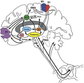
Figure 1: Cortical and subcortical brain regions related do pain perception and their connections (from Apkarian et al., 2005). Primary somatosensory cortex (S1), secondary somatosensory cortex (S2), anterior cingulate cortex (ACC), insula, thalamus, prefrontal cortex (PF), primary motor cortex (M1), supplementary motor cortex (SMA), posterior parietal cortex (PPC), posterior cingulate cortex (PCC), basal ganglia (BG), hypothalamus (HT), amygdala (AMYG), parabrachial nuclei (PB), periaqueductal gray (PAG).
28
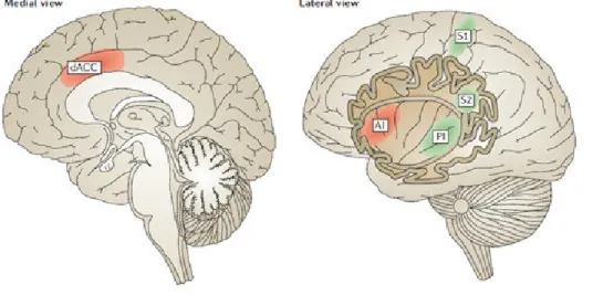
Pain pathways include two major pain components: The sensory-discriminative, involving the “lateral system” and the affective-motivational, involving the “medial pain system” (Albe-Fessard et al., 1985). The lateral pain system processes the sensory aspects of pain, such as intensity, quality and location. It comprises the somatosensory and parietal areas, and posterior insula. The medial pain system ensures the cognitive and affective dimension of pain and involves processing of unpleasantness experiences. It comprises the prefrontal cortex, the ACC and the anterior insula (AI).
Figure 2: The lateral pain system, processing sensory components of pain (green: primary somatosensory cortex, S1, secondary somatosensory cortex S2, posterior insula PI) and the medial pain system processing emotional-cognitive components of pain (red: dorsal anterior cingulated cortex dACC and anterior insula AI) (from Eisenberger, 2012).
Overall, these brainstem and forebrain regions have been considered as part of the “pain matrix” or the brain “pain signature” a set of specific brain areas activated under noxious stimulation, combining the different components of pain experience (Albe-Fessard et al., 1985; Ploghaus et al., 1999; Apkarian et al., 2005; Tracey et al., 2007). This recognition provided a huge development of pain studies but has also raised criticism
29
because it supposes a network specific for pain, failing to explain how these regions are interconnected during pain experience (Iannetti and Mouraux, 2010; Tracey, 2011). Thus, pain is not a sensory modality per se, and unlike the specific cortical areas dedicated to other sensory modalities, there is no “pain” center, rather multiple “pain matrixes” for the different pain conditions/sensations. A criticism to this approach is that several areas of the “pain matrix” are also activated as long has salient sensory (high arousal) and threatening information is conceived (Iannetti and Mouraux, 2010; Tracey, 2011).
Although these criticisms deserve consideration and highlight a need for more accurate concepts, they do not compromised the developing body of studies showing brain functioning under pain states and the relevance of different brain areas in its modulation.
1.1.3. Modulation
Melzack and Wall (1965) historical “Gate Control Pain Theory” was the first theory proposing an explanation concerning the absence of direct relation between stimulus intensity and the pain perceived. On its original work, the authors proposed that the spinal cord and brain could act has a gate, filtering the input of pain processing, modulating pain experience increasing or decreasing it. They believe that this occurred because spinal cord could open or close the gate depending of the activation of two opposing pathways: A nociceptive and non-nociceptive fibers (touch fibers). Even though following studies provided evidence that this was not an accurate mechanism, the Gate Control Theory highlighted the important notion that nociception is modulated along its processing pathways and that there is no direct relation between nociception and the pain experience. Since this initial proposal much more information was developed regarding these modulation mechanisms and it has been recognized the existence of a “Descending Pain Modulatory System”.
30 Figure 3: Development of pain modulation concept (from Bingel and Tracey, 2008).
Descending Pain Modulatory System
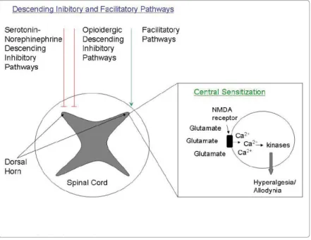
In the dorsal horn of the spinal cord (or in the trigeminal nucleus if the information is from the head and face) peripheral neurons are forming synapses with descending neurons arising from the brain and brainstem centers. One of the most interesting fields of research on pain has been the study of these networks and their ability to modulate pain: The “descending pain modulatory system” (Tracey and Mantyh, 2007; Millan, 2002). This system can have an inhibitory (anti-nociceptive) or facilitatory role (pro-nociceptive) in pain transmission.
In this system two brainstem nuclei, PAG and RVM, play a pivotal role. Several descending pathways starting in the cortex (ACC, AI, PFC), are connected to the
31
hypothalamus and amygdala, and project to PAG. PAG establishes reciprocal connections to RVM, which is directly linked to dorsal horn through the dorsolateral funiculus (DLF).
In the RVM there are three types of neurons: “ON”, “OFF” and neutral. Although the functioning of the neutral neurons is still unknown (most probably they have both type of influences), it has been argued that the OFF cells are tonically active, while the ON cells increase their action when a pain stimulus arises, facilitating pain transmission. When there is no stimulation pro-nociceptive projections are not activated and the anti-nociceptive are the most useful (and as such are under the influence of OFF cells) (Fields et al., 1983; Bederson et al., 1990). When these descending projection reach the spinal cord they modulate the activity of the wide-dynamic-range neurons (and nociceptive neurons of the trigeminal nerve) from the lamina V, neurons that can be activated by nociceptive as well as by non-nociceptive information (Millan, 1999).
Beyond the PAG and RVM, other subcortical nuclei are also important for pain modulation including the nucleus tractus solitarius, the parabrachial nucleus and the dorsal reticular nucleus. Overall, these brainstem and midbrain nuclei are key elements of the descending pain modulatory system and are part of the spino-medullar-spinal loops.
Two other pain modulation mechanisms deserve mention: One is related to the activation of motor cortex and its projection to the ventral horn of the spinal cord, and the other is the indirect impact that the sympathetic nervous system activity may have on the transmission of pain signals at the spinal cord. Thus multiple brain areas and many different descending pathways can modulate the nociception signals along its different processing stages.
The descending pain modulatory system is under serotonergic, dopaminergic and noradrenergic control and is opioid dependent (Gebhart, 2004), but many controversies still persist regarding this and other neurotransmitters functions in this system. In part this is due to the complexity of this system, expressed in its ability of both inhibit or facilitate pain signals, and also to the multiple pathways arising from different brain areas and using different neurotransmitters. Classically it has been proposed that anti-nociceptive projections to dorsal horn of the spinal cord use serotonin, dopamine, noradrenaline and opioids, while pro-nociceptive use substance P, glutamate and nerve
32
growth factor. However, the increased knowledge of the descending system provided evidence that the same neurotransmitter can participate in opposite actions, depending on the receptors involved in each mechanism (Millan, 2002). For example, glutamate, which is best known as the main excitatory neurotransmitter in the nervous system, has been related to increase pain processing through its NMDA receptors (Kawasaki et al., 2004). Nevertheless, another glutamate metabotropic receptor, mGluR1-8, has been related to increased activity in descending pain inhibitory pathway (Palazzo et al., 2011).
Increased evidence supports the view that cortical pain related areas controls spinal function using monoamines top-down projections. This has been considered a probable explanation for sleep, fatigue and emotional disorders usually comorbid of pain states due to the multiple roles that serotonin and noradrenaline can have on these functions (Bannister et al., 2009).
Figure 4: Schematic representation of the Descending Pain Modulatory System (from Bingel and Tracey, 2008).
33 Opioids
Opioids are a group of neuropeptides neurotransmitters: Enkephalins, endorphins, endomorphins, dynorphine and nociceptin. They are usually called endogenous opioids and act in several brain regions. The descending inhibitory system depends on the activity of opioid neurons, mostly through its presynaptic modulation in the spinal cord, which decreases nociceptive transmission. Inhibitory neurons arising from the PAG use opioids to induce analgesia (Park et al., 2010). Indeed, opioid injections in PAG and RVM diminishing the activity of the ON cells and increase the activity of OFF, resulting in activation of the descending inhibitory system and decrease in pain (Heinricher et al., 1992). Opioids have a major role in the placebo analgesia due to the activation of these descending inhibitory pathways (Petrovic et al., 2002). Other brain regions, such as hypothalamus, amygdala, striatum, ACC, also use opioid neurons for modulating pain signals.
Serotonin
Specifically, serotonin (5-hydroxytryptamine, 5-HT) although best known for its impact on mood, has been shown to be a key neurotransmitter in pain pathways. Due to the failure of serotonin in overcome blood-brain barrier, the peripheral and central serotonin constitutes relatively independent pools. In the periphery it is one of the elements of the “inflammatory soup”, directly contributing to its pro-nociceptive effects (Godínez-Chaparro et al., 2011), but in spinal cord and in higher brain centers is actions is less clear, due to its different receptors.
One of most relevant serotonergic projections, which was believed to play a role in reducing pain transmission starts at the RVM, particularly from the raphe nucleus (Viguier et al., 2013). However, it has been recently proposed that these serotonergic descending projections may not only contribute to anti-nociception but also to pro-nociception, if persistent pain is developed (Wai et al., 2011). Accordingly, when glutamate and GABA are in balance, serotonin that usually increase pain signals on periphery is counterbalance by inhibitory descending serotonergic projections. However, when there is an increase in
34
excitatory pain signals from periphery (due to sensitization or other processes) this balance may be lost and the serotonin descending projections may be inhibited. Some debate still persists regarding the descending facilitatory pathway for serotonin, which is complicated by the presence of other neurotransmitters, as neuropeptides (e.g. substance P, enkephalins) and the classical excitatory (glutamate) and inhibitory (GABA) neurons in the medulla that also project to dorsal horn and that may modulate pain signals (Viguier et al., 2013).
Centrally, it is believed that serotonin (specifically the 5-HT7R receptor) can reverse the dendritic dysfunctions (increased excitatory and integration activity) in pain central brain areas, as ACC, and restore the pain sensitivity in neuropathic animal pain models (Santello and Nevian, 2015). Moreover, serotonin is also interconnected to the hypothalamic–pituitary–adrenal axis, which has also a modulating role in the pain system (Andrews and Matthews, 2004).
Noradrenaline
Another key neurotransmitter in the descending pain modulatory system is noradrenaline. Although noradrenaline may have a low relevance in healthy tissues, in case of injury it may have a pro-nociceptive role due to its ability to activate motor neurons (in the ventral horn). It has also been proposed that noradrenaline may impact on the immune system. Similarly to serotonin, it may have either anti-nociceptive or pro-nociceptive roles, depending on the class of receptor that is activated (alfa1 adrenergic receptors are pro-nociceptive while alfa2 adrenergic are nociceptive). Its anti-nociceptive role occurs through presynaptic (blocking the release of excitatory neurotransmitters) or postsynaptic (hyperpolarization) actions in dorsal horn, as well as inhibiting interneurons or suppressing the action of excitatory interneurons. In higher brain centers, noradrenaline role has been not yet fully understood because, again, it may also depends on local and receptors involved (Pertovaara, 2013).
35 Dopamine
Dopaminergic system has been consistently associated to stress (Pani et al., 2010) and pain (Wood et al., 2007; Treister et al., 2013). Even though evidence has been provided regarding its involvement in the descending pain modulatory system (enhancing pain modulation) (Treister et al., 2013), much less knowledge exists about its ability to modulate pain in the descending system comparing to other neurotransmitters. Nevertheless, it has been observed that dopaminergic projections arising from hypothalamic nuclei to spinal cord have anti-nociceptive role through the D2 receptors (Wei et al., 2009; Taniguchi et al., 2011) and D1/D5 receptors (Yang et al., 2005). An increase in dopamine in mesolimbic and other pain related areas, as the medial prefrontal cortex and NA, is implicated in analgesia in animal models (Imperato et al., 1991; Sotres-Bayón et al., 2001). Similar findings are reported in animals subjected to social defeat (Tidey and Miczek, 1996). Dopamine also plays an inhibitory function on the ACC, and allows the inhibition of processing of pain mediated by NMDA receptors.
Recent evidence points toward a decrease in dopamine in the striatum in chronic pain (Martikainen et al., 2015). Indeed, patients with conditions related to a decrease in dopamine have pain complains, as FM (Wood et al., 2007) and Parkinson disease (Lee et al., 2006), and dopamine administration has been related to decreased pain in cancer (Dyckey and Minton, 1972) or diabetes (Ertas et al., 1998).
Other neurotransmitters
Other neurotransmitters may modulate pain through the descending pain modulatory system. One example is Substance P which is an important mediator of inflammation, increasing the function of cells of immune system, the production of pro-inflammatory cytokines and stimulating secretion of histamine from mast cells. It plays a significant role in pro-nociceptive pathways (De Felipe et al., 1998). Another example is Brain-derived neurotrophic factor (BDNF). It has been considered a key element for axonal growth and neuromodulation (Thoenen, 1995). Usually, neurons that express BDNF also express other pain modulators, for example, serotonin, substance P and
36
neurotensin (Yin et al., 2014). When released in the PAG, BDNF stimulates the release of neurotransmitters that increase pain signals in the RVM.
Glial Cells
Finally, it should be mentioned that the role of glial cells in pain modulation has been increasingly recognized in the later years. It has been reported an increase in cytokines, substance P, glutamate, nitric oxide and prostaglandins in microglia and astrocytes cells of the spinal cord (Watkins et al., 2001). The release of these substances by glial cells will increase the noxious transmission of nociceptor fibers in dorsal horn (Wieseler-Frank et al., 2005). Moreover, the impact of these cells at cortical level has also been found, mostly in ACC and other important brain areas of the descending pain system (Ikeda et al., 2013).
Figure 5: Neurochemical control of the descending pain modulatory system (from Lee et al., 2011).
37
In summary, the higher brain processing areas (e.g., prefrontal cortex, ACC, AI) communicate with the brainstem centers, specifically the RVM which receives projections from PAG. These brainstem nuclei are rich in opioid receptors and act through serotonin, dopamine and noradrenaline descending projections to dorsal horn. This, in turn, will influence the peripheral neuronal transmission. The descending pain modulatory system may have an anti-nociceptive or pro-nociceptive role. The network that allows this dual functioning has been extensively studied in the last decade, and it is now recognized that the inhibitory and excitatory processes are dynamic and may be flexible respond to different factors (behavioral, emotional, physiological) (Heinricher et al., 2009). The study of these factors may have an important impact in understanding pain mechanisms and in identification of potential therapeutic targets.
1.1.4. Perception
Increased body of knowledge has contributed to the recognition that pain is a complex experience, a consequence of several peripheral and central processes. Understanding how cognitive and emotional processes are affecting modulation mechanisms is, therefore, of greater interest (Price, 2000).
Cognitive Modulation
Indeed, emotional and cognitive factors can either potentiate or weaken pain perception. Pain perception depends on cognitive processes, as attention and distraction (Tracey et al., 2002). Given that cognitive resources are limited, it has been proposed that performing cognitive tasks can distract from pain, decreasing pain perception, mostly if these cognitive tasks have high attentional demands (Miron et al., 1989; Eccleston and Crombez, 1999; Good et al., 1999; Bantick et al., 2002). Indeed, some studies described that distraction activates the PAG, resulting in analgesia (Tracey et al., 2002) and correlates to increased connection between PAG and ACC and decreased activity of other brain pain-related areas (Bantick et al., 2002). Increased connectivity between ACC, orbitofrontal cortex and thalamus to PAG has also been found when distraction occurs
38
while pain stimulation is induced, suggesting the recruitment of the descending pain system (Valet et al., 2004). These results have been interpreted has indicating that pain and cognition share cognitive limited resources and that engaging attentional and control resources in a cognitive task is able to decrease the pain perception (Vohs et al., 2008).
However, other studies found different results. Seminowicz and Davis (2007) found that under intense pain stimulation, distraction tasks (high or low demanding) may not be able to significantly modulate pain. Similarly, cognitive performance may not be modulated by pain, which lead the author to argue that the brain networks for pain perception and cognitive tasks can be recruited at the same time (Seminowicz and Davis, 2007).
One possible reason for the divergent findings may be related to the differences in the cognitive tasks in use. Perhaps not all cognitive tasks significantly modulate on pain. It may depend if the task is high or low demanding, but more importantly, on cognitive processes that the task involves. Accordingly, it has been proposed that this effect may be higher for inhibitory cognitive tasks, as Stroop task, one of the most used cognitive paradigms in pain studies (Oosterman et al., 2010). This suggests that inhibitory resources are the most relevant both for cognitive tasks and pain perception. Indeed, it has been described that individuals with lower performance in inhibitory cognitive tasks were also more inefficient in the recruitment of the descending inhibitory pain system, a finding that may explain why the elderly have inefficiency in both the cognitive and pain inhibition tasks (Marouf et al., 2014).
Studies comparing these two processes found that exerting cognitive self-control in a previous high demanding tasks increase pain perception and spinal nociception (Silvestrini and Rainville, 2013). This effect occurs even after the cognitive task was finished. Using self-control resources (inhibitory) in cognitive tasks impairs the subsequent use of control resources on pain perception, even at the most basic perceptual pain physiological processes, suggesting limitation in the recruitment of the descending pain system. Further detailed studies are needed in order to fully understand how specific cognitive processes modulate pain perception.
39
Other cognitive processes, as beliefs, have been studied. For example, beliefs related to perceived control over pain stimulation have been related to diminished pain perception and anxiety (Kalisch et al., 2005; Wiech et al., 2006), processes that relay on activation of prefrontal cortex regions (VLPFC) (Wiech et al., 2006). These beliefs have been related to increased connections between the rostral part of ACC and PAG, a mechanism already described under distraction (Bantick et al., 2002) and placebo analgesia (Bingel et al., 2011). Similar findings have been found regarding catastrophization (Raczka et al., 2010).
Placebo analgesia
Placebo analgesia, that is, the relief in pain that occurs when an individual believes that is being subjected to a procedure or substance that reduces pain, has also been a matter of huge debate and provided knowledge about pain perception and the descending pain system (Price et al., 1999; Vase et al., 2005). Even though it is beyond the aims of the present work to go into details on this phenomenon, it is worth mention the dependence of the placebo analgesia on descending pain modulatory system. Molecularly, placebo analgesia has been linked to endogenous opioidergic system (Zubieta et al., 2005). Release of opioids has been found in pain modulation brain areas, as the ACC, the DLPFC and the PAG when the participant is under placebo analgesia (Bingel et al., 2008). The activation of this opioid system may inhibit the pain signals at the spinal cord, as already described. Pharmacological studies described that the use of an antagonist of mu-opioid receptors (naloxone) abolished placebo analgesia reaction. Moreover, the blockage of the opioid system was related to impaired activations DLPFC, rostral ACC, hypothalamus, PAG and RVM, and most importantly reduced the connectivity between rostral ACC and PAG, connections known to be essential for the descending inhibitory functions (Eippert et al., 2009). Interestingly, in this study it was found that the subjective analgesia was not completely blocked, as the subjects still reported decreased pain ratings under placebo analgesia manipulation. This may point toward the relevance of other non-opioidergic systems in pain perception, particularly the monoamine projections (it has been described the relevance of dopamine in placebo analgesia,
40
Schweinhardt et al., 2009) or the presence of a self-consistency bias and cognitive appraisals (Wager et al., 2006).
Emotional Modulation
Another intense aim of research is to learn about emotional modulation of pain. The effect of the emotional context on pain has been usually studied by manipulating emotions during pain induction (Villemure et al., 2003; Tang et al., 2008) based on Lang (1995) motivational priming hypothesis. This theoretical approach proposes the existence of two motivational systems: the appetitive system, which promotes positive emotions, and the defensive system, which promotes negative emotions. If a positive emotion is primed, there is higher probability of positive evaluation of an event, and if a negative state is primed, there is higher probability of a negative evaluation. In pain studies, this approach has been frequently tested using the Lang et al. (1990) International Affective Picture System (IAPS), a set of pictures with positive, negative or neutral content, that are presented while pain is induced.
Generally, it has been found that priming positive emotions decrease pain perception in healthy individuals (Meagher et al., 2001; Kamping et al. 2013; Rhudy et al., 2013). Similar findings have been reported when using different emotional stimuli, as odors (Villemure et al., 2003) or music (Roy et al., 2008). Nevertheless, results of the negative emotional priming are not so clear-cut. Although there is also a general trend toward the corroboration that negative emotions increase pain perception (e.g., Meagher et al. 2001), there has been found some interaction with arousal evoked by the situation (Rhudy et al., 2008) and many other individual differences. For example, it has been reported that under high arousal there may be a decrease in pain perception, usually called the stress-induced analgesia. Indeed, animal studies highlight that a stressful event may result in analgesia or hyperalgesia (Vidal and Jacob, 1986; Jørum, 1988) depending on a multiplicity of parameters related to individual differences (phenotypes, age, gender), the stress event (emotions induced, controllability, ability to escape) and the meaning of the environmental stress (previous experience with the stressful event) (Vidal and Jacob, 1986; Jørum et al., 1988; Butler and Finn, 2009).
41
Although the precise mechanisms that underlay the relation between emotions and pain are not completely understood, neuroimaging studies support the view that emotions impact on the cognitive-affective dimensions of pain, decreasing or increasing the unpleasantness of the pain experience. As such, Yoshino et al. (2010) found that manipulating the emotional context where electrical pain stimulation was induced could have a significant effect on neural responses to pain: The sad context was correlated to an increased activation in brain areas related to emotional aspects, as the ACC. Other neurocognitive mechanisms proposed emphasize the role of insula cortex in the integration of autonomic, pain and emotional processes, which may then activate descending projections to brainstem nuclei (Craig, 2002; Craig 2003a).
Another approach used in the study of emotional dimensions involved in pain perception is the investigation of individuals that show specific emotional traits, as those suffering psychological disorders (Wiech and Tracey, 2009). The affective-cognitive brain areas have been reported to be the key abnormally activated areas in individuals suffering from mood disorders. Depression and anxiety, have been associated with clinical pain complains and increased pain perception in experimental pain stimulation. It is well known that depressed patients describe pain complains (de Heer et al., 2014) and chronic pain patients describe mood disorders (Frank et al., 1988; Buckelew et al., 1994). Although a meta-analysis found that depression is related to lower sensitivity to pain, some inconsistencies have been reported (Dickens et al., 2003). Results from recent a case-control study comparing HCs and recently diagnosed (and non-medicated) major depression patients found the later demonstrated lower pain threshold and tolerance (Zambito Marsala et al., 2015).
1.2. Pain assessment
1.2.1 Pain Assessment in clinic
Because pain is a subjective experience, there are no objective measures to assess this experience in clinical or laboratory setting. It can only be assessed using observational or self-report methods. Observational methods comprise behavioral (posture, facial
42
expressions), functional (mobility) or vital sign assessment (respiratory and pulse rate, blood pressure, sedation) while self-report measures can inform about pain or functional impact directly asking patients.
The self-report measures include unidimensional or multidimensional measures. In the first case, the most frequently used are the numerical rating scale (NRS) and the verbal analogue scale (VAS). The numerical rating scale (NRS) simple ask a patient to choose a number that best represents its pain from “0”, “no pain”, to “10” (or 100), “the worst pain you can imagine”. The verbal analogue scale consists of continuo’s 10 cm scale and the patient can point the pain he/she experience on the scale from 0 to 10. These unidimensional scales can be used to assess pain unpleasantness ratings.
Frequently used multidimensional measures are, for example, the Brief Pain Inventory (BPI, Cleeland and Rayn, 1994) and the McGill Pain Questionnaire (MPQ), questionnaire that ask patients about several aspects of their pain: intensity, quality, relief, etc. (McGill and Togerson, 1971).
1.2.2. Pain assessment in laboratory settings
There are multiple methods to assess nociception in animal models of pain which will not be discussed herein. This section will focus on experimental pain assessment in human.
In laboratory settings pain has been studied using pain induction methods that use thermal, mechanical, electrical and chemical stimuli (Gracely, 1988). These stimuli can be applied in different body location, with variaty of durations, frequencies and intensities. For example, electrical stimulation can be applied on the skin surface or intra-cutaneous, and directed to specific nerves. Electrical stimuli can be repeated, usually have short duration and can be easily quantifiable (the electrical current can be measured). These stimuli have a clear unset and termination, but are prone to habituation effects. Another example of pain induction method in laboratory is the cold pressor test. In this test, the subjects are asked to immerse their hand in a cold water bath as much time as possible (with a safety limit). In the cold pressor test, the pain increases along the immersion. Pain