UNIVERSIDADE DE LISBOA
FACULDADE DE FARMÁCIA
Targeting Microglia Necroptosis to Arrest Neuroinflammation
Sara Rodrigues Oliveira
Dissertação
Mestrado em Ciências Biofarmacêuticas
UNIVERSIDADE DE LISBOA
FACULDADE DE FARMÁCIA
Targeting Microglia Necroptosis to Arrest Neuroinflammation
Sara Rodrigues Oliveira
Dissertação orientada pela Doutora Joana D. Amaral e co-orientada pela
Professora Doutora Cecília M. P. Rodrigues
Mestrado em Ciências Biofarmacêuticas
The studies presented in this thesis were performed within the Cellular Function and Therapeutic Targeting research group, at the Research Institute for Medicines (iMed.ULisboa), Faculty of Pharmacy, Universidade de Lisboa, under the supervision of Joana D. Amaral, PhD. and Cecília M. P. Rodrigues, PhD.
Publications
The studies included in this thesis were presented as abstracts in poster communications:
Oliveira SR, Dionísio PA, Amaral JD, Rodrigues CAB, Afonso CAM, Rodrigues CMP.
Screening for potential modulators of necroptosis in a microglial cell line. 8th iMed.ULisboa
Postgraduate Students Meeting, Lisbon, Portugal, 2016
Amaral JD, Oliveira SR, Dionísio PA, Rodrigues CAB, Afonso CAM, Rodrigues CMP. Screening for potential modulators of necroptosis in a microglial cell line. 10th FENS Forum of
Neuroscience, Copenhagen, Denmark, 2016
Dionísio PA, Oliveira SR, Afonso CA, Rodrigues CMP, Amaral JD. Using BV2 microglial cell line as a model to screen potential modulators of necroptosis. 1st Mind-Brain College
Table of Contents
Abstract ... xi
Resumo ... xiii
Agradecimentos ... xix
Abbreviations ... xxi
I. INTRODUCTION AND OBJECTIVES ... 1
1. Neurodegenerative diseases ... 3 2. Neuroinflammation ... 3 2.1. Microglia-mediated neuroinflammation ... 4 2.2. Inflammatory mediators ... 8 2.2.1. Cytokines ... 11 2.3. NLRP3 inflammasome ... 12
3. Neurodegeneration and mechanisms of cell death ... 12
3.1. Necroptosis ... 13
3.1.1. Necroptosis activation in vitro ... 14
3.1.2. Molecular mechanisms of necroptosis ... 16
3.1.3. Necroptosis and inflammation ... 21
3.1.4. Necroptosis in disease ... 24
3.1.5. Targeting necroptosis ... 26
4. Objectives ... 29
II. MATERIALS AND METHODS ... 31
1. Reagents ... 33
2. Cell culture ... 33
3. Cell treatment ... 33
3.1. Optimization of an in vitro model of microglia necroptosis ... 33
3.2. Screening of small molecules for necroptosis inhibition ... 34
4. Evaluation of cell death and viability ... 34
5. Evaluation of necroptosis signaling pathways ... 36
5.1. Total protein extraction ... 36
5.2. Soluble and insoluble fractions ... 36
5.3. Western blot analysis and densitometric analysis ... 37
6. Evaluation of inflammatory mediators ... 38
6.1. Total RNA extraction and quantitative Real-Time PCR ... 38
6.2. Enzyme-Linked Immunosorbent Assay (ELISA) ... 39
7. Microscopy ... 40
8. Statistical analysis ... 40
III. RESULTS ... 41
zVAD-fmk induces necroptosis in the BV2 microglial cell line ... 43
zVAD-fmk promotes necrosome assembly in BV2 cells ... 45
Screening for potential inhibitors of necroptosis ... 46
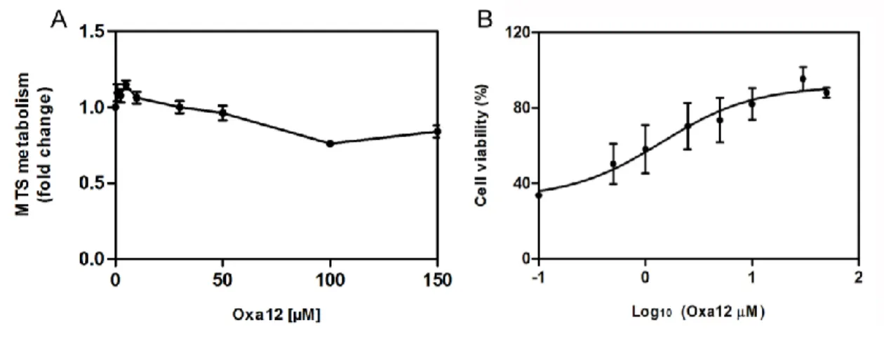
Oxa12 modulates necroptosis in the BV2 microglial cell line ... 48
Oxa12 inhibits necroptosis in the BV2 microglial cell line ... 50
Oxa12 reduces TNF-α gene expression and secretion ... 53
Oxa12 modulates zVAD-fmk-induced JNK and p38 MAPK signaling activation and increases IκB phosphorylation ... 57
IV. DISCUSSION ... 61
V. CONCLUSION AND FUTURE PERSPECTIVES ... 71
Index of Figures and Tables
Figure 1. Neuroinflammation in AD. ... 5
Figure 2. Classically activated microglia in neurodegenerative diseases. ... 8
Figure 3. Necroptosis activators ... 16
Figure 4. RIP1-mediated multimodal signaling events downstream of TNFR1. ... 19
Figure 5. Necroptotic cells release proinflammatory mediators. ... 22
Figure 6. BV2 microglial cells undergo necroptosis when exposed to LPS/zVAD-fmk or zVAD-fmk alone. ... 44
Figure 7. LPS exposure increases RIP1 and RIP3 total protein levels. ... 45
Figure 8. zVAD-fmk treatment only induces necrosome assembly at 24 h of incubation. 46 Figure 9. The first chemical library of twenty compounds does not modulate necroptosis in BV2 microglial cell line. ... 47
Figure 10. Oxa12 modulates necroptosis in the BV2 microglial cell line. ... 48
Figure 11. Oxa12 is effective at inhibiting zVAD-fmk-induced necroptosis without toxicity. ... 49
Figure 12. In silico molecular docking calculations for Oxa12 ... 49
Figure 13. Oxa12 modulates necroptosis in a microglial cell line. ... 52
Figure 14. Oxa12 reverts zVAD-fmk-induced cell morphology. ... 53
Figure 15. Oxa12 decreases TNF-α secretion levels. ... 54
Figure 16. Oxa12 decreases TNF-α gene expression. ... 55
Figure 17. Oxa12 decreases zVAD-fmk-induced JNK and p38 MAPK activation. ... 58
Figure 18. zVAD-fmk incubations does not affect Akt phosphorylation in BV2 cells. ... 58
Figure 19. Oxa12 increases IκB phosphorylation levels when compared to zVAD-fmk-treated cells. ... 59
xi
Abstract
Necroptosis is a caspase-independent form of regulated cell death executed via activation of receptor interacting protein 1 (RIP1) and RIP3, being negatively regulated by caspase-8. Neurodegenerative diseases are hereditary or sporadic conditions characterized by progressive cell death in selected areas of the nervous system, along with oxidative stress, mitochondrial dysfunction and neuroinflammation. Importantly, recent studies have demonstrated that necroptosis is involved in the pathogenesis of several disorders, including neurodegenerative diseases.
Neuroinflammation, in turn, is a response to eliminate the original insult and damaged or dead cells. However, if tissue physiology is not restored, inflammation becomes a chronic condition, where microglia plays a major role. Studies have shown that defects in caspase-8 activation in microglia may promote inflammation through activation of necroptosis.
In the present study, we used the murine BV2 microglia cell line as an in vitro model to screen a library of novel compounds, potential modulators of necroptosis, and evaluate if necroptosis modulation may contribute to attenuate microglia-mediated inflammation. Importantly, we identified one hit Oxa12 that inhibits this type of regulated cell death by preventing all necroptosis-associated events, including RIP1/RIP3 necrosome assembly, and MLKL phosphorylation. In addition, Oxa12 decreased TNF-α gene expression and secretion as well as JNK and p38 MAPK activation, while IκB phosphorylation was increased.
In conclusion, we established a robust in vitro model of microglia necroptosis and identified a strong inhibitor of this regulated form of cell death that might be a promising candidate molecule for targeting RIP1/3-driven pathologies.
xii
Keywords: Drug screening; Microglia; Molecular targeting; Necroptosis; Neuroinflammation;
xiii
Resumo
A necroptose é um tipo de morte celular regulada e independente de caspases, que ocorre quando há ativação das proteínas receptor interacting protein 1 (RIP1) e RIP3, sendo inibida pela caspase-8. As doenças neurodegenerativas são condições hereditárias ou esporádicas caracterizadas pela ocorrência de morte celular progressiva em áreas específicas do sistema nervoso, assim como pela existência de stress oxidativo, disfunção mitocondrial e neuroinflamação. É importante referir que estudos recentes têm demonstrado que a morte celular por necroptose está envolvida na patogénese de várias doenças, incluindo as doenças neurodegenerativas.
A neuroinflamação, por sua vez, visa eliminar tanto a causa inicial do dano celular, como as células mortas que resultam do insulto original. Contudo, se a fisiologia normal do tecido não for restaurada, a inflamação torna-se uma condição crónica. No sistema nervoso central (CNS), são as células da microglia que constituem a primeira linha de defesa, desempenhando um papel crucial na neuroinflamação, através da contínua produção de citoquinas pró-inflamatórias e espécies reativas de oxigénio, entre outros. Estes mediadores inflamatórios podem, por sua vez, ter efeitos neurotóxicos. De facto, a comunicação entre a microglia e os neurónios pode amplificar os sinais inflamatórios e contribuir para a patogénese das doenças neurodegenerativas. De salientar que, tal como descrito recentemente, a inativação de caspase-8 na microglia pode promover inflamação através da ativação da necroptose.
No presente estudo, foi utilizada a linha celular de microglia de murino, BV2, como um modelo in vitro para o estudo de duas bibliotecas de novos compostos, potencialmente moduladores da necroptose. Para além disso, avaliámos se a modulação da necroptose pode contribuir para atenuar a inflamação mediada pela microglia.
Em primeiro lugar, otimizou-se o modelo celular. Os resultados obtidos demonstraram que a incubação de células BV2 com o inibidor de caspases, zVAD-fmk,
xiv resultava em níveis elevados de morte celular por necroptose, tal como avaliado através de ensaios de viabilidade e citotoxicidade. Os resultados foram confirmados por Western blot, onde observámos um enriquecimento das proteínas RIP1 e RIP3 no proteoma insolúvel das células. Esta estrutura amiloide, ou necrossoma, desempenha um papel preponderante na ativação da necroptose. Pelo contrário, a incubação das células com o inibidor seletivo da atividade cinase da proteína RIP1, necrostatina-1 (Nec-1), reverteu totalmente a necroptose para níveis controlo.
Após a otimização do modelo celular, testámos duas bibliotecas de compostos de famílias diferentes, onde identificámos a molécula Oxa12 como sendo capaz de inibir a necroptose na linha celular BV2. De facto, este composto reduziu os níveis de morte celular induzida por zVAD-fmk, em cerca de 80% (p < 0.05). Mais ainda, o composto Oxa12 reprimiu todos os eventos associados à necroptose, incluindo a formação do necrossoma e, também, a fosforilação do resíduo de serina 358 da proteína MLKL pela proteína RIP3. É de salientar que a fosforilação da MLKL neste resíduo induz a sua oligomerização e consequente migração para a membrana plasmática, onde promove a rutura da mesma, sendo por isso um evento crítico na execução da necroptose. Podemos, assim, concluir que o composto Oxa12 é um inibidor forte da necroptose. De forma a melhor caracterizar a atividade do composto identificado, determinámos a concentração à qual tem metade da sua atividade máxima (EC50), assim como a sua toxicidade. Os resultados obtidos demonstraram
que o composto Oxa12 inibe a necroptose com um EC50 de 1,422 µM, revelando o seu
potencial, mesmo a concentrações reduzidas. Para além disso, verificámos que este composto não induz toxicidade em células BV2 controlo, ou seja, sem qualquer tratamento, mesmo a elevadas concentrações.
A necroptose é considerada uma forma de morte celular do tipo inflamatório, devido à libertação dos constituintes intracelulares para o espaço extracelular onde se encontram células imunitárias, tais como macrófagos, que podem potenciar a inflamação.
xv Desta forma, a nossa hipótese foi a de que o composto Oxa12, ao inibir a necroptose, contribui para uma diminuição da inflamação. Assim, para além dos níveis dos transcritos de vários mediadores inflamatórios, tais como TNF-α, IL-1β, COX2 e NLRP3, avaliados por Real-Time PCR (RT-PCR), determinámos também os níveis de secreção de TNF-α, através da utilização de um ensaio de ELISA. Uma vez que a microglia constitui a principal linha de defesa do CNS, é bem conhecida a sua capacidade de produzir e libertar várias moléculas inflamatórias. Os resultados obtidos demonstraram que a incubação das células BV2 com zVAD-fmk resulta num aumento da transcrição dos genes TNF-α e IL-1β, assim como dos níveis de TNF-α secretados. Pelo contrário, a coincubação com Nec-1 foi capaz de reverter totalmente aqueles parâmetros. De particular importância é o facto do composto em estudo, Oxa12, ter diminuído os níveis de expressão e libertação de TNF-α.
De forma a determinar quais as vias de sinalização especificamente moduladas pelo Oxa12, avaliámos os níveis de fosforilação de resíduos específicos das proteínas JNK (Treonina 183 e Tirosina 185) e p38 (Treonina 180 e Tirosina 182), componentes chave de duas vias de sinalização da família das MAP cinases. Os resultados revelaram um aumento significativo da fosforilação da proteína JNK, indicativos de ativação desta via, após incubação das células com zVAD-fmk, bem como a concomitante diminuição após adição de Nec-1 ou Oxa12. Estes resultados estão de acordo com os de outros autores, que referem a ativação da proteína JNK como um fator preponderante na indução da necroptose, nomeadamente promovendo a produção autócrina de TNF-α. Relativamente à proteína p38, a sua função na necroptose é ainda pouco conhecida, para além de contraditória. Enquanto alguns autores defendem que esta via de sinalização não interfere no mecanismo de morte, outros demonstraram que a inibição da necroptose induzida por TNF-α resulta na sua ativação. Curiosamente, os resultados obtidos neste trabalho revelaram um aumento dos níveis de p38 fosforilada após incubação das células com zVAD-fmk e, por sua vez, uma diminuição com Nec-1 ou Oxa12. É possível que este aumento de ativação com zVAD-fmk
xvi esteja relacionado com a indução de respostas inflamatórias, uma vez que tanto a via da JNK como da p38 estão envolvidas na inflamação.
Para além das vias de sinalização mediadas pela família das MAP cinases, avaliámos também os níveis de fosforilação da proteína Akt. Estudos recentes demonstraram que a inibição da proteína Akt é protetora num modelo de necroptose induzida por TNF-α em células L929. No entanto, outros estudos descrevem que o tratamento celular apenas com zVAD-fmk não é suficiente para induzir a fosforilação e, consequente, ativação da proteína Akt, sendo necessária a atividade conjunta de TNF-α e zVAD-fmk. Por outro lado, outros autores demonstraram que o zVAD-fmk é capaz de induzir a ativação da proteína Akt, sendo essa ativação dependente especificamente da fosforilação do resíduo Treonina 308. No nosso estudo, usámos um anticorpo especifico para a fosforilação do resíduo Serina 473, o que poderá justificar a ausência de diferenças significativas entre as condições estudadas. Por fim, fomos avaliar a ativação do fator de transcrição NF-κB, indiretamente, através dos níveis de fosforilação do seu inibidor IκB. Os nossos resultados demonstraram uma diminuição dos níveis de IκB fosforilado em células BV2 tratadas com zVAD-fmk e um aumento concomitante após adição de Nec-1 e Oxa12, sugerindo uma ativação daquele fator de transcrição. Estes resultados estão de acordo com os obtidos em estudos anteriores, confirmando que a via de sinalização mediada pelo NF-κB não possui um papel preponderante na ativação da necroptose ou na expressão de TNF-α. Para além disso, é ainda possível que o composto Oxa12 promova um desvio da sinalização da necroptose para vias de sobrevivência celular mediadas pelo NF-κB. No entanto, são necessários mais estudos que confirmem esta hipótese.
Em conclusão, estabelecemos um modelo celular robusto de necroptose em microglia e identificámos um forte inibidor químico deste tipo de morte celular regulada. O composto Oxa12 é um candidato promissor para ser utilizado na terapêutica de patologias associadas à ativação das proteínas RIP1 e RIP3.
xvii
Palavras-chave: Alvos moleculares; Microglia; Necroptose; Neuroinflamação; Pequenas
xix
Agradecimentos
Em primeiro lugar, gostaria de agradecer à Professora Cecília Rodrigues por me ter aceitado no seu grupo de investigação e pela confiança em mim depositada. Agradeço também a disponibilidade que teve, ao longo de todo o meu percurso, para esclarecer qualquer dúvida e me elucidar no caminho certo a seguir.
Agradeço também à Joana Amaral, por toda a paciência, compreensão e ajuda desde o primeiro ao último minuto. Obrigada por toda a orientação ao longo deste ano e por teres sempre uma solução para os problemas que foram aparecendo. Obrigada por tudo!!
Agradeço a todos os elementos do grupo CellFun, por me terem recebido com tanta simpatia e por estarem sempre disponíveis para ajudar e esclarecer qualquer dúvida. São um grupo fantástico, onde está sempre presente a boa disposição.
Um especial obrigado ao Pedro Dionísio, por me teres acompanhado ao longo de todo este ano. Obrigada, Pedro, por teres estado sempre presente e sempre disposto a ajudar em tudo. Não tenho dúvidas de que com a tua ajuda foi tudo mais fácil, obrigada!
Não podia deixar de agradecer a todas as minhas amigas. Agradeço às minhas miguitas de Coimbra, Inês, Liliana e Ana Sofia, que apesar de estarmos poucas vezes juntas, estão sempre no meu coração. Tenho muitas saudades vossas! Agradeço também às minhas mais recentes amigas de Mestrado, Ana Cachucho, Rute e Ana Antão, por todos os momentos passados ao longo destes dois anos de Mestrado e por toda a vossa boa disposição. Um especial obrigado a ti, Cachucha, por teres estado sempre pronta a ouvir os meus desabafos ao longo deste percurso e por toda a ajuda com os últimos pormenores (tu sabes, ahah :p). Um grande obrigado também a ti, Rute, por todas as conversas, por vezes um bocado em desespero, durante este ano.
Um enormíssimo obrigado ao Pedro, por teres estado sempre comigo, sempre disponível para me ouvir, mesmo não percebendo metade do que digo (:p). Obrigada por
xx todo o incentivo e por acreditares tanto em mim, por vezes até mais do que eu. Mil obrigadas é pouco!
Por último, agradeço a toda a minha família mais próxima por todo o apoio. Mas, acima de tudo, agradeço à minha Mãe por todo o amor e toda a dedicação ao longo de toda a minha vida. Obrigada por nunca me deixares ir abaixo, obrigada por toda a paciência, obrigada por me ouvires sempre que preciso, obrigada por seres a melhor do mundo!!
xxi
Abbreviations
ABL – abelson leukemia AD – Alzheimer’s disease
ALS – amyotrophic lateral sclerosis ANOVA – analysis of variance AP1 – activating protein 1
ASC – apoptosis-associated speck-like protein containing CARD Aβ – amyloid-β
BCR – breakpoint cluster region CD – cluster of differentiation
cFLIP – cellular FLICE-like inhibitory protein cIAP – cellular inhibitor of apoptosis
CNS – central nervous system COX2 – cyclooxygenase 2 CYLD – cylindromatosis
DAMPs – damage-associated molecular patterns ELISA – enzyme linked immunosorbent assay ERK – extracellular signal-regulated kinase FADD – FAS-associated death domain protein HD – Huntington’s disease
HMGB1 – high-mobility group box 1
HOIL1 – hem-oxidized iron-regulatory protein 2 ubiquitin ligase-1 HOIP – HOIL-1 interacting protein
xxii
IKK – inhibitor of κB kinase IL – interleukin
iNOS – inducible nitric oxide synthase IRAK4 – IL-1R-associated kinase 4 IκB – inhibitor of κB
JNK – c-Jun N-terminal kinase LDH – lactate dehydrogenase LPS – lipopolysaccharide
LUBAC – linear ubiquitin chain assembly complex MAPKs – mitogen-activated protein kinases MCMV – murine cytomegalovirus
MCP1 – monocyte chemoattractant protein 1 MLKL – mixed lineage kinase like
MS – multiple sclerosis
mTOR – mechanistic target of rapamycin
MyD88 – myeloid differentiation primary response 88 Nec-1 – necrostatin-1
NEMO – nuclear factor-κB essential modulator NF-κB – nuclear factor-κB
NLRP3 – NLR protein pyrin domain containing 3
NLRs – nucleotide-oligomerization binding domain proteins NO – nitric oxide
NSA – necrosulfonamide
xxiii
PAMPs – pathogen associated molecular patterns PCD – programmed cell death
PD – Parkinson’s disease
PGAM5 – phosphoglycerate mutase family member 5 PI – propidium iodide
PINK1 – PTEN-induced putative kinase 1 PKC – protein kinase C
PRR – pattern recognition receptors PS – phosphatidylserine
qRT-PCR – quantitative real-time PCR
RAGE – receptor for advanced glycation end-products RHIM – RIP homotypic interaction motif
RIP – receptor-interacting protein ROS – reactive oxygen species SEM – standard error of the mean
SHARPIN – SHANK-associated RH domain-interacting protein SIRS – systemic inflammatory response syndrome
SP1 – specificity protein 1
TAK1 – transforming growth factor beta-activated kinase 1 TGF-β – transforming growth factor β
TLR – toll-like receptor TNFR – TNF-α receptor
TNF-α – tumor necrosis factor α
xxiv
TRAF2 – TNFR-associated factor 2
TRAIL-R – TNF-related apoptosis-inducing ligand receptor
TRIF – Toll-IL-1 receptor domain-containing adaptor inducing IFN-β TWEAK – TNF-related weak induced of apoptosis
XIAP – X-linked IAP
zVAD-fmk – Z-Val-Ala-Asp-fluoromethylketone α-syn – α-synuclein
3 Dementia is a clinical syndrome characterized by progressive deterioration in cognitive ability and capacity for independent living (Prince M et al, 2013). The global incidence of dementia places a considerable burden on society, since overall life expectancy has greatly increased. Indeed, aging populations worldwide face a growing problem of neurodegenerative diseases (Organization WH, 2012).
1. Neurodegenerative diseases
Neurodegenerative diseases are defined as hereditary or sporadic conditions characterized by progressive nervous system dysfunction. However, only an extremely small proportion (less than 5%) are caused by genetic mutations, with the remaining proportion being sporadic. These disorders are often associated with atrophy of the affected central or peripheral structures of the nervous system and include diseases such as Alzheimer’s disease (AD), Parkinson’s disease (PD), amyotrophic lateral sclerosis (ALS) and multiple sclerosis (MS), but also epilepsy, stroke and others. AD, PD and ALS are the major diseases in terms of mortality, morbidity, and health care costs. However, their triggering mechanisms are far from fully deciphered, and effective diagnosis and treatment are still highly demanded.
Overall, the hallmarks of neurodegenerative diseases include cellular death in selected areas of the nervous system, as well as abnormal protein assemblies, oxidative stress, mitochondrial dysfunction and neuroinflammation.
2. Neuroinflammation
In addition to neurodegeneration, also neuroinflammation has been implicated in many central nervous system (CNS) diseases including acute brain damage and chronic
4 neurodegenerative disorders (Banati RB et al, 1998; Raine CS, 1994). Neuroinflammation is a complex cellular and molecular response to stress that attempts to contain the injury by way of clearing pathogens, dead or damaged host cells, and aid in returning the damaged area to its normal state. If tissue physiology is not restored, inflammation becomes a chronic condition. Of note, a characteristic feature of chronic inflamed tissues is the presence of an increased number of monocytes, as well as monocyte-derived tissue macrophages, called microglia cells in the CNS, in addition to an increased expression of acute phase proteins and proinflammatory cytokines which are hardly evident in the normal brain (Rubio-Perez JM et al, 2012). Microglia, astrocytes, neurons and the complementary system are all involved in the inflammatory reaction in the CNS. Although it was previously thought that the CNS was an immune-privileged site, due to its isolation from the immune system by the blood–brain barrier, the lack of draining lymphatics, and the apparent immunoincompetence of microglia, now it is known that certain features of the inflammatory process occur normally in response to injury, infection or disease (Rubio-Perez JM et al, 2012). More specifically, in response to brain injury, an inflammatory response mediated by microglia and astrocytes is induced, thus activating signaling pathways that may lead to neurodegeneration (Meraz-Ríos MA et al, 2013) (Figure 1). Whether brain inflammation is a cause or a consequence of neurodegeneration is still a matter of debate.
2.1. Microglia-mediated neuroinflammation
Microglia are resident brain cells derived from monocyte precursors during embryogenesis. They constitute the main line of the innate immune defense in the CNS (Meraz-Ríos MA et al, 2013). In humans, microglia constitute up to 16% of the CNS cellular population, where they play a central role in neuroinflammation (Norden DM et al, 2013).
5 Figure 1. Neuroinflammation in AD. Amyloid-β (Aβ) aggregates activate microglia through toll-like receptors (TLRs) and receptors for advanced glycosylation end products (RAGE). These receptors, in turn, activate nuclear factor-κB (NF-κB) transcription factor, which induces reactive oxygen species (ROS) production and expression of proinflammatory cytokines interleukin (IL)-1β, IL-6, and tumor necrosis factor α (TNF-α). These inflammatory factors act on neurons and stimulate astrocytes, which amplify proinflammatory signals, inducing neurotoxic effects. Adapted from Meraz-Ríos MA et al, 2013.
Normally, microglia exist in an inactive or resting state, characterized morphologically by a small soma with branching processes (Norden DM et al, 2013). However, some studies have reported that microglia in the healthy CNS are not truly “resting”, since they are engaged in environment surveillance, constantly sampling areas around them in efforts to maintain homeostasis (Nimmerjahn A et al, 2005). Under pathological conditions, such as neurodegenerative diseases, these cells become activated,
6 migrate, surround damaged or dead cells, and subsequently clear cellular debris from the injured area (Rubio-Perez JM et al, 2011).
Microglia possess several families of pattern recognition receptors (PRR), including specific toll-like receptors (TLRs) and receptors for advanced glycosylation end products (RAGE) that upon binding of conserved motifs of microbial and viral-derived molecules classified as pathogen-associated molecular patterns (PAMPs), activate downstream signaling cascades that culminate in microglial activation. In addition, several of these receptors are also involved in the recognition of distinct molecules released from endogenous compartments or modified in structure, such as abnormal protein aggregates, that constitute damage-associated molecular patterns (DAMPs) (Landreth GE et al, 2009; Heneka MT et al, 2014). Upon activation, microglia suffer visible morphological changes, with acquisition of an amoeboid form consisting in decreased branching and soma growth, and expression of a wide variety of specific cellular surface receptors and key enzymes, presenting increased phagocytic capabilities and secretion of inflammation-related molecules (Cherry JD et al, 2014). In the brain, this inflammatory response is fundamental to protect the CNS. However, uncontrolled or prolonged neuroinflammation is potentially harmful and can result in cellular damage, thus contributing to neurodegeneration (Frank-Cannin TC et al, 2009).
Several studies have led to the concept that, phenotypically, microglia can present different activation stages, ranging from “classical” activation (M1 activation) to “alternative activation” (M2 activation) (Cherry JD et al, 2014). Classically activated microglia are most commonly associated with disease states that are driven by inflammation, such as chronic inflammatory diseases, and leads to increased release of reactive oxygen species (ROS) and nitric oxide (NO), as well as production of proinflammatory mediators such as cytokines interleukin (IL)-1β, IL-6 and tumor necrosis factor α (TNF-α), which in turn are responsible for autocrine and paracrine inflammatory signaling in other glial cells and
7 neurons. Additionally, there is also increased production of chemokines such as IL-8 and monocyte chemoattractant protein 1 (MCP1), which can recruit other microglia and peripheral macrophages to the injury site. In contrast, alternative activation states are associated with protection from diseases and are classically stimulated by IL-4 and IL-13, being characterized by the repression of proinflammatory gene expression and increased production of neurotrophic factors and anti-inflammatory cytokines such as IL-10, transforming growth factor β (TGF-β), IL-4 and IL-13. This phenotype reduces inflammation, promotes phagocytosis and potentiates tissue repair (Lyman M et al, 2013; Meraz-Ríos MA et al, 2013).
Microglia activation by abnormal protein aggregates, such as amyloid-β (Aβ) and α-synuclein (α-syn) aggregates, or by lipopolysaccharide (LPS), or even injury, promotes the stimulation of nuclear factor-κB (NF-κB)-dependent pathway that, in turn, is required for cytokine production. The subsequent activation of mitogen-activated protein kinase (MAPK) pathways may induce proinflammatory gene expression leading to the production of proinflammatory cytokines, chemokines and ROS (Rubio-Perez JM et al, 2011). Indeed, several studies have shown that the three MAPK pathways, extracellular signal-regulated kinase (ERK)1/2, c-Jun N-terminal kinase (JNK) and p38 are activated in stimulated-microglia, thus contributing to TNF-α and IL-1β production (Kim EK et al, 2010). Conversely, others have demonstrated that microglia-induced production of ROS and proinflammatory cytokines can also stimulate MAPK signaling pathways, constituting a positive feedback loop (Kim EK et al, 2010). The proinflammatory mediators produced by classically activated microglia can then activate astrocytes and the products released by both cell types may exert neurotoxic effects. Therefore, communication between microglia and astrocytes and/or neurons can amplify the proinflammatory signals and contribute to the pathobiology of neurodegenerative diseases (Saijo K et al, 2011) (Figure 2). In fact, long-term inflammation can have disastrous consequences in the CNS, ranging from loss of synapses to impaired
8 cognition and overt neurodegeneration (Rao JS et al, 2012; Hein AM et al, 2009). In this regard, the therapeutic modulation of microglia-induced inflammation in the CNS may be a useful approach to prevent or attenuate disease progression.
Figure 2. Classically activated microglia in neurodegenerative diseases. Several disease-associated factors such as DAMPs and neurodegenerative disease-specific protein aggregates activate microglia through PRR to establish an M1-like microglial cell phenotype. Proinflammatory mediators produced by classically activated microglia activate astrocytes, and the products released by activated microglia and astrocytes may exert neurotoxic effects. Communication between microglia and astrocytes and/or neurons may therefore amplify proinflammatory signals initially sensed by microglia, and contribute to pathology of neurodegenerative diseases (Saijo K et al, 2011).
2.2. Inflammatory mediators 2.2.1. Cytokines
Cytokines are a group of small and nonstructural soluble proteins with molecular weights ranging from 8 to 40 kDa secreted not only by a variety of immune cells but also by
9 nonimmune cells (e.g., Schwann cells or fibroblasts) (Rubio-Perez JM et al, 2012). Cytokines include TNFs, ILs and interferons (IFNs), among other molecules. These molecules play an important role in CNS development during embryogenesis, being involved in the stimulation and inhibition of cell growth and differentiation, apoptosis, antiviral activity, upregulation of surface membrane proteins and inflammatory responses (Meraz-Ríos MA et al, 2013; Rubio-Perez JM et al, 2012). However, they are also involved in the pathogenesis of several disorders, especially neurodegenerative diseases (Meraz-Ríos MA et al, 2013). Indeed, elevated levels of proinflammatory cytokines such as IL-1β, IL-6, and TNF-α have been reported in mouse models of AD, as well as in PD, ALS and MS patients, where they contribute to neurodegeneration (Jiang H et al, 2011; Patel NS et al, 2005).
In other cases, proinflammatory cytokines might favor microglial and astrocytic activation to ultimately promote phagocytosis and neuronal survival. Nevertheless, chronic cytokine production is strongly related to neurodegeneration and strategies to tackle neuroinflammation are needed, possibly to be used as a therapeutic approach to attenuate neuronal death.
TNF-α
TNF-α is a pleiotropic cytokine that is present at increased levels in several neurodegenerative diseases (Montgomery SL et al, 2012; Liu S et al, 2014). It plays a central role in initiating and regulating the cytokine cascade during the inflammatory response (Rubio-Perez JM et al, 2012). Although astrocytes and neurons are able to produce TNF-α, it is assumed that microglia cells are the major source of this cytokine during neuroinflammation. However, since activated microglia may trigger different signaling pathways, including NF-κB and MAPKs, depending on the inflammatory stimuli, it is difficult to determine which pathway is indeed implicated in the induction of TNF-α expression (Olmos G et al, 2014). Moreover, TNF-α secreted by glial cells can activate them in an
10 autocrine manner and sustain cytokine production and astrogliosis. TNF-α is also involved in other cellular responses besides inflammation, including apoptosis and necrosis (Liu S et al, 2014). Curiously, a recent study on MS showed that TNF-α released by necroptotic microglia is able to further induce necroptosis in oligodendrocytes in vitro, thus contributing to disease progression (Ofengeim D et al, 2015).
IL-1
The IL-1 family is a group of 11 cytokines basally expressed in the healthy CNS. IL-1α and IL-1β are the most studied members, because they were first discovered and also for their strong proinflammatory effects. Microglia and astrocytes are the primary source of IL-1β in the CNS (Toda Y et al, 2002); however, oligodendrocytes and neurons can also synthetize IL-1β and its signaling receptor (IL-1R) (Fogal B et al, 2008).
IL-1β is implicated in the progression of chronic neurodegenerative diseases, such as AD and PD. In fact, this cytokine increases rapidly in response to stress or pathogenic invasion of the CNS (Rothwell NJ et al, 2000). Once secreted, IL-1β can stimulate its own production in an autocrine/paracrine manner and increase the expression of other relevant cytokines and proteins, such as IL-6, TNF-α and S100B (Kim SH et al, 2004). IL-1β has been reported to play an important role in neuronal degeneration through S100B present in reactive astrocytes. IL-1β also induces ROS and IL-6 production in astrocytes, thus stimulating inducible nitric oxide synthase (iNOS) activity that culminates in neuroinflammation and neuronal damage (Meraz-Ríos MA et al, 2013; Rubio-Perez JM et al, 2012).
IL-6
IL-6 is a potent pleiotropic cytokine mainly produced by activated microglia in different brain regions that can induce a strong inflammatory response (Lyman M et al,
11 2013). Importantly, significant high levels of IL-6 were found in patients with AD, PD, and MS. Like IL-1β, IL-6 can also stimulate the release of other proinflammatory cytokines and acute phase proteins, in microglia and astrocytes (Meraz-Ríos MA et al, 2013). Finally, this cytokine also plays a complex role in regulating cognitive function (Yirmiya R et al, 2011), since increased levels of IL-6 were associated with age-related cognitive decline (Weaver JD et al, 2002).
2.3. NLRP3 inflammasome
Inflammasomes are multimeric protein complexes that assemble in the cytosol after sensing PAMPs or DAMPs (Lamkanfi M et al, 2014; Martinon F et al, 2002). Activation of the inflammasome is a key function mediated by the innate immune system. The NOD-like receptor (NLR) protein pyrin domain containing 3 (NLRP3) inflammasome is the most extensively studied and it is formed after oligomerization of NLRP3 and subsequent recruitment of adaptor protein apoptosis-associated speck-like protein containing CARD (ASC) and pro-caspase-1 (Takeuchi O et al, 2010). Upon activation of NLRP3, ASC proteins assemble into fiber-like structures culminating in the production of a large protein aggregate that amplifies the activation of caspase-1 (Martinon F et al, 2002; Sutterwala FS et al, 2014). The NLRP3 inflammasome is activated in response to a variety of infectious stimuli or to cellular stress caused by various danger signals, including misfolded protein aggregates and aberrant accumulation of certain metabolites (Takeuchi O et al, 2010).
Importantly, inflammasomes have been linked to several disorders, including neurodegenerative diseases. For instance, it was recently shown that Nlrp3 gene expression increases in the spinal cord during experimental autoimmune encephalomyelitis progression, an animal model that mimics MS. In contrast, Nlrp3-deficient mice present less severe disease, accompanied by a reduction in inflammatory cells infiltration and astrogliosis (Gris D
12 et al, 2010; Inoue M et al, 2012). Moreover, in AD, fibrillary Aβ induces NLRP3 inflammasome-dependent activation of caspase-1, suggesting a link between inflammasome activation and AD (Halle et al, 2008). Similarly, also fibrillary α-syn fully activates the NLRP3 inflammasome in PD by inducing caspase-1 activation and mature IL-1β production (Codolo G et al, 2013).
Other proinflammatory mediators, apart from cytokines and inflammasomes, such as iNOS and cyclooxygenase-2 (COX2) are also involved in the inflammatory response mediated by activated microglia, being highly regulated by diverse adaptor molecules.
3. Neurodegeneration and mechanisms of cell death
Programmed cell death (PCD) include apoptosis and autophagic cell death and it is crucial for normal neural development. PCD regulates the number and types of cells in the developing brain and spinal cord, and plays a key role in constructing an efficient neuronal network (Miura M, 2011). Under pathological conditions, PCD is also responsible for the loss of neurons associated with neurodegenerative diseases (Tendi EA et al, 2010).
Apoptosis is a form of regulated cell death executed by a group of intracellular cysteine proteases, namely caspases (Thornberry NA et al, 1998). This type of cell death occurs by two well-characterized pathways: the extrinsic pathway, which is activated through the binding of various death-inducing ligands to their respective receptors; and the intrinsic pathway that is activated after mitochondrial intermembrane space proteins are released into the cytosol (Du C et al, 2000; Peter ME et al, 2003). Ultimately, apoptosis leads to phosphatidylserine (PS) exposure on the cell surface, nuclear condensation, membrane blebbing and genomic DNA fragmentation (Elmore S, 2007). Importantly, this type of cell death is frequently implicated in neurodegeneration. For instance, studies in the brain of AD
13 patients showed a strong upregulation of apoptosis when compared to healthy age-matched controls, with activation of caspase-3 being detected in more than 50% of hippocampal neurons (Su JH et al, 2011; Colurso GL et al, 2003). Similarly, in PD, apoptosis is also considered the dominant mechanism for neurodegeneration (Kountouras J et al, 2012).
Conversely, necrosis is characterized by a rapid loss of plasma membrane integrity, in addition to organelle swelling and ensuing inflammatory responses (Festjens N et al, 2006; Vandenabeele P et al, 2010). This type of cell death frequently occurs when cells are challenged with excessive external stress, such as heat, ischemia, and pathogen infection (Zhou W et al, 2014). For a long time, necrosis was considered as an accidental and passive cell death mechanism. Interestingly, the purely unregulated nature of necrosis was questioned in 1988, when some authors reported that distinct cell types died in response to the same trigger, TNF-α, while manifesting either the features of apoptosis or a morphology without nuclear disintegration (Laster SM et al, 1988). Since then, studies have reported that a subset of necrosis, known as necroptosis, may be a form of regulated cell death (Degterev A et al, 2005). Importantly, some studies have shown that necroptosis is involved in the pathogenesis of several diseases, including neuronal damage induced by ischemic insults, as well as Huntington’s disease (HD), ALS and MS (Vandenabeele P et al, 2010).
3.1. Necroptosis
During a long time, apoptosis was considered the sole form of regulated cell death during development, homeostasis and disease, whereas necrosis was regarded as an unregulated and uncontrollable process. However, evidence now reveal that some subtypes of necrosis such as necroptosis can also be regulated (Vandenabeele P et al, 2010).
14 Necroptosis has been described as an alternative cell death pathway triggered by death receptor signaling in multiple cell types (Degterev A et al, 2005). In addition, it was already demonstrated that necroptosis involves not only the early loss of cytoplasmic membrane integrity and then a loss of mitochondrial membrane potential, but also the activation of autophagy, probably as a self-clearance mechanism (Degterev A et al, 2005; Holler N et al, 2000). Nevertheless, necroptotic cell death in vitro is confirmed by plasma membrane rupture and lack of specific apoptotic markers such as caspase activation, chromatin condensation or nucleosomal-sized DNA fragmentation. By flow cytometry, necrotic cells can be distinguished by the concomitant inclusion of fluorescent DNA dyes such as propidium iodide (PI) and positive Annexin V staining, whereas live cells and early apoptotic cells are impermeable to PI (Krysko DV et al, 2008).
3.1.1. Necroptosis activation in vitro
Necroptosis was first recognized in 1998 as a caspase-independent form of regulated cell death that can be triggered by treatment with TNF-α only in the presence of a pan-caspase inhibitor, such as Z-Val-Ala-Asp-fluoromethylketone (zVAD-fmk) (Vercammem D et al, 1998). Since then, necroptosis has been intensively studied upon stimulation of death receptors, particularly tumor necrosis factor receptor 1 (TNFR1). However, some studies have shown that the death receptor cluster of differentiation (CD)95 and tumor necrosis factor (TNF)-related apoptosis-inducing ligand receptor (TRAIL-R)1/R2 are also inducers of necroptosis (Fouqué A et al, 2014). Currently, it is known that necroptosis can be initiated not only by ligands of the death receptor family but also by a variety of extracellular and intracellular stimuli that induce their expression and/or activation (Upton JW et al, 2012). Throughout the years, several authors have reported that various cell types can be sensitized to death receptor-induced necroptosis by inhibition of caspases with
15 pharmacological compounds, such as zVAD-fmk. Indeed, the murine fibroblast-like cell line L929 undergo spontaneous or TNF-induced necroptosis after treatment with zVAD-fmk (Vercammen D et al, 1998). Later studies demonstrated that this necroptotic response is, at least in part, mediated via autocrine TNF-α secretion (Wu YT et al, 2011). Importantly, various cellular stimuli can also induce activation of the proinflammatory and cell death protective NF-κB that, when activated, might lead to the production and autocrine secretion of TNF-α, thus enabling autocrine TNF receptor (TNFR) stimulation. In addition, other reports have also shown that T cells are sensitized for TNF-induced, TRAIL-induced or CD95-induced necroptosis by zVAD-fmk (Holler N et al, 2000).
TLRs are key sensors of the innate immune system that can recognize endogenous DAMPs to activate inflammatory signaling. During TLR2, TLR3, TLR4, TLR5 or TLR9 stimulation, caspase-8 inhibition promotes necroptosis that is dependent on adaptor molecules such as myeloid differentiation primary response 88 (MyD88) in macrophages and other cell types (Kaiser WJ et al, 2013; Schworer SA et al, 2014). Importantly, signaling through MyD88 also induces autocrine secretion of TNF-α and further signaling via TNFR (Kaiser WJ et al, 2013). On the other hand, cellular inhibitor of apoptosis protein (cIAP)1/2 antagonist, TLR3 agonists and IFN-γ may trigger necroptosis independently of death receptors (Feoktistova M et al, 2011; Kim SJ et al, 2013).
Murine cytomegalovirus (MCMV) can also activate DNA-dependent activator of interferon regulatory factors (DAI), therefore inducing necroptosis (Upton JW et al, 2012), and finally, a number of cell-intrinsic stimuli, such as genotoxic stress may activate necroptosis by downregulation of IAPs (Tenev T et al, 2011) (Figure 3).
16 Figure 3. Necroptosis activators. Many triggers of necroptosis have been identified and include death receptors TNFR, CD95, TRAIL, TNF-related weak induced of apoptosis (TWEAK), as well as genotoxic stress and PAMPs, such as LPS. Recently, IFNs were also shown to induce necroptosis (Berghe TV et al, 2014).
3.1.2. Molecular mechanisms of necroptosis
Although there is a large variety of necroptosis activators, the members of the TNFR superfamily, are still the major mediators of this type of cell death. At the molecular level, after TNFR triggering, receptor-interacting protein 1 (RIP1) ubiquitination controls the switching between pro-survival signaling, and apoptotic and/or necroptotic cell death. Briefly, after ligand binding to TNFR1, two complexes with opposing signaling can be formed. The pro-survival complex, also called complex I, contains the adaptor protein TNFR1-associated death domain protein (TRADD), RIP1, TNF-associated factor 2 (TRAF2), E3-ubiquitin ligases
17 cIAP-1, cIAP2, and hem-oxidized iron-regulatory protein 2 ubiquitin ligase-1 (HOIL-1) that, along with HOIL-1L interacting protein (HOIP) and SHANK-associated RH domain-interacting protein (SHARPIN), form the linear ubiquitin chain assembly complex (LUBAC) (Haas TL et al, 2009) (Figure 4). In complex I, RIP1 is rapidly modified by ubiquitination, which includes Met1-linear and Lys63-linked polyubiquitination mediated by E3 ligases LUBAC and cIAP1, respectively. Ubiquitination of RIP1 functions as a scaffold for the recruitment of NF-κB essential modulator (NEMO) and transforming growth factor beta-activated kinase 1 (TAK1), the critical mediators of NF-κB and MAPK pathways, respectively. In fact, RIP1 recruitment to complex I and its polyubiquitination by TRAF2/5, cIAP1 and cIAP2 promotes the activation of ERK, JNK and p38 signaling pathways, which leads to cell survival and inflammation (Mahoney DJ et al, 2008). Complex I remains associated with TNFR1 (Bertrand MJ et al, 2008). Importantly, RIP1, but not its kinase activity, has an important role in the survival-promoting activity mediated by complex I (Degterev A et al, 2005). By contrast, deubiquitination of RIP1 by cylindromatosis (CYLD), a Lys63-specific deubiquitinating enzyme, promotes apoptosis or necrosis through the formation of the cytoplasmic death-inducing signaling complex termed complex II (Hitomi J et al, 2008) (Figure 4). Endosomal internalization of TNFR1 is accompanied by the release of the complex from TNFR1 and recruitment of caspase-8, FAS-associated death domain protein (FADD), TRADD, and cellular FLICE-like inhibitory protein (cFLIP), resulting in the formation of complex IIa. This complex can trigger apoptosis through activation of downstream caspase-3 and -7, by FADD and caspase-8, and inhibit necrosis by cleavage of RIP1, RIP3, and CYLD (O’Donnell MA et al, 2011). On the other hand, complex IIb initiates necroptosis. If recruitment of caspase-8 is prevented, RIP1 and RIP3 accumulate and become phosphorylated, leading to necroptosis (He S et al, 2009; Zhang DW et al, 2009) (Figure 4). RIP1 and RIP3 kinases contain small protein domains called rip homotypic interaction motifs (RHIMs), by which they interact. Indeed, active RIP1 can move into alternative protein complexes, where it can activate RIP3
18 via RHIM-RHIM interactions, leading to necroptosis (Sun X et al, 2002). More recently, it was demonstrated that interaction between RIP1 and RIP3 forms a large amyloid-like structure, named necrosome, which is a key event in the necroptotic signaling pathway (Li J et al, 2012). The physical interaction between both proteins allows for their auto- and trans-phosphorylation, which is important for proper necrosome activity (Li J et al, 2012; Ofengeim D et al, 2013). What determines the switch from complex IIa to IIb is still unclear.
Although less explored, necroptosis can also be initiated by members of the PRR family, including TLR that are expressed by cells of the innate immune system, such as microglia cells in the CNS, as previously described. The TLR-activated microglia undergo downstream Toll-IL-1 receptor domain-containing adapter-inducing interferon-β (TRIF) activation, leading to RIP3-dependent necroptosis, when exposed to caspase inhibitors (Fricker M et al, 2013). Indeed, after TLR3/TLR4 stimulation in combination with caspase inhibition, TRIF, which is an adaptor-signaling molecule for receptors possessing a RHIM sequence, interact with RIP1/RIP3 to activate necroptosis (He S et al, 2011; Kaiser WJ et al, 2013).
19 Figure 4. RIP1-mediated multimodal signaling events downstream of TNFR1. Upon binding to TNF-α, the cytoplasmic death domain of trimerized TNFR1 recruits a membrane-associated complex, named complex I, which comprises TRADD, RIP1, TRAF2, cIAP1, cIAP2 and the complex LUBAC. In complex I, RIP1 is polyubiquitinated, which mediates the recruitment and activation of TAK1 and inhibitor of κB (IκB) kinase (IKK) complex by ubiquitin binding. The phosphorylation and ubiquitin proteasome system-mediated degradation of IκB leads to activation of NF-κB. In contrast, alternative cytosolic complexes, complex IIa or complex IIb can also be formed. Complex IIa includes the adaptor protein FADD, caspase-8 and RIP1 and mediates the activation of caspase-8 and consequently apoptosis. When the activation of caspase-8 is inhibited, RIP1 kinase is activated and binds to RIP3 to form complex IIb, initiating necroptosis (Vandenabeele P et al, 2010).
20 More recently, mixed lineage kinase like (MLKL) has been reported as a key player in the necroptotic process. It appears that necrosome assembly leads to recruitment of MLKL (Conrad M et al, 2016). MLKL consists on a carboxy-terminal pseudokinase domain that is connected to an amino-terminal four-helix bundle (4HB) domain by a two-helix linker. This 4HB domain is the cell death executioner domain that is kept in an inactive state by the pseudokinase domain. Moreover, MLKL is phosphorylated by RIP3 at Thr357 and Ser358 that are located in the pseudokinase domain, thus allowing MLKL oligomerization and migration to the plasma membrane (Cai Z et al, 2014; Chen X et al, 2014; Hildebrand JM et al, 2014). The precise mechanism by which MLKL induces membrane rupture is controversial, with some reports implicating disruption of calcium or sodium ion channels (Cai Z et al, 2014; Chen X et al, 2014), and others showing direct binding to membrane phospholipids and disruption of membrane integrity (Dondelinger Y et al, 2014).
Importantly, under specific cellular contexts, increased levels of RIP3 or overexpression of a RIP3 phospho-mimetic mutant can trigger necroptosis in the absence of RIP1, suggesting RIP3 as the master regulator of necroptotic cell fate (Moujalled DM et al, 2013; Upton JW et al, 2010; Zhang DW et al, 2009).
Finally, the mitochondrial protein phosphoglycerate mutase family member 5 (PGAM5) was also suggested to interact with necrosome components. In fact, this protein was initially thought to recruit RIP1/RIP3 to the mitochondria to induce cell necroptosis (Wang Z et al, 2012). However, now it is known that the influence of PGAM5 on RIP1/RIP3 recruitment is very low. Moreover, PGAM5 appear to have a necroptosis protective function, both in vitro and in vivo, possibly due to its ability to promote PTEN-induced putative kinase 1 (PINK1)-dependent mitophagy of damaged mitochondria (Lu W et al, 2016). In addition, overproduction of ROS has also been described as a possible contributing factor in some cellular contexts (Degterev A et al, 2005; Zhang DW et al, 2009). Indeed, RIP3 can activate
21 multiple metabolic enzymes, thus increasing energy metabolism-associated ROS production (Zhang DW et al, 2009).
3.1.3. Necroptosis and inflammation
Necrosis is generally considered a proinflammatory type of cell death when compared to apoptosis. During necrosis, the intracellular constituents, or DAMPs, are released to the extracellular space where innate immune cells such as macrophages are located, thus promoting inflammation (Moriwaki K et al, 2013). Necroptosis, similar to necrosis, is also thought to result in the release of DAMPs into the extracellular space, including molecules with cytokine-like properties such as IL-1, ATP, high-mobility group protein B1 (HMGB1) and heat shock proteins, thus implicating necroptosis as a highly inflammatory type of cell death (Davidovich P et al, 2014) (Figure 5).
The effect of necroptotic cell death in inflammation is now a topic of strong research. For instance, studies using mice with epidermis-specific FADD deletion demonstrated that these animals presented spontaneous necroptosis of keratinocytes and developed severe inflammatory skin disease. The development of this inflammatory phenotype was dependent on RIP3-mediated necroptosis, since RIP3 deficiency fully prevented keratinocyte necroptosis and skin inflammation (Bonnet MC et al, 2011). Other studies reported that mice with epidermal keratinocyte restricted caspase-8 ablation developed severe inflammation, suggesting that caspase-8 deficiency triggered inflammation by a mechanism that involves p38-dependent upregulation of pro-caspase-1, resulting in inflammasome-mediated release of IL-1β (Kovalenko A et al, 2009). Interestingly, TLR4 signaling pathway appears to be involved in HMGB1 secretion from macrophages by a mechanism dependent on IL-1R-associated kinase 4 (IRAK4), indicating that HMGB1 might be released during TLR4-mediated necroptosis (Wang et al, 2010). Additionally, it has also
22 been proposed that TLR signaling contributes to the severe inflammatory response in ischemia-reperfusion injury models, indicating that, in this case, inflammation may be induced by DAMPs released by necroptotic cells (Wu et al, 2010).
Figure 5. Necroptotic cells release proinflammatory mediators. Membrane rupture, an initial step of necroptosis, leads to the release of intracellular DAMPs to the extracellular space where innate immune cells are located, thus activating them to elicit a secondary inflammatory response. This inflammatory response is dependent on MyD88 and NF-κB signaling: HMGB1 via TLR4 and RAGE; IL-1 and IL-33 via IL-1R. For instance, HMGB1-TLR4 can be internalized into the endosome to initiate two different TRIF-dependent pathways: the first, a RIP1-dependent pathway, leading to NF-κB-induced proinflammatory cytokine production; and the second that involves the induction of interferon regulatory transcription factors (IRFs) and production of IFN-β. In addition, TNF-α triggers NF-κB signaling that is regulated by RIP1. Loss of cIAPs can lead to TNF- α-induced phosphorylation of RIP3
23 or activation of caspase-8. Phosphorylation of RIP3 can promote formation of the NLRP3 inflammasome, which activates caspase-1 generating mature IL-1β, or proinflammatory cytokine production (Silke J et al, 2015).
The role of RIP1 and RIP3 in animal models of human diseases has been highly associated with necroptosis. However, accumulating evidence indicates that RIP1 ubiquitination, deubiquitination and phosphorylation are the key events that determine whether a cell survive and activate an inflammatory response, or die through apoptosis or necroptosis (Christofferson DE et al, 2010; O’Donnell MA et al, 2011). In this regard, RIP1 was shown to have an important role in modulating NF-κB activation (Ting AT et al, 1996). Further, RIP1 was also implicated in TLR-mediated inflammation. After TLR3 and TLR4 activation, RIP1 modulates the downstream intracellular response, which involves two parallel pathways mediated by MyD88 and TRIF (Cusson-Hermance N et al, 2005; Meylan E et al, 2004) (Figure 5).
Other studies have shown that RIP1 kinase has an important role in regulating the inflammatory response in primary cell types such as macrophages and dendritic cells. In dendritic cells, deletion of caspase-8 results in increased production of proinflammatory cytokines TNF-α, IL-1β, IL-6, and IL-12, which is facilitated by RIP1 kinase activity (Moriwaki K et al, 2014), suggesting that caspase-8 is an important suppressor of the inflammatory response. Also, macrophages lacking cIAP1, cIAP2 and X-linked IAP (XIAP) secreted elevated levels of cytokines and chemokines, such as TNF-α and IL-6, which involves a mechanism that depends on RIP1 and RIP3, but not MLKL (Wong WW et al, 2014). Importantly, RIP1 knock-out mice die at birth due to severe systemic inflammation, whereas knock-in of RIP1 kinase-dead are viable and healthy, indicating that RIP1 has indeed a third function beyond its NF-κB-inducing activation and kinase function in necroptosis (Berger SB et al, 2014; Kaiser WJ et al, 2014).
24 Recently, it was also reported that RIP1 and RIP3 can regulate the inflammasome activation. In fact, caspase-8 deficiency in dendritic cells enhanced TLR4-induced formation and activation of the NLRP3 inflammasome by a mechanism dependent on RIP1, RIP3, MLKL and PGAM5, thus resulting in increased expression of mature IL-1β. Interestingly, these effects appear to be independent of necroptosis (Vince JE et al, 2012). Similarly, in IAP-competent cells, caspase-8 attenuates the assembly and function of NLRP3 inflammasome primed by TLR4 or TLR2 engagement, and the consequent IL-1β production is largely dependent on both RIP1 kinase activity and RIP3 (Kang TB et al, 2013).
3.1.4. Necroptosis in disease
The physiological relevance of necroptosis has been demonstrated in a variety of paradigms. In particular, necroptosis appears as a critical event in the pathogenesis of several inflammatory diseases, including neurodegenerative diseases. Indeed, increased expression of RIP1 and RIP3, two critical necroptosis mediators, is observed in many pathological conditions (Roychowdhury S et al, 2013; Vitner EB et al, 2014).
Necroptosis is involved in the pathogenesis of pancreatitis (He S et al, 2009), chronic inflammation of gut (Gunther C et al, 2011; Welz PS et al, 2011), skin inflammation (Bonnet MC et al, 2011), as well as lung (Rodrigue-Gervais IG et al, 2014), kidney (Tristão VR et al, 2012), heart (Smith CC et al, 2007), liver (Afonso MB et al, 2015; Takemoto K et al, 2014) and hematopoietic system injuries (Roderick JE et al, 2014). Importantly, in all cases, necroptosis inhibition resulted in disease improvement.
Regarding the CNS, other studies reported that necroptosis plays a critical role in the pathogenesis of cell death during traumatic brain injury, ischemic stroke, and neonatal hypoxia-ischemia brain injury. The link between necroptosis and neuronal damage has been suggested by studies demonstrating a protective effect of necroptosis inhibition in these
25 diseases. Indeed, in traumatic brain injury, mice administered with RIP1 kinase inhibitor necrostatin-1 (Nec-1) had reduced brain damage and improved motor and cognitive performance. In addition, Nec-1 also reduced brain neutrophil infiltration and suppressed microglial activation (You Z et al, 2008). Similarly, in a mouse model of transient focal cerebral ischemia, Nec-1 markedly decreased the infarct volume (Degterev A et al, 2005). Moreover, Nec-1 blocked RIP1/RIP3 interaction in neurons of a mouse model of ischemia brain injury. Importantly, necroptosis inhibition by Nec-1 also suppressed IL-1β, IL-6 and TNF-α expression, and NF-κB activation, thus highlighting the relevance of necroptosis inhibition to attenuate neuroinflammation (Northington FJ et al, 2011).
Inhibition of RIP1 kinase activity by Nec-1 was also protective against excitotoxicity in a primary rat cortical culture (Li Y et al, 2008) and in a mouse hippocampal cell line (Xu X et al, 2007). In addition, RIP3 deficiency alleviated the loss of mouse hippocampal neurons after stimulation with TNF-α (Liu S et al, 2014), as well as the onset and progression of disease in a transgenic mouse model of HD (Vandenabeele P et al, 2010; Zhu S et al, 2011), suggesting that necroptosis inhibition may have a beneficial effect in these conditions. Studies using co-cultures of primary motor neurons with astrocytes showed that astrocytes compromise neuronal survival in a RIP1 kinase- and MLKL-dependent manner. Importantly, Nec-1 rescues the death of motor neurons, indicating RIP1 inhibition as an important therapeutic strategy in ALS therapeutics (Re DB et al, 2014).
The contribution of necroptosis to neurodegeneration has been recently questioned by several authors that interpret microglia necroptosis as a strategy for preserving neurons. More specifically, it was shown that caspase-8 inhibition in mixed cerebellar cultures of primary neurons, astrocytes, and microglia prevented LPS-induced neuronal loss (Fricker M et al, 2013).
The contribution of necroptosis to inflammation is currently the focus of much attention. Of particular importance, caspase-8 is predominantly expressed in the microglial
26 lineage and defects in its activation have been associated with inflammation by engaging the necroptotic machinery (Wallach D et al, 2014). In this respect, a recent study showed elevated levels of inactive caspase-8 in MS patients, suggesting that a defect in caspase-8 activity in microglia in MS cortical lesions may activate necroptosis and promote inflammation (Ofengeim D et al, 2015). The same authors demonstrated that RIP1 and RIP3 were present at higher levels in an amyloid-like conformation in microglia, thus confirming necroptosis activation. Importantly, microglia necroptosis appears to promote inflammation through the release of TNF-α, which in turn further activates necroptosis in oligodendrocytes, thus contributing to disease progression (Ofengeim D et al, 2015).
3.1.5. Targeting necroptosis
RIP1 inhibitors
In 2005, a phenotypic screen for small molecules that inhibit TNF-induced necrotic cell death in human monocytic U937 cells identified the first RIP1 kinase inhibitor, Nec-1, which is an allosteric inhibitor of RIP1, stabilizing a specific inactive conformation of the kinase domain (Degterev A et al, 2008; Takahashi N et al, 2012). Nec-1 has increased specificity for RIP1, while it has no effect on RIP2 and RIP3, two RIP proteins with 33% sequence identity in the kinase domain. Therefore, Nec-1 is considered a potent necroptosis inhibitor (Degterev A et al, 2008). However, this molecule is a far-from-ideal drug, having a very short in vivo half-life and non-specific activity against indoleamine 2,3-dioxygenase (IDO), an enzyme also involved in the inflammatory response (Vandenabeele P et al, 2013). Importantly, it has been shown that Nec-1 protected mice and rats against neurodegenerative conditions, such as AD, HD, and stroke, as well as retinal degeneration and inflammatory diseases (Degterev A et al, 2005; Degterev A et al, 2008; Jouan-Lanhouet S et al, 2014). Other studies have also demonstrated that Nec-1 analog, Nec-1 stable
(Nec-27 1s), is > 1000-fold more selective than Nec-1, in addition to increased potency as necroptosis inhibitor (Degterev A et al, 2005; You Z et al, 2008). This molecule can prevent against TNF-induced lethality in a mouse model of systemic inflammatory response syndrome (SIRS) (Northington FJ et al, 2011). By contrast, Nec-1 inactive (Nec-1i) showed to be 100x less effective than Nec-1 and 10x less potent than Nec-1 and Nec-1s, and it is often used as an inactive control in studies using Nec-1 to exclude nonspecific off-target effects inherent to inhibitors. Recently, also the anti-leukemic agents and breakpoint cluster region-abelson leukemia (BCR-ABL) inhibitors ponatinib and pazopanib were indicated as inhibitors of both RIP1 and RIP3 proteins (Fauster A et al, 2015; Najjar M et al, 2015). Indeed, ponatinib and pazopanib inhibit RIP1- and RIP3-dependent cell death as well as transcription of TNF-α, thus indicating their cytoprotective and anti-inflammatory properties (Fauster A et al, 2015). Furthermore, fusion of the scaffold of ponatinib and Nec-1s generated a hybrid molecule, named PN10, a highly potent and selective RIP1 inhibitor, which are able to protect against TNF-induced SIRS in vivo (Najjar M et al, 2015).
RIP3 inhibitors
Among the years, the recognition that necroptosis could be activated independently of RIP1 and the observation that RIP3 knockout mice are viable led to the development of RIP3 inhibitors. Mandal et al. have shown that three selective small compounds GSK’840, GSK’843 and GSK’872 are able to inhibit RIP3-dependent necroptosis with high specificity by interacting with RIP3 to activate caspase-8 (Mandal P et al, 2014). Importantly, these compounds are able to prevent LPS-induced death of mouse macrophages, as well as cell death induced by TNF-α plus zVAD-fmk and poly(I:C)-triggered death of interferon-β-sensitized cells in vitro. However, these RIP3 kinase inhibitors have an unexpected caspase-independent cytotoxicity (Mandal P et al, 2014).