Rômulo Braga Areal
Dissertation presented to obtain the Ph.D degree in Immunology
Instituto de Tecnologia Química e Biológica António Xavier | Universidade Nova de LisboaInsert here an image
with rounded corners
Reciprocal interactions between
Helicobacter hepaticus
and the
Rômulo Braga Areal
Dissertation presented to obtain the Ph.D degree in Immunology
Instituto de Tecnologia Química e Biológica António Xavier | Universidade Nova de Lisboa
Oeiras, October, 2016
Reciprocal interactions between
Helicobacter hepaticus
and the
mouse immune system
The work presented on this thesis was supported by Fundação para a Ciência e a Tecnologia – FCT, grant SFRH/BD/51883/2012
Table of Contents
Acknowledgements ... 7
Summary ... 9
Sumário ... 11
List of Abbreviations ... 13
List of Figures and Tables ... 14
Chapter 1 : Introduction ... 17
Reciprocal interactions between the host and the microbiota ... 17
1 – Immunological impact of the host on the microbiota ... 17
2 - Effects of the microbiota on the host ... 21
2.1 - Age independent effects of the microbiota on the host .. 21
2.2 - Age dependent effects of the microbiota on the host .... 23
Helicobacter hepaticus ... 25
Chapter 2 : IL-10 promotes Helicobacter hepaticus persistence in the gut, while Antibodies, T and B cells contribute equally to bacteria elimination. 29 Preliminary notes ... 30
Abstract ... 31
Introduction ... 32
Results ... 34
Impact of the adaptive immunity on H. hepaticus in the gut ... 34
Impact of T cells on the response to H. hepaticus ... 36
Role of B cells and IgA on the response to H. hepaticus ... 40
5
Materials and Methods ... 46
Mice ... 46
Helicobacter hepaticus culture and mice colonization ... 46
Helicobacter hepaticus quantification ... 47
Total and specific Immunoglobulin quantification by ELISA ... 48
Fecal Lipocalin-2 quantification ... 49
IgA FACS on live H. hepaticus ... 49
Data analysis ... 49
Supplementary Material for Chapter 2 ... 51
Chapter 3 : Perinatal transmission of Helicobacter hepaticus consolidates symbiosis through induction of life-long immune tolerance mediated by Foxp3+ regulatory T cells ... 57
Preliminary notes ... 58
Abstract ... 59
Introduction ... 60
RESULTS ... 62
Mother to pup transmission confers long-lasting tolerance that benefits H. hepaticus ... 62
The age but not the mode of primo-exposure to H. hepaticus conditions the host response ... 64
Neither maternal antibodies nor the microbiota are required for perinatally induced tolerance to H. hepaticus ... 66
Long lasting immune tolerance to H. hepaticus upon mother to pup transmission is Treg mediated ... 68
Perinatal transmission of H. hepaticus shapes the microbiota of the
host in immuno-dependent and independent ways ... 75
Discussion ... 78
Materials and Methods ... 82
Mice ... 82
Helicobacter hepaticus culture and mice colonization ... 82
Helicobacter hepaticus quantification ... 83
Total and specific Immunoglobulin quantification by ELISA ... 84
Induction of acute colitis with DSS ... 86
Serological Analysis ... 86
Fecal Lipocalin-2 quantification ... 86
Drug Treatments ... 86
Lamina Propria Lymphocytes Isolation ... 87
IgA FACS on live H. hepaticus ... 87
16S rRNA analysis ... 88
Data analysis ... 89
Supplementary Material for Chapter 3 ... 91
Chapter 4 : Discussion ... 105
7
Acknowledgements
I would like to thank Jocelyne Demengeot, for your dedication and enthusiasm with the work, for being an example of commitment and determination, and for your guidance and patience. Your support and kindness were crucial in the difficult situations, both personal and work related, and your incredible ability to find solutions for complex problems was decisive for the outcome of this thesis.
I am very grateful to all the present and past members of the FL lab, especially Marie Louise Bergman, Ana Catarina Martins, Margarida Araújo, Vasco Correia and Inês Cabral, that were directly involved in this work. Thank you for the commitment and support! Thanks as well to Vânia Silva, for the most helpful discussions about work, science and the universe, to Francisca Fontes, for the great support, to Íris Caramalho, for your encouragement and the most clear-minded and objective opinions, and to everybody else for creating a great work environment.
Thanks to Ana Regalado, for sharing many of the sad and happy moments of this doctoral work. Thank you for your help with protocols, experiments and existential crisis. Your kindness and wise advice helped me get through everything.
I am very grateful to my thesis committee, Michael Parkhouse and Luis Teixeira, for helpful criticism and great advice. Thanks to Jorge Carneiro, who inspired me to use R and learn how to do a better job in data analysis. Thanks to Isabel Gordo, Karina Xavier and Miguel Soares, for helpful discussions in our “Mouse Microbiota Meetings”. Thanks as well to João Batista, Jorge Sousa, Jessica Thompson, Ana Rita Oliveira, Bahtiyar Yilmaz and Sofia Rebelo for all the help with experiments and discussions.
Thanks to Manuela Cordeiro, for all the help along the way, and to Élio Sucena, for the good advice and willingness to help.
I am in great debt to Manuel Rebelo, Joana Bom, Sofia Leocadio, Karen Berman, Ana Lúcia Ribeiro, Adérito Vieira, Lévi Pires and many other members of the IGC’s animal house facility. Thanks for the patience and all the help structuring complex experiments along this project.
Thanks to Margarida Parente and Lígia Saraiva for help with the culture of Helicobacter hepaticus and antigen preparations.
Thanks to my friends Arnon Juberg, Tiago Macedo and Rodrigo Oliveira, for helping me keep in touch with Brazil, sharing many great moments of festivity and joy, and also for encouraging me in hard times. Thanks as well to Luciana Moraes, for the encouragement and all the hilarious discussions on the corridor, and to Thiago Carvalho, for good advice and for all the important papers you kept sending me over the years. Thanks to all my colleagues of the PhD program, especially PIBS 2011, for the great discussions during the classes and the great fun in the AMeeGuS meetings. Thanks to the “bastardos” Özhan Özkaya, Jarek Surkont and Rafał Gumienny, for all the great times and also for the long conversations about the most random subjects. Thanks for your friendship and encouragement.
Obrigado a meus pais, Gildete e José Antônio, e a meu irmão Romero, pelo apoio nos momentos difíceis e entusiasmo nos momentos alegres.
Thanks to my wife, Juliane Menezes, for being my partner and my best friend for the last 8 years. Thanks for the great support and encouragement, for helping me on long weekend hours in the lab, for sharing the good and the bad moments, and for inspiring me to always give my best. And thanks to our son, Antônio, for helping me finish this thesis on time :)
9
Summary
Vertebrates are host for a very large number of bacteria, notably in the intestinal lumen. This complex microbiota encompasses microorganisms that can cause pathology in immunocompromised individuals, but not in healthy hosts who remain silent carriers. The reciprocal interactions between the host and such microbes must involve components of active immune tolerance maintaining at check protective immune responses of the ridding type. We addressed this hypothesis by studying mouse-Helicobacter hepaticus interactions. H. hepaticus is a gut bacteria commonly found in mouse facilities and in the wild. Mice can be persistently colonized with this microbe, even from the first days of life, without developing signs of pathology or decrease in breeding efficiency. However, immunocompromised animals can develop colitis when colonized with H. hepaticus. In this work, we sought to identify the immunological mechanisms triggered upon colonization that ensure a stable relationship between healthy mice and H. hepaticus.
Despite abundant evidence that H. hepaticus induces a strong adaptive immune response in mice, the impact of this response on the bacteria load is unknown. To directly address this issue, a reference H.
hepaticus strain was used to colonize mice lacking various components of
the adaptive immune system, and H. hepaticus load in the gut was monitored along time. We found that both B and T lymphocytes contribute to reduce the bacterial load, and that antibodies, notably of the IgA type, participate in this control, but do not account for the whole effect. We next evidence that IL-10 deficient animals mount an exacerbated immune response, promoting H. hepaticus elimination. We conclude that T and B cells limit the bacterial load in both an antibody-dependent and independent
manner and that regulatory mechanisms dampen this effect to promote the persistence of H. hepaticus in the gut.
H. hepaticus is transmitted through coprophagy in adults, and also
from mother to pups around birth. Given the major physiological changes associated with post-natal maturation, we next asked whether and how the age of the host at primo-exposure affects its reciprocal interactions with H.
hepaticus. We analyzed adult mice that were colonized with H. hepaticus
either during the perinatal period or at 8-10 weeks of age. When compared with animals colonized at adult age, mice that were infected early in life present with increased load of H. hepaticus in the ileum and colonic mucosa. This increment associates with a total absence of fecal IgA specific to H. hepaticus, indicating that primo-exposure during the perinatal period leads to long lasting immune tolerance. Performing kinetic and limiting dilution analysis, and using loss-of-function approaches, we reveal that immunological tolerance to H. hepaticus is induced during the first two weeks of life by a mechanism that requires neither maternal Immunoglobulins nor the microbiota, and that its long lasting maintenance
is mediated by Foxp3+ regulatory T cells. Tolerant mice show normal
serology for various physiological markers and no predisposition to induced gut pathology. Finally, 16S analysis of fecal bacterial content reveals that
H. hepaticus shapes the microbiota composition both in a lymphocyte
independent and dependent manner, and that in the latter case, age at primo-exposure matters.
In conclusion, our study provides new insights into how lymphocytes control bacteria in the microbiota and maintain intestinal homeostasis. Furthermore, they evidence a developmental time window favoring life-long symbiosis, through early education of the immune system.
11
Sumário
Os organismos vertebrados hospedam uma grande quantidade de bactérias, notavelmente no intestino. Esta microbiota complexa contém microorganismos que podem causar patologia em indivíduos imunocomprometidos mas não em hospedeiros saudáveis, que permanecem como portadores assintomáticos. As interações recíprocas entre o hospedeiro e tais microrganismos devem incluir componentes de tolerância imunológica que inibem respostas com vista à sua eliminação. Nós abordamos esta hipótese estudando as interações entre o ratinho e
Helicobacter hepaticus. O H. hepaticus é uma bacteria intestinal
encontrada comumente em biotérios e na natureza. Os ratinhos podem ser colonizados com este microrganismo, mesmo desde os primeiros dias de vida, sem apresentar sinais de patologia ou decréscimo de eficiência reprodutiva. No entanto, animais imunocomprometidos podem desenvolver colite quando colonizados com H. hepaticus. Neste tabalho procuramos identificar os mecanismos imunológicos ativados pela colonização que garantem uma relação estável entre o ratinho e H. hepaticus.
Várias evidências indicam que o H. hepaticus induz uma forte resposta imune adaptativa em ratinhos, porém o impacto desta resposta na carga bacteriana é desconhecido. Para testar este ponto diretamente, uma estirpe de referência de H. hepaticus foi usada para colonizar ratinhos deficientes em vários componentes do sistema imune adaptativo, e a quantidade desta bactéria no intestino foi medida ao longo do tempo. Os nossos resultados demonstram que tanto os Linfócitos B como os Linfócitos T contribuem para a redução da carga bacteriana com eficiência comparável. Apesar dos Anticorpos participarem neste mecanismo, estes não são responsáveis por todo o efeito, indicando que células T e B também contribuem para a eliminação de bactérias via um mecanismo independente de anticorpos. Em contrapartida, a ausência de IL-10 aumenta a resposta imune, levando à eliminação da bactéria. Assim, enquanto a citocina imunoregulatória IL-10 promove a persistência de H.
hepaticus no intestino, os Linfócitos T e B controlam o número de
bactérias.
O H. hepaticus é transmitido por coprofagia em adultos e também de mães para filhos no período logo após o nascimento. Devido às grandes mudanças fisiológicas associadas à maturação pós-natal, nós testámos se e a idade aquando da primeira exposição afeta as relações recíprocas entre o ratinho e o H. hepaticus. Para este fim, foram analisados animais adultos previamente colonizados com uma estirpe de referência durante o período neonatal ou com 8 a 10 semanas de vida. Nós primeiro observamos que a quantidade de H. hepaticus estava elevada na mucosa do ileum e do colon de animais colonizados no período neonatal, quando comparados com animals colonizados quando adultos. Isto estava acompanhado de uma ausência de resposta de IgA fecal contra H.
hepaticus em animals adultos colonizados em idade neonatal,
contrastando com uma forte resposta de IgA em animais colonizados quando adultos. Portanto, a colonização com H. hepaticus no período neonatal promove uma tolerância de longa duração em ratinhos. Através de análise cinética e diluição limitante, e utilizando estratégias de perda de função, revelamos que a indução de tolerância imunológica a H. hepaticus durante o período neonatal é independente de imunoglobulinas maternas e da microbiota, sendo a sua manutenção mediada por células T reguladoras
Foxp3+. Demonstramos ainda que animais colonizados perinatalmente
apresentam serologia normal e nenhuma predisposição a patologia intestinal. Por último, a análise de 16S em amostras de bactéria fecal revelou que o H. hepaticus molda a composição da microbiota de maneira independente e dependente de linfócitos, e neste último caso, a idade de exposição primária desempenha um efeito.
Em conclusão, os nossos resultados providenciam novos conhecimentos de como os linfócitos controlam bactérias da microbiota e mantêm a homeostase intestinal e revelam uma janela do desenvolvimento que favorece a simbiose vitalícia, via educação precoce do sistema imune.
13
List of Abbreviations
Adcol Adult-colonized
AMP Antimicrobial Peptide
CSR Class Switch Recombination
DCs Dendritic Cells
DEREG mice expressing DTR under the promoter of Foxp3
DT Diphtheria Toxin
DTR Diphtheria Toxin Receptor
GALT Gut Associated Lymphoid Tissue
GF germfree
Hh Helicobacter hepaticus
i.p. intraperitoneal
IgA Immunoglobulin A
IL Interleukin
ILF Isolated Lymphoid Follicles
MDS Multidimensional scaling
MLN Mesenteric Lymph Nodes
Nbcol Newborn-colonized
OTU Operational Taxonomic Unit
PCoA Principal Coordinates Analysis
PCR Polymerase Chain Reaction
PP Peyer’s Patches
Q-PCR Quantitative PCR
SPF Specific Pathogen Free
TLR Toll Like Receptor
List of Figures and Tables
Figure 2.1 - Impact of the adaptive immunity on H. hepaticus in the gut ... 35
Figure 2.2 - Impact of αβT cells and IL-10 on the response to H. hepaticus ... 37
Figure 2.3 - Impact of B cells and IgA on the response to H. hepaticus .... 41
Figure 2.4 - Effect of secreted Immunoglobulin on H. hepaticus load in the gut ... 42
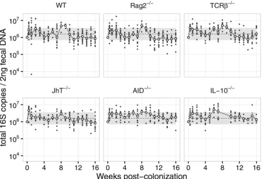
Figure S2.1 - Total 16S levels in fecal DNA after H. hepaticus colonization (complementary to Figures 2.1A, 2.2A and 2.3A). ... 51
Table S2.1 - Linear regression coefficients (Fig 2.2A and Fig 2.3A) ... 52
Figure S2.2 - Linear regression residuals plot (Fig 2.2A and Fig 2.3A) ... 52
Table S2.2 - Linear regression coefficients (Fig 2.4A) ... 53
Figure S2.3 - Linear regression residuals plot (Fig 2.4A) ... 53
Figure S2.4 – Representative individual plots (Fig 2.2A and Fig 2.3A) ... 54
Figure S2.5 – Lipocalin-2 correlation to IgA and load (complementary to Fig. 2.2) ... 55
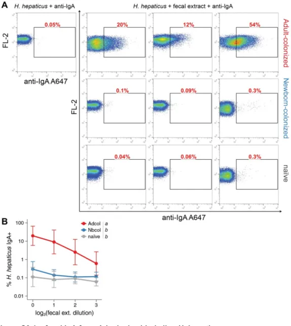
Figure S2.6 – IgA from colonized mice binds to the surface of H. hepaticus ... 56
15
Figure 3.1 - Mother to pup transmission confers long-lasting tolerance to H.
hepaticus ... 63
Figure 3.2 - The age but not the mode of primo-exposure to H. hepaticus conditions the host response ... 65 Figure 3.3 - Neither maternal antibodies nor the microbiota are required for perinatally induced tolerance to H. hepaticus ... 67 Figure 3.4 - Long lasting immune tolerance to H. hepaticus upon mother to pup transmission is Treg mediated ... 69 Figure 3.5 – Tolerance to H. hepaticus does not affect the health of the host ... 73 Figure 3.5 – Tolerance to H. hepaticus does not affect the health of the host ... 74 Figure 3.6 - Perinatal transmission of H. hepaticus shapes the microbiota of the host in immuno-dependent and independent ways ... 76 Table S3.1 – IgM and IgG analysis by ELISA on fecal extracts ... 91 Figure S3.1 – fecal IgA from Adcol mice binds live H. hepaticus ... 92 Figure S3.2 – IgA coated bacteria in the feces of SPF, Nbcol and Adcol mice ... 93 Figure S3.3 – Very low amount of H. hepaticus from feces is able to colonize SPF WT mice ... 94
Figure S3.4 – H. hepaticus fecal load 10 weeks post-colonization (Figure 3.2 B - D) ... 95 Figure S3.5 – Complementary to Figure 3.4 A and B ... 96 Figure S3.6 – Complementary to Figure 3.4 C ... 97 Figure S3.7 – Tregs are depleted in MLN, PP and Large Intestine Lamina Propria upon DT treatment in DEREG mice ... 98 Figure S3.10 – Anti-H. hepaticus mucosal IgA is T-cell dependent ... 101 Figure S3.11 – anti-IL10R treatment is able to break tolerance to H.
hepaticus ... 102
Figure S3.12 – Complementary to Figure 3.6 ... 103 Figure S3.13 – Complementary to Figure 3.6 ... 104 Figure 4.1 - Reciprocal interactions between H. hepaticus and the host immune system on different developmental stages ... 110
17
Chapter 1
: Introduction
Helicobacter hepaticus is a gut bacterium commonly found in animal
facilities and in the wild. This bacterium can cause pathology in immunocompromised animals, however Wild Type (WT) strains are not affected, and can be persistently colonized with this microbe, even from the first days of life, without signs of pathology or decrease in breeding efficiency. The goal of this doctoral work was to identify the immunological mechanisms that maintain such stable relationship between WT mice and
H. hepaticus, and how each organism affects the other. In order to
approach this problem, we explored different components of the mucosal immune system, and their relationship with the intestinal microbiota. The following chapter is an overview of the literature related to microbiota-immunity interactions relevant to this work.
Reciprocal interactions between the host and the
microbiota
1 – Immunological impact of the host on the microbiota
The amazing diversity of bacteria in the mammalian gut poses a very particular challenge to the host, that of discriminating between beneficial and deleterious microbes, as some of the latter could even be pathogenic. During co-evolution of the host and the microbiota many mechanisms arose to control the bacteria in the gut (and other surfaces of the body), which guarantee the protection of the host and the stability of the bacterial community (Hooper et al., 2012).
In the gut, bacteria are stratified and isolated from the host tissue by redundant mechanisms. The epithelial cell layer is the ultimate barrier, above which there is the mucus layer, present in the small and large
intestine (Johansson et al., 2008; Vaishnava et al., 2011). Mucus proteins are secreted by intestinal goblet cells, and self-assemble to form a tightly packed inner layer in the colon, with a thicker and looser external layer above it. Only a loose layer is present in the small intestine, but in both places a 50µm region in the mucus, just above the epithelium, is found almost devoid of bacteria, in contrast to the bacteria-rich region above it. This inner layer is rich in antimicrobial peptides and IgA, which prevents bacterial contact with the epithelium (Vaishnava et al., 2011).
Antimicrobial peptides (AMPs) are effector molecules produced and secreted at the epithelial surface. They form a broad class of molecules, some constitutively expressed, like α-defensins and lysozyme, and others induced by microbial sensing, like cryptdin-related sequences (CRS)-peptides and RegIIIγ (Mukherjee and Hooper, 2015). Expression and secretion of RegIIIγ by epithelial cells is induced by microbial products via Toll-Like Receptors (TLR), and impairment of TLR signaling leads to reduced expression of RegIIIγ, which in turn abrogates the bacteria-free zone above the epithelium (Vaishnava et al., 2011). AMPs can be broadly expressed, or produced by specialized cell types, like Paneth cells, which are mostly found in the crypts of the small intestine, and release granules containing AMPs like lysozyme and α-defensins. Degranulation of Paneth cells is a highly controlled process, and occurs mostly through sensing of Interferon-γ (IFN-γ) (Farin et al., 2014).
Despite the mechanisms in place to confine bacteria above the epithelium, the intestinal tissue is not sterile. Bacteria that cross the epithelium are usually phagocytosed and killed by macrophages in the Lamina Propria (Kelsall, 2008). In addition, these bacteria can be engulfed by Dendritic Cells (DCs), which also sample microbes from the lumen (Farache et al., 2013). DCs carrying live bacteria can present their antigens to T and B cells in the Mesenteric Lymph Nodes (MLN), activated
19
lymphocytes will spread through all mucosal surfaces (Macpherson and Uhr, 2004). Microbes with higher propensity to cross the intestinal barrier, or living in close proximity to it, are more likely to get sampled and further controlled by activated T and B cells and targeted by IgA (Mirpuri et al., 2013).
IgA is the most common antibody in mucosal surfaces (Mantis et al., 2011). In newborns, before the Gut Associated Lymphoid Tissue (GALT) is mature, IgA in the maternal milk provides protection and helps shape the microbiota that colonizes the neonatal intestine (Rogier et al., 2014). After the maturation of the GALT, and in response to the microbiota, endogenous IgA is produced in great amounts, binding to bacteria inside the lumen and conferring protection against invasion (Bunker et al., 2015; Macpherson and Uhr, 2004; Mantis et al., 2011). IgA is secreted by plasma cells in the Lamina Propria, predominantly as dimers, which are transported through the Polymeric Immunoglobulin Receptor (pIgR) to the gut lumen (Kaetzel, 2014). There, IgA can bind to antigens and prevent their negative effects on the host, for example by neutralizing cholera toxin (Lycke et al., 1999). IgA can also affect the motility and invasiveness of pathogens (Forbes et al., 2008, 2011), for instance by binding to molecules on the bacteria surface used to attach to the mucus (Peterson et al., 2007). Indeed, it was found that the appearance of IgA that could bind to Segmented Filamentous Bacteria (SFB) in the gut correlated with a decrease in bacterial load (Jiang et al., 2001). Furthermore, absence of IgA results in expansion of opportunists contained in the microbiota (Mirpuri et al., 2013; Suzuki et al., 2004), which illustrates the role of this antibody in the maintenance of homeostasis in the gut.
The differentiation of B lymphocytes into IgA secreting cells (plasma cells) can occur with or without T cell help, as mice without T cells can produce IgA that bind to bacteria in the gut (Macpherson, 2000). More
potent immune responses occur though when both T and B cells are activated and help each other. Antigen presentation by B cells increases T cell responses (Merkenschlager et al., 2016), and T cell help in germinal centers allows B cells to undergo class-switch recombination (CSR) and somatic hypermutation. This, through the process of affinity maturation, will improve the binding of the secreted antibody to the inducing antigen (Fagarasan et al., 2010). Bacteria that induce strong responses in the host usually trigger T cell dependent IgA production, as is the case of SFB (Lécuyer et al., 2014).
T cells are major players in the immune system, and make up another layer of effector response. T cells can kill infected cells, improve antibody responses through the promotion of germinal center reactions, CSR and affinity maturation on B cells, and they can modify and regulate the innate immune response through the production of cytokines (Khader et al., 2009). Upon activation, T cells can differentiate into different profiles, that will dictate how they respond to further stimulation and where in the body they will migrate to (Bromley et al., 2008). Th2 cells produce Interleukin-4 (IL-4) and promote immunity against parasite infections. Th1 cells produce Interferon-γ (IFN-γ), which activates macrophages and promotes resistance to many infections (Khader et al., 2009), and can also induce the secretion of AMPs by Paneth Cells (Farin et al., 2014). IL-17 producing T cells (Th17) have been implicated in the protection against many pathogens and in the control of the microbiota composition, as the cytokines they produce can recruit neutrophils to the gut and also induce the production of AMPs by epithelial cells (Khader et al., 2009).
21
2 - Effects of the microbiota on the host
The gut microbiota also modifies and is essential for the normal development of many tissues in the host, including the immune system. Mice born and raised in the absence of microbes (germfree - GF) exhibit a series of defects when compared to conventionally raised animals. GF mice have enlarged ceca, reduced intestinal motility, longer villi and shorter crypts in the intestine, reduced amount of AMPs, absence of Isolated Lymphoid Follicles (ILF) in the Lamina Propria, reduced number of intraepithelial lymphocytes, and smaller Peyer’s patches (PPs) and MLN (Gensollen et al., 2016). All these differences evidence the impact the microbiota has on the maturation of the host, and particularly of the GALT. Most of these problems can be reverted by colonization of GF mice with a conventional microbiota, but some cellular defects cannot be corrected once these mice age past a critical time window.
2.1 - Age independent effects of the microbiota on the host
Lymphocytes expressing αβ T cell receptors, CD4+ and CD8+, are
greatly decreased in numbers in the Lamina Propria and Intraepithelial compartments in the intestines of GF compared to conventionally raised mice. This phenomenon can be reverted if adult GF mice are colonized with normal mouse microbiota, but not completely upon colonization with human or rat gut microbes, showing how the intricate relationship of the host immunity and microbiota was shaped by co-evolution (Chung et al., 2012).
Lamina Propria Lymphocytes produce less 10, IFN-γ, 4 and IL-17 in GF compared to conventional mice, which could be reverted by adult-colonization with conventional flora. Particularly Th17 cells, which are important for the production of IgA in the gut, can be restored to normal
levels in the Lamina Propria of adult GF mice with monocolonization with SFB (Gaboriau-Routhiau et al., 2009).
The homeostasis in the gut is dependent on effector mechanisms, which should be controlled to prevent exacerbated immune reactions. CD4+
αβT cells expressing the transcription factor Foxp3 can suppress immune reactions and are designated Regulatory T cells (Treg). Treg are the major source of the immunoregulatory cytokine IL-10 in the gut (Rubtsov et al., 2008) and can respond to commensal antigens (Lathrop et al., 2011). The frequency of Foxp3+ cells inside the CD4+ αβT cell population reaches
some of the highest levels in the colonic Lamina Propria of conventionally raised mice. Furthermore, Treg levels are reduced upon antibiotic treatment and are dramatically decreased in GF animals (Atarashi et al., 2011). Colonization of adult GF mice with conventional microbiota, or with a combination of commensals of the genus Clostridium, can restore Treg levels in the colonic Lamina Propria. Other groups of bacteria were also shown to induce Treg in mice, like Bacteroides fragilis (Round and Mazmanian, 2010), which is a human commensal, and Lactobacillus
murinus (Tang et al., 2015).
The levels of IgA-producing B cells in the Lamina Propria of GF mice are also greatly reduced (Hapfelmeier et al., 2010). This phenotype can also be reverted by colonization of adult GF mice with a conventional microbiota (Gensollen et al., 2016), and could also be achieved by monocolonization, with the number of IgA+ plasma cells being maintained
increased in the Lamina Propria even after bacteria elimination (Hapfelmeier et al., 2010).
23
2.2 - Age dependent effects of the microbiota on the host
Strong immune responses to infection usually require Th1 differentiation and IFN-γ production. In neonates however, Th1 responses are usually impaired. Neonatal B cells produce high amounts of IL-10 upon TLR stimulation (Walker and Goldstein, 2007; Zhang et al., 2007), which prevents DC activation and IL-12 production (Sun et al., 2005). Murine neonatal T cells produce high levels of IL-4 and IL-13 upon activation, which favor Th2 polarization (Zaghouani et al., 2009). Th17 development is also impaired in neonates, since IL-6 stimulation is essential for this type of polarization and newly formed T cells (Recent Thymic Emigrants - RTEs) are less sensitive to IL-6 than mature T cells (Paiva et al., 2013). Additionally, neonatal T cells have a higher propensity to become Tregs than adult T cells, converting in high frequency to Foxp3+ Tregs upon TCR
stimulation (Wang et al., 2010). All these properties make neonates very susceptible to infections, but at the same time, they offer the best environment to forge symbiotic relationships through the development of tolerance. Therefore, the colonization with the microbiota in the neonatal stage shapes the immune system of the host in unique ways.
In GF mice, absence of microbiota causes accumulation of invariant Natural Killer T cells (iNKT) with a pro-inflammatory phenotype in the lung and intestine, leading to susceptibility to airway hyperresponsiveness and colitis. Colonization with a conventional microbiota during the first weeks of life, but not at adult age, could prevent iNKT accumulation and susceptibility to disease (An et al., 2014; Gensollen et al., 2016; Olszak et al., 2012). Similarly, disturbance of the microbiota by antibiotic treatment early in life causes increased susceptibility to Inflammatory Bowel Disease (IBD) in humans (Kronman et al., 2012; Shaw et al., 2010).
Another phenotype observed in GF mice is a high level of serum IgE and increased susceptibility to Antigen-Induced Oral Anaphylaxis. Colonization early in life with a complex microbiota could revert the hyper IgE phenotype in GF animals, yet this was not the case if colonization was performed in adults (Cahenzli et al., 2013).
Specific Pathogen Free (SPF) mice, which have a complex microbiota but are free of known pathogens, show accumulation of Helios
-Foxp3+ Treg (supposedly converted outside of the thymus) in the lungs in
the first week of life, a phenotype that was not observed in GF animals. Treatment of SPF neonates for two weeks with anti-PD-L1 could prevent the Helios- Treg accumulation in the lungs and increased susceptibility to
airway inflammation (Gollwitzer et al., 2014). This indicates that the lung microbiota induces the generation of Tregs early in life, which confers a long-lasting protective effect on the lungs. Furthermore, neonatal mice treated with antibiotics have increases susceptibility to allergic asthma later in life, which does not happen if treatment was performed in adult animals (Russell et al., 2012). A similar association between early life antibiotic treatment and the risk of asthma development was also reported in humans (Risnes et al., 2011).
Accumulation of Tregs on the neonatal skin has been shown to be essential for the generation of immune tolerance to commensal at this site. Tregs specific for commensal antigens were found in high frequency in the skin of newborn-colonized mice, and shown to prevent experimentally induced skin inflammation (Scharschmidt et al., 2015). In addition, mice treated in the neonatal period with antibiotics displayed increased susceptibility to experimental psoriasis in adulthood (Zanvit et al., 2015). This could be partially reverted by co-housing the treated animals with non-treated controls, providing evidence that the disturbance in the microbiota caused the increased susceptibility to disease.
25
Helicobacter hepaticus
Helicobacter hepaticus was first isolated from the liver and mucosal
scrapings of SCID/NCr and A/JCr mice (Fox et al., 1994), because it was causing liver pathology in these strains. It was also found later to cause colitis in B6.IL-10-/- and 129SvEv.Rag2-/- mice. Notwithstanding, H. hepaticus was found to colonize most Wild Type strains in animal facilities
worldwide (Whary and Fox, 2006), without any sings of pathology or decrease in breeding efficiency (Solnick and Schauer, 2001). It was also found to be widespread in the wild (Wasimuddin et al., 2012), along with many other bacteria of the genus Helicobacter. The absence of disease in normal animals and the high prevalence in the wild suggest that H.
hepaticus does not fit the normal classification of pathogen. Rather, the
evidence suggests that most of the described pathologies associated with this microbe come from exacerbated immune responses and not from actual invasion and damage directly caused by the bacteria. Because of its resemblance to IBD in humans, colitis induced by H. hepaticus became a model to study pathological immune responses and immune regulation in the gut.
H. hepaticus triggers T cell-dependent colitis in IL-10-/- mice and in
alymphatic mice transferred with naïve T cells (Cahill et al., 1997; Kullberg et al., 1998). This pathology can be reproduced by transfer of a CD4+ T cell
clone specific for a bacterial epitope, and only H. hepaticus-colonized recipients develop the disease, suggesting that an uncontrolled T cell response to H. hepaticus, rather than a breach in tolerance to self, causes immunopathology in this model (Kullberg et al., 2003). Transfer of CD4+
CD45RBlow cells, which were previously shown to be enriched in T
regulatory cells (Tregs) (Annacker et al., 2001; Read et al., 2000), can prevent colitis, but only when these cells come from colonized donors
(Kullberg et al., 2002). Tregs from H. hepaticus-colonized mice can suppress IFN-γ production, and secrete IL-10 when stimulated in vitro with
H. hepaticus antigens (Kullberg et al., 2002). Taken together, these reports
support the idea that H. hepaticus has the capacity to induce an antigen-specific regulatory T-cell response in mice, and when this regulatory component is absent or compromised, a strong T-cell mediated inflammatory response is mounted, mediated by IFN-γ and IL-17 (Morrison et al., 2013).
Despite the abundance of information on the immune response triggered by H. hepaticus, particularly in the T cell compartment of the gut, very few information is available on the actual impact of this response on the bacteria. Transfer of CD4+ CD45RBhigh (predominantly naïve) T cells to
Rag-/- mice colonized with H. hepaticus causes colitis, which can be
suppressed by co-colonization with the human commensal Bacteroides
fragilis. However, H. hepaticus intestinal load was found to be the same in
both sick and healthy animals, suggesting that increased inflammation by itself is not enough to influence H. hepaticus numbers in the gut (Mazmanian et al., 2008). Similarly, gut pathology in 129SvEv.Rag2-/- mice
colonized with H. hepaticus can be suppressed by the transfer of CD4+
CD25+ T cells (enriched in Treg), but the reduced inflammation also does
not influence H. hepaticus intestinal load in this system (Maloy et al., 2002). On the other hand, in animals deficient for the innate immunity adaptor Nod2, which show decreased expression of α-defensins in the gut, H.
hepaticus numbers were found to be elevated when compared to WT
controls (Petnicki-Ocwieja et al., 2009). Additionally, the transgenic expression of the human α-defensin 5 in Nod2 deficient mice was sufficient to rescue this phenotype, reducing H. hepaticus load in the gut (Biswas et al., 2010). This suggests that innate immunity can control H. hepaticus in
27
the gut, but the role of the adaptive immune response in this control is largely unknown.
H. hepaticus transmission occurs easily between adults, mainly
through coprophagy (Livingston et al., 1998). Possibly for that reason, H.
hepaticus became widespread in animal facilities worldwide, which lead to
the development of several methods for elimination of this bacteria from experimental mice. Since antibiotic treatments largely failed to eradicate infection (Solnick and Schauer, 2001), and rederivation techniques are often difficult to perform, fostering of pups born from H. hepaticus positive dams by H. hepaticus-free foster mothers was a great promise. However, in fostering attempts it was found that only transfers in the first day of life were successful, because if fostering was done from the second day of age onwards a large percentage of the pups was already positive for H.
hepaticus (Singletary et al., 2003). This shows that H. hepaticus is
transmitted from mothers to newborns, in the first few days of life, possibly through pups contact with maternal feces. Due to the intrinsic differences between the adult and neonate immune system, it is conceivable that mother to pup transmission shapes the interactions between the host and
H. hepaticus differently than upon colonization at adult age.
Taken together, our analyses of the literature lead us to identify two sets of unanswered questions related to the interactions between the mouse and H. hepaticus, namely:
- whether the immune response in adult mice affects the bacteria, and which components of the immune system would be involved in this response;
- whether the age at colonization conditions the immune response to this bacteria and whether this would affect the health and the microbiota composition of the host.
In this work, we directly addressed these questions by analyzing various mice upon colonization with a reference strain of H. hepaticus.
29
Chapter 2
:
IL-10 promotes Helicobacter hepaticus
persistence in the gut, while Antibodies, T and B
cells contribute equally to bacteria elimination.
Preliminary notes
The author of the thesis participated in the planning, execution and analysis of all the experiments presented in this chapter
31
Abstract
Helicobacter hepaticus is a pathobiont that lives in close association
with the mouse intestinal epithelium. Despite abundant evidence that H.
hepaticus induces a strong adaptive immune response in the mice, the
impact of this response on the bacteria living in the gut is largely unknown, as well as why this bacterium is not eliminated by this response. We tested if the adaptive immunity impacts H. hepaticus in the intestine by colonizing mice lacking different components of the adaptive immune system, and verifying the effect on the bacterial numbers in the gut. We found that B and T lymphocytes impact bacterial load in the gut, with a similar contribution. Antibodies participate in this effect, but do not account for the whole outcome, which indicates that T and B cells also contribute to bacteria elimination in an antibody-independent manner. Absence of IL-10 results in an increased response, which can lead to bacteria elimination. We conclude that IL-10 promotes the persistence of H. hepaticus in the gut, while T and B cells control bacterial numbers. Our study provides new insights into how lymphocytes control bacteria in the microbiota and maintain intestinal homeostasis, which could help in the understanding and prevention of gut immunophatologies.
Introduction
Some of the large amount of bacteria in the microbiota lives in close association with the host. One of those is Helicobacter hepaticus, which is broadly spread in the wild and animal facilities (Wasimuddin et al., 2012), and was found to be associated with the host epithelium (Chow and Mazmanian, 2010). It would be expected, hence, that the host would produce an immune response to avoid uncontrolled expansion of this bacterium, so close to the surface of the body. If this response exists, however, it is not strong enough to eliminate this microbe, since mice are positive for live upon colonization (Solnick and Schauer, 2001; Wasimuddin et al., 2012; Whary and Fox, 2006).
It is clear that H. hepaticus induces a T cell response in mice, particularly in the absence of immune regulation (Cahill et al., 1997; Kullberg et al., 1998), yet the information on the gut response to this bacterium remains limited, though it is known to induce a fecal IgA response (Whary et al., 1998).
Secretory IgA is thought to act as a barrier, regulating the microbiota and restricting the entrance of intestinal antigens into the blood (Pabst, 2012). Some reports have highlighted the importance of IgA on the host protection (Forbes et al., 2008, 2011; Lycke et al., 1999), and although there is evidence that T cell (particularly Tregs) are necessary for optimal IgA production and homeostasis of the microbiota (Kawamoto et al., 2014), others have claimed that only part of the IgA in the gut is T cell-dependent (Bunker et al., 2015) and that it only targets particular groups of bacteria in the gut (Mirpuri et al., 2013).
Here, we sought to understand the impact of the immune response on H. hepaticus load in the gut. We found that specific IgA to H. hepaticus
33
is T-cell dependent and negatively regulated by IL-10. In the absence of IgA, specific secretory IgM is found in the feces, but at a much lower absolute concentration. B and αβT lymphocytes, together with IgA, seem to contribute similarly to H. hepaticus load reduction, as the latter increases gradually with the removal of each component separately. These results evidence how complex and multilayered the immune response to bacteria is in the gut.
Results
Impact of the adaptive immunity on H. hepaticus in the gut
Mice are easily colonized with H. hepaticus through contact with contaminated feces, majorly through coprophagy (Livingston et al., 1998). To examine if colonization by this normal route results in a specific response in the gut, and to test if this response affects the bacteria inside the intestine, we colonized adult (10 weeks old) B6 WT and Rag2-/- mice by
gavage with H. hepaticus-positive feces, and evaluated the bacterial load in the feces of these mice on the course of 16 weeks by qPCR. H. hepaticus colonization occurs in all animals, with a peak in bacterial load between 1 and 2 weeks post-colonization (Fig. 2.1A). However, while the bacterial
load was considerably stable in Rag2-/- animals after this initial shift, a
steady decrease in bacterial load was observed in WT mice, reaching levels almost 2 logs lower than those in Rag2-/- mice (Fig. 2.1A). This
difference was not due to variations in the microbiota of WT and Rag2
-/-mice, as co-housing of animals of these two genotypes from 4 to 9 weeks of age before colonization produced very similar results (Fig. 2.1B). These
data indicate that the colonization with H. hepaticus induces an adaptive immune response in the mice, and that this response impacts the bacteria in the gut.
35
Figure 2.1 - Impact of the adaptive immunity on H. hepaticus in the gut
Adult mice (10 weeks old) WT and Rag2-/-, separately raised (A) or co-housed for 5
weeks (B), were colonized with H. hepaticus by feces oral gavage. Bacterial load was
measured on feces by qPCR up to 16 weeks post-colonization, and analyzed as relative abundance = (H. hepaticus 16S / eubacteria 16S). p < 001, linear regression (week 1 - 16). Colored dots = individual mice, white circles = mean, error bars = SEM, dotted line = linear regression from 1-16 weeks, shade = linear regression confidence interval. N = 10 per group, pooled from two independent experiments. ND = not detected.
Impact of T cells on the response to H. hepaticus
Because it is known that H. hepaticus induces a potent T cell response in the gut (Cahill et al., 1997), and that this response is even stronger in the absence of IL-10 (Kullberg et al., 1998), we next tested the impact of the T cell response on H. hepaticus in the intestine using mice deficient in either αβT cells (TCRβ-/-) or IL-10 (IL-10-/-). We observed a
higher H. hepaticus load in the absence of T cells, compared to WT mice, but still lower than the levels in Rag2-/- mice (Fig. 2.2A). Accordingly, the
response in IL-10-/- mice had a large effect on the H. hepaticus load, with
levels dropping below the detection limit in most of the animals after 11 weeks of colonization (Fig. 2.2A). Taken together, these results evidence
that T cells play a key role in the eliminating response to H. hepaticus in the gut.
WT mice displayed high titers of anti-H. hepaticus IgA on feces throughout the colonization (Fig. 2.2B), and this IgA was able to bind to the
surface of live H. hepaticus (Fig. S2.6), hence suggesting that it can affect
the bacteria in the gut. Meanwhile, IL-10-/- mice showed fecal IgA titers
almost 2 logs higher than WT (Fig. 2.2B), which correlated with the H.
hepaticus load reduction (Fig. 2.2D). Conversely, specific IgA was almost
absent in animals lacking T cells, showing that the induction of anti-H.
hepaticus mucosal IgA is T cell dependent (Fig. 2.2B). An increase of
about 1 log in total fecal IgA production was seen in WT and IL-10
-/-animals 1 week after colonization, but also in TCRβ-/- with a delay (Fig.
2.2C). This was correlated with the levels of lipocalin-2 (lcn-2), a biomarker
of inflammation in the gut, in IL-10-/- and TCRβ-/- mice, but not in WT
animals (Fig. 2.2E and F and Fig. S2.5A). Lcn2 levels did not correlate
with the reduction in H. hepaticus levels in the gut (Fig. S2.5C) or with
specific IgA levels (Fig. S2.5B), suggesting that the inflammation triggered
37
Fig.2.2 - Impact of αβT cells and IL-10 on the response to H. hepaticus
A) Adult mice (10 weeks old) TCRβ-/- and IL-10-/- were colonized with H. hepaticus by
feces oral gavage. Bacterial load was measured on feces by qPCR as described in
Fig.1. WT and Rag2-/- load (same as in Fig.1 A) is shown for comparison. Groups with
the same letter are not significantly different at p = 0.05 (pairwise comparisons of linear regressions, p values corrected using single-step method). Colored dots = individual mice, white circles = mean, error bars = SEM, dotted line = linear regression from 1-16 weeks, shade = linear regression confidence interval. N = 10 per group,
pooled from two independent experiments. B and C) Anti-H. hepaticus IgA titer (B)
and total fecal IgA (C) were evaluated by ELISA on WT, TCRβ-/- and IL-10-/- mice
shown in A, from 0 – 16 weeks post-colonization. Statistical comparison was
performed using Area Under the Curve (AUC), calculated per mice with trapezoid method and averaged per week. Groups with the same letter are not significantly different at p = 0.05 (Wilcoxon test with BH correction). Colored dots = individual mice, white circles = mean, error bars = SEM, dotted line = local polynomial regression
fitting (loess), shade = loess confidence interval. D) Comparison of specific IgA titers
and bacterial load in mice shown in A. Colored dots = individual samples, line = linear
regression, shade = linear regression confidence interval, p value and R2 for the linear
regression shown on the graph. E and F) Fecal Lipocalin-2 levels were evaluated by
ELISA in the feces of the mice shown in A, from 0 – 16 weeks post-colonization (2 –
16 weeks for Rag2-/- mice). E) white circles = mean, error bars = SEM, lines connect
the means. F) Dots represent individual mice. Groups with the same letter are not
significantly different at p = 0.05 (Wilcoxon test with BH correction). AUC = Area Under the Curve, calculated per mice with trapezoid method and averaged per week.
G and H) Anti-H. hepaticus serum Ig titer (G) and total serum Ig concentration (H)
were measured in WT, TCRβ-/- and IL-10-/- mice shown in A, at 16 weeks
post-colonization. Dots represent individual mice, groups with the same letter are not significantly different at p = 0.05 (Wilcoxon test with BH correction).
39
Analysis at 16 weeks post-colonization showed high titers of anti-H.
hepaticus IgG and IgA in the serum of WT and IL-10-/- animals, suggesting
that this bacterium crosses the intestinal barrier, eliciting a systemic immune response (Fig. 2.2G). Similarly to the mucosal response, specific
IgA levels were higher in IL-10-/- animals compared to WT, but not IgG,
suggesting that IL-10 could impact specifically the IgA production. Similar levels of specific IgM were found in WT, IL-10-/- and TCRβ-/- mice, but
specific IgG and IgA were not found in the latter (Fig. 2.2G). Total
Immunoglobulin levels were similar between the groups analyzed, with a small increase in total IgA levels in IL-10-/- mice (Fig. 2.2H).
Role of B cells and IgA on the response to H. hepaticus
Given the negative correlation between the IgA response and H.
hepaticus load, we tested if the response to H. hepaticus would be affected
by the removal of antibodies from the system, using mice devoid of B cells (JhT-/- mice), or by only removing IgA, using AID-/- mice (which can’t
perform class-switch recombination, hence only IgM can be produced). In the absence of B cells the H. hepaticus fecal load was higher than in WT animals, but lower than in Rag2-/- (Fig. 2.3A), suggesting that B cells
impact the bacterial load, but do not account for the whole effect. Interestingly, the absence of IgA in AID-/- results in an intermediate
phenotype between the B cell deficient and WT (Fig. 2.3A), indicating that
IgA affects the bacterial load but also that B cells have other effects besides antibody production, since the IgM levels, specific and total, were very low in AID-/- after colonization (Fig. 2.3B and C). Fecal lcn-2 levels in
JhT-/- and AID-/- (Fig. 2.3D) were very similar to the values found in WT
mice (Fig. 2.2E), indicating no evident increase in gut inflammation.
To directly test the impact of secreted antibodies on the bacterial load, excluding any possibly effect of IgM in AID-/- mice, we used AID-/-uS
-/-animals, which have B cells but cannot secrete antibodies. We found that the H. hepaticus fecal levels were higher in AID-/-uS-/- mice compared to
WT, but lower than those in Rag2-/- mice (Fig. 2.4A). To confirm this effect,
we analyzed the H. hepaticus mucosal load in the ileum and colon of the same mice, after 20 weeks of colonization. No difference between the groups was observed in the ileum, where H. hepaticus was found in lower frequencies (Fig. 2.4B). However, the bacterial load reached high levels in
the colon of Rag2-/- animals, about 2 logs higher than in WT mice, with
intermediate values in AID-/-uS-/- (Fig. 2.4B). These data show that secreted
antibodies can impact directly the bacterial load in the gut, but do not account for the whole effect.
41
Figure 2.3 - Impact of B cells and IgA on the response to H. hepaticus
A) Adult mice (10 weeks old) JhT-/- and AID-/- were colonized with H. hepaticus by
feces oral gavage. Bacterial load was measured on feces by qPCR. WT and
Rag2-/- load (same as in Fig.1 A) is shown for comparison. Groups with the same
letter are not significantly different at p = 0.05 (pairwise comparisons of linear regressions, p values corrected using single-step method). Colored dots = individual mice, white circles = mean, error bars = SEM, dotted line = linear regression from 1-16 weeks, shade = linear regression confidence interval. N = 9
per group, pooled from two independent experiments. B and C) Specific and Total
fecal IgM in the AID-/- mice shown in A. D) Fecal Lipocalin-2 levels were evaluated
by ELISA in the feces of the mice showed in A, from 0 – 16 weeks
post-colonization (2 – 16 weeks for JhT-/- mice). White circles = mean, error bars =
Figure 2.4 - Effect of secreted Immunoglobulin on H. hepaticus load in the gut
WT, Rag2-/- and AID-/-µS-/- mice were colonized by feces gavage with H. hepaticus. A)
Bacterial load was measured on feces by qPCR from 0 – 20 weeks post-colonization. Relative abundance = (H. hepaticus 16S / eubacteria 16S). Colored dots = individual mice, black circles = mean, error bars = SEM, dashed line = linear regression, shade = linear regression confidence interval. Groups with the same letter are not significantly different at p = 0.05 (pairwise comparisons of linear regressions, p values corrected using
single-step method). B) H. hepaticus mucosal load, measured by qPCR, in the ileum and
colon of the mice shown in A, >20 weeks post-colonization. Data was normalized to host
DNA, quantified using 18S primers. Dots represent individual mice and groups with the same letter are not significantly different at p = 0.05 (Wilcoxon test with BH correction).
43
Discussion
The gut microbiota is composed of a great diversity of bacterial species, and the control of those, particularly the ones close to the epithelium, is a great challenge on the host immune system (Hooper and Macpherson, 2010). Using Helicobacter hepaticus, a pathobiont that lives in close association to the host epithelium (Chow and Mazmanian, 2010), we could explore how the host adaptive immunity influences the number of bacteria in the gut, exposing the essential components of this response.
We first showed that lymphocytes are essential to the optimal control of the bacterial load, as alymphatic (Rag2-/-) mice present with higher levels
of colonization (Fig. 2.1). Then, we demonstrated that αβT and B cells are
both necessary for this effect, as mice without either one of these cell types presented with similar bacterial load, which was higher than observed in WT animals and lower than what was observed in Rag2-/- animals (Fig.
2.2A and Fig. 2.3A). As we verified that αβT cells are required for a
sustained anti-H. hepaticus IgA production (Fig. 2.2B), and antibodies are
absent in B cell-deficient mice, we tested if antibodies were solely responsible for the anti-bacterial effect, using mice deficient in IgA (AID-/-)
and in secreted antibodies (AID-/-uS-/-). With these models, we could verify
that secreted antibodies do impact the bacterial load in the gut (Fig. 2.4)
and that IgA makes the biggest difference (Fig. 2.3A and B), since a
significant compensatory IgM response was not observed in the absence of IgA. However, antibodies do not seem to account for the whole effect of the adaptive immunity on H. hepaticus in the gut, since in their absence the bacterial levels do not reach the load observed in Rag2-/- mice (Fig. 2.3A
and Fig. 2.4). Furthermore, although T and B cells cooperate to mount a
robust antibody response, we found evidence of an antibody-independent effect from these two cell types on bacteria in the gut. First, a T cell effect
could explain the difference in load between JhT-/- and Rag2-/- mice (Fig.
2.3A). Second, the differences between the H. hepaticus load in TCRβ
-/-and Rag2-/- (Fig. 2.2A) and between JhT-/- and AID-/- (Fig. 2.3A) could be
explained by an antibody-independent B cell effect.
T cell derived cytokines could indirectly impact bacteria in the gut through the control of antimicrobial release at the epithelial level. As an example, Paneth cells, which are largely important for the maintenance of the homeostasis at the intestinal host-microbial interface (Vaishnava et al., 2008), release antimicrobial products only when stimulated by immune cell-derived IFN-γ, but not bacterial products (Farin et al., 2014). Antigen presentation at the epithelial level could then assure the specificity of the T cell response, guaranteeing that it would only occur if a particular group of bacteria were increased. B cells, on the other hand, have other ways to impact bacteria in the gut besides the production of antibodies. It was
demonstrated that IgA+ plasma cells can acquire a multifunctional
phenotype in the gut, with expression of iNOS and TNFα, which was important for bacterial control (Fritz et al., 2012). Moreover, B cells can facilitate the expansion of T cells (Merkenschlager et al., 2016), and possibly increase the T cell-mediated anti-bacterial response.
We also demonstrated that an exacerbated response, in the absence
of IL-10, impact directly the bacteria in the gut (Fig. 2.2A), with a
concomitant increase in specific intestinal IgA production (Fig. 2.2B). This
response could even lead to elimination of this bacteria in some animals (Fig. 2.2A), which allow us to conclude that IL-10 promotes H. hepaticus
persistence in the gut. Indeed, it was demonstrated that this bacterium induces a regulatory T cell (Treg) response, which produce IL-10 upon stimulation with H. hepaticus antigens (Kullberg et al., 2002). However, these Treg are not able to prevent the immune response to this bacterium, as we observed a steady decrease in H. hepaticus levels in WT mice,
45
which also reached undetected levels in some animals (Fig. 2.1 and Fig. 2.4A). Still, the Treg response could avoid rapid elimination, allowing this
bacteria to spread to other individuals and possibly reach the next generation, being also maintained later by coprophagy between different mice.
Collectively, our studies demonstrate that the adaptive immune response impacts H. hepaticus in the gut directly, through IgA but also by a mechanism involving B and T cells, possibly antibody-independent. Further analysis using antibody-deficient mice will help to understand this phenomenon. The exploration and understanding of these mechanisms are important for the comprehension of what maintains homeostasis in the gut, and how to preserve and restore it in pathological conditions.
Materials and Methods
Mice
All the mice were in a C57BL/6 background, bread in our specific pathogen free facility, which excludes Helicobacter hepaticus. All experiments involving H. hepaticus colonization were performed in a dedicated room. AID-/-uS-/- mice were obtained by inter-crossing AID-/- and
uS-/- mice in our facility, as described elsewhere (Lino et al., 2013).
Helicobacter hepaticus culture and mice colonization
Helicobacter hepaticus reference strain (CIP 104102) was obtained
from the Biological resources center of the Pasteur Institute. Growth was performed in 10% horse blood-agar (HBA, Oxoid) plates, supplemented with 12.5 mg/L vancomycin, 0.3 mg/L polymyxin B, 6.3 mg/L trimethoprim and 5.0 mg/L amphotericin B, at 37°C, under microaerophilic conditions (6% O2, 7% CO2, 3.5% H2 and 83.5% N2) generated by an Anoxomat
system (MART Microbiology). Bacteria were harvested after 5 days of cultured in PBS solution and numbers were estimated by OD quantification. Mice were colonized by oral gavage with 100µL of bacteria suspension at 109 CFU/mL in PBS, using an animal feeding needle with silicone tip, size
20G x 1.5’’ (Cadence Science). Colonization was confirmed by PCR on fecal DNA using H. hepaticus 16S specific primers (Petnicki-Ocwieja et al., 2009). To reproduce colonization through coprophagy, mice were colonized using a preparation of H. hepaticus positive feces, from mice previously colonized with cultured H. hepaticus. Fecal pellets were homogenized in PBS at a ratio of 1 pellet to 400µL (~ 100mg/mL) and filtered in 100µm mesh. Each mouse received 100µL of this fecal preparation by oral gavage.
47
Helicobacter hepaticus quantification
The following primers were used in qPCR reactions: H. hepaticus 16S
fwd: GCATTTGAAACTGTTACTCTG, H. hepaticus 16S rev:
CTGTTTTCAAGCTCCCCGAAG, 417bp product (Petnicki-Ocwieja et al., 2009); Eubacteria 16S fwd: ACTCCTACGGGAGGCAGCAGT, Eubacteria 16S rev: ATTACCGCGGCTGCTGGC, ~180bp product, 18S fwd: CATTCGAACGTCTGCCCTAT, 18S rev: CCTGCTGCCTTCCTTGGA, 137bp product (Vaishnava et al., 2011). H. hepaticus (Hh) 16S copy number was estimated using plasmid standard curve, total bacteria (eubac) 16S copy number was estimated using a standard curve constructed with
E. coli K12 DNA, and host DNA amount in ng was estimated using a
standard curve constructed with intestinal tissue DNA. All reactions contained standard curves for absolute quantification. Fecal H. hepaticus amount is expressed as log10 of the ratio Hh 16S copies/ eubac 16S copy.
Mucosal H. hepaticus amount is expressed as log10 of the ratio Hh 16S
copies/ ng of host DNA. DNA from fecal pellets of ~40mg (average 40.2mg ± 10.3, measured from 100 samples) and from intestinal tissue (3 cm piece of ileum and colon, after washing luminal content with PBS) was extracted using NZYTech Tissue gDNA extraction kit (NZYTech), with an initial step at 95°C. For quantification, 2ng of fecal DNA and 10ng of mucosal DNA were used. Primers were used at 0.5µM in the reaction, containing 5µL of iTaq Universal SYBR Green Supermix (Bio-Rad), 2µL of sample and water to a final volume of 10µL. qPCR conditions: 95°C - 3min; 45 cycles of 95°C - 30s, 60°C - 30s, 72°C - 36s; followed by a melting curve of 65 - 95°C with 0.5°C increase every 5s. All reactions were prepared in 384 well hard shell plates (Bio-Rad), on ice, in less than one hour, to minimize primer dimer formation, and run immediately after preparation on a CFX384 real-time PCR detection system (Bio-Rad).
Total and specific Immunoglobulin quantification by ELISA
To quantify total and H. hepaticus specific fecal antibodies, fecal extracts were prepared from freshly collected or -80°C stored feces. Fecal pellets of ~50mg were collected from each mice (average 56.6mg ± 15.4, measured from 100 samples), and homogenized in 500µL of PBS containing a protease inhibitor cocktail (P8340, Sigma-Aldrich), for a solution of about 100mg/mL. Homogenates were centrifuged at 16,000G for 10min at room temperature, and supernatants were stored at -20°C until further use.
Soluble H. hepaticus antigens (SHelAg) were prepared as previously described (Kullberg et al., 1998). After cultured for 5 days in blood-agar plates, bacteria were harvested in PBS solution, washed and lysed in a French Press. The solution was centrifuged at 8,000G for 30 min at 4°C, the supernatant was filtered sterile with a 0.22µm pore size filter and protein content was determined using BCA protein assay. The preparation was stored at -80°C.
ELISA was performed using high binding 384 well ELISA plates (UltraCruz). For total Ig quantification, plates were coated with Goat anti-Mouse IgM, IgG or IgA polyclonal antibodies (Southern Biotech), and for anti-H. hepaticus Ig quantification, coating was done using 10µg/mL of SHelAg. Plates were coated overnight at 4°C, washed in PBS 0.05% Tween20 and blocked with 2% BSA solution in PBS. Fecal extracts were added undiluted (specific Ig) or 10 fold diluted (total Ig) to plates and diluted 7 times, 3 fold each time. For specific fecal IgA quantification, a reference sample pooled from 5 adult colonized B6 was used in all assays. For specific serum IgM, IgG and IgA, a reference serum sample of colonized IL-10-/- diluted 1:210 was used in all assays. After overnight incubation at 4°C,
anti-49
Mouse IgM, IgG or IgA conjugated with HRP. Reactions were revealed with TMB (BD) and stopped with 0.1M H2SO4.
Fecal Lipocalin-2 quantification
Quantification of fecal Lipocalin-2 was performed on fecal extracts, obtained as described above, using the kit manufacturer instructions (Mouse Lipocalin-2/NGAL DuoSet ELISA, catalog number: DY1857, R&D).
IgA FACS on live H. hepaticus
H. hepaticus was cultured as described above, and 106 CFU were
used per assay. Bacteria was washed once in PBS, pelleted (12,000G, 5min), resuspended in fecal extract (prepared as described for ELISA assay) from naïve or 16 weeks colonized Adult B6 mice, and incubated on ice for 30min. Bacteria were then washed in PBS and incubated for 15min on ice with Goat anti-Mouse IgA (Southern Biotech), labeled using Alexa647 Labeling Kit (Thermo Fisher Scientific). The bacteria was washed again and resuspended in a PBS solution containing 5µM of SYTO 9 (Molecular Probes). Events were acquired on a CyAn ADP Analyzer (Beckman Coulter) and analyzed on FlowJo Software (Tree Star). Analysis was performed gating on live bacteria, which were SYTO 9 Bright, as confirmed by counterstaining with 45µM of propidium iodide (Molecular Probes), and excluding events with high pulse width.
Data analysis
Data and statistical analysis were performed using R software version 3.2.5 (R Core Team, 2014). Multiple comparisons were done using Kruskal-Wallis and Mann–Whitney–Wilcoxon test with the Benjamini and Hochberg p value correction. Multiple comparisons of linear regressions were performed using linear model and the function glht (General linear hypotheses and multiple comparisons), using single-step procedure for p
value correction, in package multcomp (Hothorn et al., 2008). Graphs were made in R software using the package ggplot2 (Wickham, 2009).
51
Supplementary Material for Chapter 2
Figure S2.1 - Total 16S levels in fecal DNA after H. hepaticus colonization (complementary to Figures 2.1A, 2.2A and 2.3A).
Total 16S (quantified using primers that recognize a conserved region in all bacteria 16S) in DNA extracted from feces of mice of the indicated genotype from 0 – 16 weeks post-colonization with H. hepaticus. Total 16S values were used to normalize the number of H. hepaticus 16S presented as relative abundance in
figures 2.1A, 2.2A and 2.3A. Dots = individual mice, white circles = mean, error
bars = SEM, dotted line = overall mean, shade = overall standard deviation. N = 10
for WT, Rag2-/-, TCRβ-/- and IL-10-/-, N = 9 for JhT-/- and AID-/-, pooled from two