Effect of Transthyretin in the brain vasculature
Implications in physiology and pathology
José Ricardo da Cruz Vieira
Master in Neurobiology
José Ricardo da Cruz Vieira
Effect of Transthyretin in the brain vasculature
Implications in physiology and pathology
Tese de candidatura ao grau de Mestre em
Neurobiologia, submetida à Faculdade de Medicina
da Universidade do Porto.
Orientadora – Doutora Isabel Cardoso
Categoria – Investigadora Principal
Afiliação – Instituto de Biologia Molecular e Celular (IBMC) e
Instituto de Investigação e Inovação em Ciência (I3S)
A todas as pessoas que de alguma
forma ajudaram a tornar-me
no cientista que hoje sou.
“If you can't be a pine on the top of the hill Be a scrub in the valley-but be The best little scrub by the side of the rill; Be a bush if you can't be a tree.
If you can't be a bush be a bit of the grass, And some highway some happier make; (…) If you can't be a highway then just be a trail, If you can't be the sun be a star; It isn't by size that you win or you fail- Be the best of whatever you are!” [Be the best of whatever you are, Douglas Malloch]
Embora esta secção não esteja diretamente envolvida com o trabalho científico, e talvez não tenha qualquer significado para outras pessoas para além de mim, é uma das partes mais importantes deste trabalho. Ao longo de todo o percurso académico de um estudante, existem vários “pilares” que permitem que tudo pareça mais fácil, com quem sabemos que podemos contar, e que estão ao nosso lado para nos apoiar, incondicionalmente. Sem eles, de certeza que este ano não teria sido tão fácil, enriquecedor e marcante. Sinto que aprendi bastante e, principalmente, que evolui tanto como cientista como pessoa. A esses “pilares”, o meu imenso obrigado. Não podendo deixar de agradecer individualmente a algumas pessoas:
Um especial agradecimento à minha orientadora, Dra. Isabel Cardoso, por me ter aceitado e recebido tão bem neste pequeno “grande” grupo MiND. E, principalmente, por me ter acompanhado ao longo de todo este ano, estando sempre disponível para responder a todas as minhas dúvidas, orientando-me e transmitindo-me todo o conhecimento essencial para me tornar num melhor cientista.
Ao Luís Miguel Cardoso, por todo o tempo e paciência que teve comigo, por me ter integrado tão rapidamente, por toda a alegria e boa disposição partilhada, pela dança de abertura do I3s retreat (que show!)…e principalmente por se ter tornado num grande amigo. Acredito que um dia nos iremos cruzar e voltaremos a trabalhar juntos.
Ao nosso pequeno grupo de amigos: À Jéssica Eira, por todas as conversas, por todos os bons conselhos partilhados e por me encorajar a pensar “grande”. À Marina Silva, pelas conversas partilhadas e por toda a alegria e energia que enaltece, não é fácil acompanhar-te Sissi. Ao Pedro Rodrigues, companheiro de ginásio, pelos momentos de boa-disposição que partilhámos.
À Mobina Alemi, Sara Silva e Ângela Oliveira, por toda a paciência que tiveram para aguentar a minha energia e as minhas “músicas” intermináveis. Sei que (às vezes, claro) não é fácil aturar-me.
À Joana Bravo, colega de mestrado e grande amiga, por todo o tempo passado juntos ao longo deste ano a discutir sobre a Ciência e sobre a vida, por todos os cafezinhos partilhados e por me ter acompanhado ao longo da escrita desta dissertação.
A todo o grupo Neurobiologia Molecular pela simpatia, boa disposição e por me integrarem tão facilmente no grupo.
Aos meus grandes amigos que já me acompanham há bastante tempo, o meu grande obrigado por todo o apoio e ajuda.
Por último e mais importante, à minha família: Ao meu irmão, mãe e pai por todo o apoio incondicional dado em todas as minhas decisões, encorajando-me a seguir o que eu acredito, sem nunca duvidarem de mim. Obrigado por serem parte de mim e estarem sempre presentes.
The blood-brain barrier (BBB) is an important barrier that separates and protects the brain from the peripheral circulation. This BBB is formed by special tight junctions, a feature of the brain endothelium that is responsible for the BBB integrity and its low permeability. In several diseases, including Alzheimer’s disease (AD), BBB is impaired and changes in some important proteins are found. AD, the most common type of dementia, is characterized by an abnormal extracellular deposition of amyloid-β peptide (Aβ), which is believed to be a consequence of a failure in the balance between its production and clearance. Several molecules have been proposed as Aβ carriers and among them transthyretin (TTR) has been shown to have the capacity to bind and cleave Aβ, preventing its deposition and promoting its clearance, being considered as a neuroprotective protein. Moreover, previous reports highlighted the importance of TTR in the regulation of the expression of central nervous system (CNS) genes, pro-angiogenic genes in the eye and in the umbilical cord and blood-brain barrier genes.
In this project, we aimed at further investigating the participation of TTR in the brain vasculature, in biologic as in pathologic environments.
In order to achieve our objectives, we began by evaluating the effect of TTR in the expression of tight junctions (TJs) - related genes, at the transcript and protein levels, using hCMEC/D3 cells. Our initial results obtained by qRT-PCR, using a PCR array kit, suggested that TTR was modifying the expression of several genes, but further individual qRT-PCR for the most affected genes did not confirm the PCR array results. Moreover, three other genes not evaluated in the PCR array, were assayed by qRT-PCR and no effect of TTR was detected. Next, we decided to advance to the analysis of gene expression at the protein level of two important TJs proteins, occludin (OCLN) and Claudin-3 (CLDN3), by immunocytochemistry. Here, we observed that also protein levels of these TJs were not altered by TTR, as seen at the transcript level. Altogether, our results suggest that TTR is not capable to modulate the gene expression of TJs-related genes.
Furthermore, to evaluate the role of TTR in brain vessel angiogenesis, we examined the expression of genes known to be imported in angiogenesis, using two different endothelial cell lines, hCMEC/D3 and bEnd.3 cells. The qRT-PCR results showed that both cell lines presented similar transcript levels in the absence or presence of TTR variants, indicating that TTR presence also does not influence the expression of angiogenesis-related
genes. Additionally, using an in vitro wound healing assay, we investigated if the endothelial cell lines presented a different migration response, an important process of angiogenesis, when incubated in the presence or absence of TTR variants. However, the migratory response of the endothelial cells was similar in both conditions, as no significant differences were detected in the recovery of the wounded area in cells treated with TTR compared to control. These results supported our qRT-PCR results, demonstrating that TTR variants do not have a modulatory role on angiogenesis in the brain.
Finally, we intended to understand if TTR is involved in the regulation of collagen IV levels in brain vessels. Using AD mice with different TTR backgrounds (TTR+/+, TTR+/- and TTR-/-), by immunohistochemistry, we showed that the collagen IV layer was increased in AD/TTR+/- and in AD/TTR-/-, as compared to AD/TTR+/+ animals. To further understand if TTR is directly responsible for this collagen IV alteration, we measured the collagen IV levels in non-transgenic mice with different TTR backgrounds. Here, we observed that collagen IV levels were similar in all animals, indicating that TTR is not directly responsible for the differences observed in the AD mice. Thus, we hypothesized that Aβ was the responsible for the increase of collagen IV levels in AD animals and, since TTR is capable of binding Aβ promoting its clearance, the differences in collagen IV measured in AD animals may result from an indirect action of TTR. In fact, incubation of bEnd.3 cells with Aβ species induced increased collagen IV which was counteracted by TTR, when co-incubated with Aβ. TTR alone did not alter collagen IV in bEnd.3 cells, further corroborating our observations in the non-transgenic animals, and supporting the explanation that in AD mice, Aβ is responsible for the alterations in collagen IV layer and that TTR, indirectly, delays or partially avoids this alteration by sequestering the peptide, avoiding its aggregation and accumulation, and by enhancing its elimination from the brain.
Previous results from our group also indicated that the protective effect of TTR on Aβ can be improved by stabilizing the tetrameric structure of this protein. With this in mind, also in a previous project, iododiflunisal (IDIF), a potent TTR stabilizer, was administrated to AD mice, resulting in cognitive improvement, as well as in decreased Aβ brain levels. To further investigate the relation between collagen IV levels and the protective role of TTR in AD, we now evaluated its levels in IDIF-treated and in control animals. Our results showed
that levels of collagen IV were decreased upon IDIF treatment, strengthening the stability hypothesis.
Altogether, our results support a neuroprotective role for TTR in AD, mainly by acting directly on Aβ and avoiding, at least some of, the downstream events caused by this peptide. We showed that alterations at the vascular level, such as the thickness of the basement membrane, i.e. collagen type IV layer, can be prevented by TTR. Given that the vascular alterations are thought to be part of the initial alterations in AD, it is of the outmost importance to explore therapeutic strategies aiming at increasing TTR levels, known to be decreased in this disorder.
Key concepts: Blood-brain barrier; Tight-Junctions, Angiogenesis, Alzheimer’s disease, amyloid-β peptide, Collagen IV, Transthyretin.
A barreira hemato-encefálica (BHE) é uma barreira essencial que tem como função separar e proteger o cérebro da circulação periférica. Esta barreira é conseguida através da existência de junções de oclusão, características do endotélio vascular cerebral, e que são responsáveis por manter tanto a integridade como a baixa permeabilidade da BHE. Em várias doenças, incluindo a doença de Alzheimer (DA), a BHE encontra-se comprometida, sendo possível detetar alterações em diversas proteínas importantes para o funcionamento desta barreira. A DA, o tipo de demência mais comum, é caracterizada por uma deposição extracelular anormal do péptido beta-amilóide (βA), pensando-se que esta deposição se deve a uma falha no balanço entre a produção e a remoção do péptido. Várias moléculas capazes de transportar βA têm sido propostas, entre elas a transtirretina (TTR), proteína capaz de ligar e clivar o βA, prevenindo a sua deposição e promovendo a sua remoção, atuando assim como uma proteína neuroprotetora. Para além desta sua capacidade, publicações anteriores sublinharam a importância da TTR na regulação da expressão de genes no sistema nervoso central, genes pró-angiogénicos no olho e no cordão umbilical e também de genes importantes para a BHE.
Neste projeto, o nosso objetivo foi investigar a participação da TTR a nível vascular no cérebro, tanto em condições fisiológicas como patológicas.
Para atingir este objetivo, começámos por avaliar o efeito da TTR na expressão de genes relacionados com as junções de oclusão, a nível da transcrição e tradução, utilizando a linha celular hCMEC/D3. Os nossos resultados obtidos inicialmente por reação em cadeia da polimerase em tempo real (RCP-TR), utilizando um kit contendo um painel de genes, sugeriu que a TTR estaria a alterar os níveis de expressão de diversos genes, mas após se realizar RCP-TR individuais para os genes mais alterados, os resultados do kit não foram confirmados. Além disso, três genes que não tinham sido avaliados no kit foram analisados individualmente, sendo que também não foram detetadas diferenças. De seguida, decidimos avançar para a análise dos níveis proteicos de duas junções de oclusão importantes, a ocludina e a claudina-3. Aqui, observámos que também os níveis proteicos destas duas junções de oclusão não sofreram alterações pela TTR, resultado idêntico ao que foi observado para os níveis de transcrição. Em suma, estes resultados sugerem que a TTR não é capaz de modular a expressão destes genes relacionados com as junções de oclusão.
De seguida, para avaliar o papel da TTR no processo de angiogénese em vasos cerebrais, avaliámos a expressão de genes que promovem a angiogénese, usando duas linhas celulares diferentes, as hCMEC/D3 e as bEnd.3. Os resultados da RCP-TR demonstraram que, em ambas linhas celulares, os níveis de transcrição dos genes pró-angiogénicos mantiveram-se semelhantes na presença e ausência das variantes da TTR, indicando que a presença da TTR não influencia a expressão destes genes envolvidos na angiogénese. De seguida, utilizando o in vitro healing assay, investigámos se as linhas celulares apresentavam diferenças na sua capacidade de migração, um passo importante do processo de angiogénese, quando incubadas na presença ou na ausência das variantes da TTR. Os nossos resultados demonstraram que a migração das células foi semelhante em ambas condições, uma vez que não foram detetas diferenças no encerramento do golpe nas diferentes condições. Estes resultados, em conjunto com os obtidos nas RCP-TR, demonstram que as variantes da TTR não têm uma função moduladora do processo de angiogénese a nível cerebral.
Como último objetivo, pretendíamos perceber se a TTR poderia estar envolvida na regulação dos níveis de colagénio IV nos vasos cerebrais. Para isso, usando murganhos DA e com diferentes genótipos para a TTR (TTR+/+, TTR+/- e TTR-/-), demonstrámos por imunohistoquímica, que a camada de colagénio IV estava aumentada nos animais DA/TTR+/- e DA/TTR-/-, quando comparados com os animais DA/TTR+/+. De seguida, para perceber se a TTR estaria envolvida diretamente nestas alterações do nível de colagénio IV, quantificámos os níveis desta proteína em animais não-transgénicos com diferentes genótipos para a TTR. E, aqui, observámos que em ambos os genótipos, os níveis de colagénio IV eram semelhantes, indicando que a TTR não é diretamente responsável pelas diferenças observadas nos animais DA. Tendo em conta este resultado, colocámos a hipótese de que o βA poderia ser o responsável pelo aumento de colagénio IV nos animais DA e, uma vez que a TTR é capaz de ligar-se ao βA promovendo a sua remoção, as diferenças no nível de colagénio IV observadas nos animais DA podiam ser devido a esta sua ação no βA. E, de facto, ao incubar as células bEnd.3 com diferentes espécies de βA, observou-se um aumento de colagénio IV, aumento este contrariado quando βA e TTR foram incubados simultaneamente. Adicionalmente, quando as células foram incubadas só com TTR, os níveis de colagénio IV mantiveram-se inalterados, corroborando os resultados observados com os animais não transgénicos e apoiando a explicação de que nos animais DA, a TTR é
indiretamente responsável pelas alterações do colagénio IV, através da sua capacidade de sequestrar o βA, evitando não só a sua agregação e acumulação, como também por promover a sua eliminação do cérebro.
No nosso grupo, resultados anteriores indicaram que o efeito protetor da TTR no βA pode ser melhorado através da estabilização da sua estrutura tetramérica. Com isto em mente, também num projeto anterior, um potente estabilizador da TTR, o iododiflunisal (IDIF), foi administrado em animais DA, tendo originado melhorias cognitivas e uma diminuição dos níveis de βA no cérebro. Para investigar uma relação entre o colagénio IV e a função protetora da TTR na DA, avaliámos os níveis de colagénio IV em animais controlo e animais tratados com IDIF. Os resultados desta experiência demonstraram que após o tratamento com IDIF, os níveis de colagénio IV diminuíram, fortalecendo a importância da estabilização da TTR.
Em suma, os nossos resultados apoiam o papel neuroprotetor da TTR na DA, principalmente por atuar diretamente no βA, evitando assim as consequências negativas dos seus efeitos. Demonstrámos que alterações a nível vascular, tal como o aumento da espessura da membrana basal, especificamente da camada de colagénio IV, podem ser prevenidas pela TTR. Uma vez que se pensa que as alterações vasculares podem fazer parte das alterações que ocorrem inicialmente na DA, é extremamente importante explorar estratégias terapêuticas que tenham como objetivo aumentar os níveis de TTR, que se encontram diminuídas nesta doença.
Conceitos chave: Barreira hemato-encefálica; Junções de oclusão; Angiogénese; Doença de Alzheimer; péptido beta-amilóide; colagénio IV; transtirretina
Agradecimentos ... 5 Abstract ... 8 Resumo ... 12 Table of Contents ... 16 List of figures ... 20 Abbreviations ... 22 Introduction ... 25 1. Blood-Brain Barrier ... 26 1.1. Neurovascular Unit ... 28 1.1.1. Endothelial cells ... 28 1.1.2. Basal membrane ... 29 1.1.3. Astrocytes ... 29 1.1.4. Pericytes ... 30
1.2. Junctional complexes at the BBB ... 31
1.2.1. Tight junctions, adherens junctions and associated proteins ... 31
1.3. Transport at BBB ... 33
1.4. Development of new vessels ... 36
1.4.1. Extracellular Matrix functions during Angiogenesis ... 38
1.4.1.1 Collagen IV as a player in angiogenesis ... 38
2. Alzheimer’s Disease ... 39
2.1. Genetics behind Alzheimer’s disease ... 39
2.2. Pathophysiology of Alzheimer’s disease ... 40
2.2.1. Neuropathology ... 40
2.2.1.1. Neurofibrillary tangles ... 41
2.3. Biochemistry of Aβ ... 43
2.3.1. APP processing: non-amyloidogenic and amyloidogenic pathways ... 44
2.3.2. Amyloid cascade hypothesis ... 45
2.3.3. Aβ clearance ... 46
2.4. BBB dysfunction and AD ... 47
2.4.1. Cerebral amyloid angiopathy – Aβ type ... 48
3. Transthyretin ... 49
3.1. TTR – Structure and functions... 49
3.2. TTR as a cause of disease ... 50
3.3. TTR as a neuroprotective protein in AD ... 51
3.3.1. Importance of TTR stabilization ... 52
3.4. TTR as a CNS gene modulator ... 53
Objectives ... 56
Material and Methods ... 59
1. Recombinant TTR production and purification ... 60
2. Preparation of Aβ species ... 61
3. Transmission electron microscopy ... 61
4. Cell culture ... 61
5. Gene expression – qRT-PCR ... 62
6. Immunocytochemistry ... 64
7. Wound Healing Assay ... 64
8. Animals ... 65
9. Tissue processing ... 66
10. Immunohistochemistry ... 66
12. Statistical Analysis... 68
Results ... 69
1. Effect of TTR on tight junction-related genes ... 70
1.1. At the transcript level ... 70
1.2. Effect at the protein level ... 71
2. Effect of TTR on the angiogenic capacity of endothelial cells ... 74
2.1 Effect of TTR variants in the expression of pro-angiogenic genes in hCMEC/D3 cells ... 74
2.2 Effect of TTR variants in the hCMEC/D3 cells migration capacity. ... 75
2.3. Impact of TTR variants in the migration capacity of bEnd.3 cells ... 75
3. Effect of TTR in collagen IV levels in AD ... 79
4. Isolation of brain microvessels ... 86
Discussion ... 88
Conclusions and Perspectives... 93
List of figures
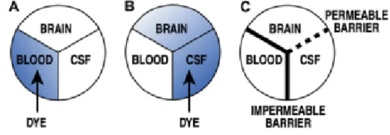
Figure 1 - Schematic representation of the diffusion of a dye according to the injection place
………...26
Figure 2 - History of the blood-brain barrier research and discoveries ...27
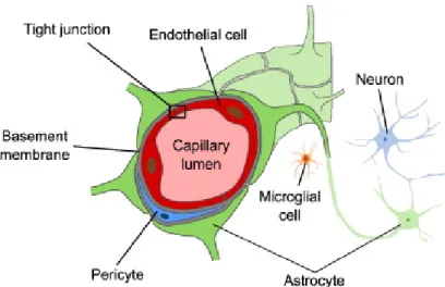
Figure 3 - Representation of the neurovascular unit ...30
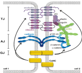
Figure 4 - Representation of the interendothelial junctions network...31
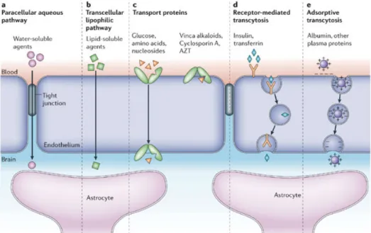
Figure 5 - Scheme of transport mechanisms across the BBB, depending on the compound nature...34
Figure 6 - Overview of sprouting angiogenesis...36
Figure 7 - Angiogenesis regulation...37
Figure 8 - Representation of microscopic hallmarks of AD...41
Figure 9 - Representation of NFTs formation. ...42
Figure 10 - Representation of APP processing. ...44
Figure 11 - Representation of Aβ assembly...45
Figure 12 - Alterations leading to BBB dysfunction in aging and in AD...47
Figure 13 - Representation of brain blood vessels with CAA...48
Figure 14 - Representation of TTR-mediation in Aβ clearance...54
Figure 15 - PCR array analysis of gene expression of several TJs-related genes...70
Figure 16 - qRT-PCR analysis of TJs-related genes using “in-house” primers...71
Figure 17 - Analysis of protein level of tight junctions in hCMEC/D3 cells without or with incubation of TTR for 24h...72
Figure 18 - qRT-PCR analysis of pro-angiogenic genes in hCMEC/D3 cells...74
Figure 19 - Effect of TTR variants on the migration of hCMEC/D3 cells...76
Figure 20 - Effect of WT TTR concentration on the migration of hCMEC/D3 cells...77
Figure 21 - Effect of WT TTR on the angiogenic capacity and on gene expression of pro-angiogenic genes in bEnd.3 cells...78
Figure 22 - Effect of TTR genetic reduction in collagen IV levels in AD mice...80
Figure 1 - Effect of TTR genetic reduction in collagen IV levels in brain vessels...81
Figure 24 - Morphological characterization of Aβ species by TEM analysis...82
Figure 25 - Analysis of collagen IV levels in bEnd.3 cells without or with incubation of Aβ species for 24h...83
Figure 26 - Analysis of collagen IV levels in bEnd.3 cells with incubation of TTR alone, Aβ alone or both together...84
Figure 27 - Effect of IDIF treatment in collagen IV levels of AD/TTR+/- mice brain...85
Abbreviations
ABC ATP-binding cassette tranporters AD Alzheimer’s disease
AICD APP intracellular domain AJ Adherent junctions
AMT Adsorptive-mediated transcytosis ANGPT Angiopoietin
APOA1 Apolipoprotein A1 APOE Apolipoprotein E
APP Amyloid-beta precursor protein Aβ Amyloid-beta peptide
BBB Blood-brain barrier
BCSFB Blood-cerebrospinal fluid barrier
bEnd.3 Immortalized mouse brain endothelial cell line bFGF Basic fibroblast growth factor
BM Basal membrane
BSA Bovine serum albumin CAA Cerebral amyloid angiopathy CLDN Claudin
CNS Central nervous system CSF Cerebrospinal fluid CTF C-terminal fragment DEAE Diethylaminoethyl
DMEM Dulbecco’s modified eagle medium DMSO Dimethyl sulfoxide
DPBS Dulbecco’s phosphate-buffered saline DR Diabetic retinopathy
EC Endothelial cell ECM Extracellular matrix
FAD Autosomal dominant familial AD FAP Familial amyloidotic polyneuropathy FBS Fetal bovine serum
GDNF Glial-derived neurotrophic factor GLUT Glucose transporters
hCMEC/D3 Immortalized human cerebral microvascular endothelial cell line HEPES 4-(2-hydroxyethyl)-1-piperazineethanesulfonic acid
HFIP Hexafluoro-2-propanol
hRECs Human retinal microvascular endothelial cells HUVECs Human umbilical vein endothelial cells IDIF Iododiflunisal
IGF-I Insulin-like growth factor I
IGF-IR Insulin-like growth factor I receptor IPTG Isopropyl β-D-1-thiogalactopyranoside JAM Junctional adhesion molecules
kDA Kilodalton
LB Lubia-Bertani
LRP1 Low-density lipoprotein receptor-related protein 1 LRP2 Low-density lipoprotein receptor-related protein 2 NCBI National Center for Biotechnology Information NFT Neurofibrillary tangles
NVU Neurovascular unit
OCLN Occludin
O/N Overnight
PBS Phosphate-buffered saline PCR Polymerase chain reaction PFA Paraformaldehyde
Pgp P-glycoprotein
PMSF Phenylmethylsulfonyl fluoride PSEN1 Presenilin 1
qRT-PCR Real-time polymerase chain reaction
RAGE Receptor for advanced glycation end products RBP Retinol binding protein
RT Room temperature
sAPP Soluble amyloid-beta precursor protein SD Standard deviation
SEM Standard error of the mean SLC Solute carrier
sLRP Soluble low-density lipoprotein receptor-related protein
SP Senile plaques
TEM Transmission electron microscopy TGF-β Transforming growth factor beta TGFB2 Transforming growth factor beta 2
TJ Tight junctions
Tris Tris(hydroxymethyl)aminomethane TTR Transthyretin
T4 Thyroxine
VE-Cadherin Vascular endothelial cadherin VEGF Vascular endothelial growth factor
VEGFR1 Vascular endothelial growth factor receptor 1 VEGFR2 Vascular endothelial growth factor receptor 2
WT Wild-type
Thesis focus
The main goal of this thesis is to explore the neuroprotective role of a specific protein, called transthyretin (TTR) in one of the most important networks of our brain, the brain vasculature, both in physiology and in pathology. The lack of previous studies on this relation, TTR-brain vasculature, was the initial motivation to start this work, and thus, the next section will provide an overview of relevant topics, introducing key concepts, for a better comprehension of the research project.
1. Blood-Brain Barrier
When we talk about the brain, the first thing that comes to our mind is the complexity of this organ, being active in every second. Although it accounts only for ≈ 2% of the body weight, the brain consumes a large amount of energy, requiring around 20% of an individual’s resting metabolic rate (Attwell and Laughlin 2001). To ensure that enough oxygen, glucose and other nutrients are adequately delivered to the brain, it is necessary a large blood vessel and microvessel network, being estimated that the total length of capillaries in the human brain is about 600 km, and the capillary surface area available for molecular transport is about 20 m2 (Begley and Brightman 2003). But this comes with a problem: despite the
advantage of having this large network, undesirable compounds that could be toxic to the brain would be in higher concentrations, which is a problem since the brain is the most critical and sensitive organ in our
body (Mahringer, Ott et al. 2014). However, our body is meticulously made, existing a special protection in the central nervous system (CNS) microvessels, the so called
blood-Figure 2. Schematic representation of the diffusion of a dye according to the injection place. (A) and (B) Results of Ehrlich and Goldman experiments,
respectively. (C) Assumption of the existence of an impermeable barrier between the CNS and the blood, and a permeable barrier between the brain and the CSF (Zlokovic 2008).
brain barrier (BBB), an exclusive anatomical and physiological barrier separating the CNS and the peripheral circulation. Its existance was firstly hypothezised after Paul Ehrlich has observed that a peripherally injected dye stained peripheral organs but not the brain and the spinal cord (figure 1) (Ehrlich 1885).
Few years later, Goldman performed a staining experiment, similarly to Paul Ehrlich, injecting a dye directly into the cerebrospinal fluid (CSF), after which he observed that the brain and the CSF stained blue, but not the blood (Goldman 1913). These two experiments were essential for the suggestion of the existance of the BBB and, since these discoveries, new information about the BBB has been published for more than 100 years (figure 2).
Figure 3. History of the blood-brain barrier research and discoveries (Mahringer, Ott et
The BBB is responsible for several roles, as controlling cerebral homeostasis, providing protection against toxic xenobiotics and pathogens (Weiss, Miller et al. 2009), mediating the efflux of waste products and restricting ionic and fluid movements between the blood and the brain (Abbott, Ronnback et al. 2006).
A second “barrier” is formed by the epithelial cells of the choroid plexus, which constitute the blood-cerebrospinal fluid barrier (BCSFB). This one will not be explored, since it was not the focus of our work.
1.1. Neurovascular Unit
The blood-brain barrier exists primarily as a barrier constituted by the cerebral microvascular endothelium, a thin layer of simple cells called endothelial cells (ECs), which are significantly different from those in the periphery (Hawkins and Davis 2005). In close proximity to these brain ECs, other cells are indirectly involved in the development and maintenance of the BBB. All these cells together with the basal lamina, constitute the neurovascular unit (NVU). The first concept of NVU was introduced by Harder (Harder, Zhang et al. 2002), describing a relation between neurons, astrocytes and capillaries. These three types of cells, along with pericytes and basal membrane (BM), create the NVU.
1.1.1. Endothelial cells
Brain ECs are the primary element of the BBB, creating the walls of the capillaries, where a cerebral microvessel is enclosed by a single endothelial cell. These brain ECs are significantly different from non-brain ECs by:
Absence of fenestrations (Fenstermacher, Gross et al. 1988);
Presence of intercellular tight junctions (TJs) (Kniesel and Wolburg 2000); Enhanced mitochondrial content, associated with a strong metabolic activity
(Oldendorf, Cornford et al. 1977);
Low level of non-specific transcytosis (pinocytotic) (Sedlakova, Shivers et al. 1999);
Prevented free exchange of solutes between blood and brain (Ohtsuki and Terasaki 2007).
All features together reveal the hallmark of the brain endothelium: a restrict and controlled permeability to plasmatic compounds and ions, protecting the brain from an imbalance in the homeostasis. Endothelial cells are one of the most important components of the NVU, being influenced by the others NVU’s cells/BM, as described next.
1.1.2. Basal membrane
Endothelial cells and pericytes are surrounded by the BM, which is constituted by laminin, collagen type IV, proteoglycans, fibronectin and other extracellular matrix proteins produced by both cell types of the NVU (Farkas and Luiten 2001). The BM contribution to the NVU was often underestimated, but it must be considered as an important part of the BBB regulation (Berzin, Zipser et al. 2000). Besides that, disruption of this extracellular matrix is associated with BBB breakdown and increase of its permeability (Rascher, Fischmann et al. 2002), since collagen IV is involved in the regulation of endothelial tight junction protein expression (Savettieri, Di Liegro et al. 2000). Moreover, collagen IV is capable of regulating angiogenesis, which will be discussed in the subchapter 1.4.
1.1.3. Astrocytes
Astrocytes are one of the glial cells that are present in the neurovascular unit. As can be seen in figure 3, these cells communicate with pericytes, neurons and capillary endothelial cells via their several foot processes (Abbott, Ronnback et al. 2006). It has been well documented the importance of astrocytes in the induction and maintenance of BBB integrity (Janzer and Raff 1987, Kuchler-Bopp, Delaunoy et al. 1999). When capillaries are co-cultured with astrocytes, TJs (one of the most important feature in brain endothelial cells) are enhanced in length, width, and complexity, indicating a role in the formation and
configuration of these TJs (Tao-Cheng, Nagy et al. 1987). Also, astrocytes and endothelial cells are capable to communicate through calcium signals, influencing some aspects of the BBB functioning and transport, such as its permeability (Braet, Paemeleire et al. 2001). Moreover, astrocytes synthesize some biologically active molecules as the transforming growth factor-β (TGF-β), glial-derived neurotrophic factor (GDNF) and basic fibroblast growth factor (bFGF), which may influence endothelial cells (Wilhelm and Krizbai 2014).
1.1.4. Pericytes
Pericytes, also called Rouget cells after their discovery by Charles Rouget, are randomly distributed along the brain and non-brain microvessels, being surrounded (together with ECs) by the basal membrane composed of collagen type IV and other extracellular matrix proteins mentioned above (Bergers and Song 2005). These cells produce substances such as TGF-β, angiopoetin-1 and vascular endothelial growth factor (VEGF), influencing the endothelial function (Wilhelm and Krizbai 2014). Pericytes are important in the formation and maintenance of the BBB since it was shown that pericytes deficiency leads to an increase in the permeability of the BBB (Armulik, Genove et al. 2010), endothelial hyperplasia and abnormal vascular morphogenesis (Hellstrom, Gerhardt et al. 2001). Moreover, the integrity of the wall of the capillaries is compromised by the loss of pericytes (Lindahl, Johansson et al. 1997). It was also shown that pericytes are modulators of blood flow (Peppiatt, Howarth
Figure 4. Representation of the neurovascular unit. The BBB is composed
by microvessel endothelial cells which are surrounded by pericytes, astrocytes end-feet, neurons and the basal lamina, all essential for the development and maintenance of the BBB (Heye, Culling et al. 2014).
et al. 2006) and pericytes-derived angiopoietin-1 induces TJs expression, specifically occludin, confirming that pericytes influence positively the maintenance of BBB (Hori, Ohtsuki et al. 2004).
1.2. Junctional complexes at the BBB
As mentioned above, one of the main roles of the BBB is to act as physical barrier, separating the brain from the blood, with a restricted paracellular diffusion and a low permeability across the endothelium. This feature is accomplished by a network of interendothelial junctions as TJs, adherens junctions (AJs) and gap junctions (when present) – figure 4 (Bazzoni and Dejana 2004).
1.2.1. Tight junctions, adherens junctions and associated proteins
TJs in the brain ECs are very similar to epithelial TJs. In this group we can find a large amount of proteins involved, but it is possible to highlight three major transmembrane proteins (or families): occludin (OCLN), claudins (CLDNs) and junction associated molecules (JAMs). Linked to these proteins we can find different cytoplasmic proteins asFigure 5. Representation of the interendothelial junctions network. Adjacent endothelial cells are connected by different
zonula occludens (ZO) family, vinculin and cingulin which are in contact with transmembrane proteins and to the actin cytoskeleton, creating a multi-protein complex (Wolburg and Lippoldt 2002).
The first transmembrane protein identified, firstly in chickens (Furuse, Hirase et al. 1993) and then in mammals (Ando-Akatsuka, Saitou et al. 1996), was OCLN, a 65 kDa protein with four transmembrane domains, whose expression is developmentally regulated. OCLN is highly expressed in the brain endothelium but less (or even absent) in non-neural endothelial cells (Daneman, Zhou et al. 2010). In OCLN deficient mice, TJs are morphologically indistinguishable from the wild-type (wt) controls, indicating that OCLN is not so important for TJs formation (Saitou, Furuse et al. 2000). But, other studies have shown the opposite (Balda, Whitney et al. 1996, Wong and Gumbiner 1997). Still associated with this role, OCLN seems to be an important protein that can alter paracellular permeability (Hirase, Staddon et al. 1997). This protein is mostly associated with the TJ formation but its functions are not limited to this role. As example, OCLN can be involved in epithelial differentiation (Schulzke, Gitter et al. 2005).
With molecular weighs between 20-34 kDa, the CLDN family is constituted by more than 22 members that can be found in different tissues, but only a few are expressed at the BBB (Mahringer, Ott et al. 2014). These are important for the formation and maintenance of the BBB, and their breakdown lead to the disruption of the BBB. In CLDN-5 deficient mice (CLDN5-/- mice), molecules with <800 Da were capable to cross the BBB, even if the
development and morphology of blood vessels were not compromised (Nitta, Hata et al. 2003). Moreover, in the same study, CLDN5 downregulation was shown to be related with BBB breakdown via VEGF, although reversible when recombinant CLDN5 was expressed, leading to the rescue of low paracellular permeability and thus, decreasing the BBB breakdown (Argaw, Gurfein et al. 2009).
The other relevant TJs involved in the BBB integrity are the JAM family. There are three known JAMs: JAM-A, JAM-B and JAM-C (or JAM-1, JAM-2 and JAM-3, respectively). These proteins are present in endothelial and epithelial cells, leukocytes, platelets and erythrocytes (Mandell and Parkos 2005). JAM proteins are involved in different
processes, including the regulation of tight junction assembling (Liu, Nusrat et al. 2000) and angiogenesis signaling (Naik, Mousa et al. 2003).
As a part of the junctional complex, it is possible to find other proteins at cell-cell junctions, forming adhesive contacts between cells, the so called adherens junctions (also known as zonula adherens). The AJs complex are mainly composed by the interaction of the cadherin protein family, such E-cadherin and vascular endothelial (VE)-cadherin, and the catenin family including β-catenin, α-catenin and p-120-catenin, which interact with actin (Hartsock and Nelson 2008). Similar to TJs, these proteins are important for the BBB integrity and to reduce its permeability (Corada, Mariotti et al. 1999, Navaratna, McGuire et al. 2007). VE-cadherin, besides its role as a tight connection between cells, is also associated with processes like angiogenesis (Wallez, Vilgrain et al. 2006) and cell proliferation (Caveda, Martin-Padura et al. 1996).
Equally important, transmembrane proteins are associated with cytosolic proteins, some already mentioned above: ZO family, cingulin, vinculin. ZO-1, probably one of the most studied proteins, was shown to be involved in cell-cell tension, angiogenesis, barrier formation (Tornavaca, Chia et al. 2015), and its dissociation is followed by an increase of permeability (Abbruscato, Lopez et al. 2002). Actin is also an important component of this junctional complex. It permits the anchorage of transmembrane proteins (using intermediated proteins) to the actin cytoskeleton, essential for the barrier stability/permeability and cell movement (Lai and Kuo 2005, Dejana, Tournier-Lasserve et al. 2009).
Altogether, we can say that a complex network of proteins is vital for the formation and maintenance of BBB, with innumerous functions, and the failure of one part might be enough to compromise this important barrier.
1.3. Transport at BBB
The transport across the BBB is very controlled, since the maintenance of the homeostasis is crucial for the brain, which is achieved through numerous mechanisms, depending on the nature of the substance crossing the barrier (figure 5). These include
transmembrane diffusion, saturable transporters, receptor-mediated transcytosis and adsortive transcytosis (Banks 2009).
Due to its tight barrier properties, only a few small polar compounds are able to cross the BBB via diffusion across TJs (paracellular transport), including urea and water, since almost no space exists between adjacent cells.
The main type of transport is the transmembrane diffusion (or passive diffusion) (Oldendorf 1974), where molecules can enter inside endothelial cells via a non-saturable mechanism, simply depending on the capacity of the compound to melt into the cell membrane. Small and lipophilic molecules can easily cross the cell membrane, but other factors as charge and tertiary structure also affect this type of transport (Banks 2009). Despite the ability of these molecules to cross the luminal cell membrane, not all will reach the brain. In fact, to regulate passive transport into the brain, endothelial cells have efflux pumps that are responsible to return many unwanted molecules back to the blood (Wong, Ye et al. 2013). This return is made by the ATP-binding cassette tranporters (ABC). At the BBB, the most important ABC transporters for efflux transport are the P-glycoprotein (Pgp), the multidrug resistance-associated proteins and breast cancer resistance protein (Abbott, Patabendige et al. 2010) which remove potentially neurotoxic endogenous or xenobiotic molecules from the
Figure 6. Scheme of transport mechanisms across the BBB, depending on the compound nature
brain, protecting and detoxifying this organ (Dallas, Miller et al. 2006). Oxygen and carbon dioxide are examples of molecules using passive diffusion as the way of crossing the BBB.
Another way for compounds to reach the brain is via carrier-mediated transport. Essential compounds, such as glucose, aminoacids and nucleosides, can enter in the brain using specific transporters. The most common transporter belong to the solute carrier proteins (SLC) family. SLC2A1, more known as glucose transporter-1 (GLUT1), is responsible for the facilitation of glucose transport, following its concentration gradient (Hediger, Romero et al. 2004). Other transporters from the SLC family are involved, such as the monocarboxylate transporters, which transport short-chain monocarboxylic acids (as lactate and pyruvate), and the SLC7 which transport cationic aminoacids (arginine, lysin and ornithine) (Mahringer, Ott et al. 2014). These transporters are crucial for the brain activity since it requires a lot of energy to work, thus, it also needs these metabolic substrates (specially glucose). Ions transporters, such as sodium pump and sodium-hydrogen exchanger, are other type of transporters that are essential for the maintenance and regulation of intracellular pH in the endothelium (Taylor, Nicola et al. 2006).
Proteins and peptides are solutes with a large size and mass and cannot cross the endothelium by passive transport. These molecules reach the brain via vesicular transport across BBB – transcytosis. Two ways of transcytosis can occur: either by specific receptor-mediated transcytosis or nonspecific adsorptive-receptor-mediated transcytosis (AMT). The first one is made by specific interaction between the molecules and receptors localized on ECs membrane. Proteins as insulin, leptin and transferrin are examples of proteins transported using this way (Pardridge 2003, Herve, Ghinea et al. 2008). In the AMT, positively charged peptides in the blood interact with the negatively charged phospholipid head groups of the cell membrane, inducing membrane invagination and vesicle formation. After entering the intracellular space, vesicles fuse with the abluminal side of endothelium and are released to the brain. Briefly, it consists in an elctrostatic interaction between molecules and cell membrane (Jones and Polt 2015).
1.4. Development of new vessels
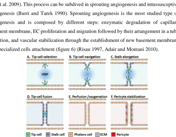
The development of blood vessels can be made via two different processes: angiogenesis and vasculogenesis. In the last one, blood vessels have as origin the differentiation of hemangiogenic stem cells and angioblasts from pluripotent mesenchymal cells (Demir, Kayisli et al. 2006). Angiogenesis, the formation of vessel branches from pre-existing vessels, is the predominantly process of blood vessel formation in the brain (Lee, Han et al. 2009). This process can be subdived in sprouting angiogenesis and intussusceptive angiogenesis (Burri and Tarek 1990). Sprouting angiogenesis is the most studied type of angiogenesis and is composed by different steps: enzymatic degradation of capillary basement membrane, EC proliferation and migration followed by their arrangement in a tube formation, and vascular stabilization through the establishment of new basement membrane and specialized cells attachment (figure 6) (Risau 1997, Adair and Montani 2010).
In more detail, the beginning of blood vessels formation starts with the degradation of the BM by enzymes secreted by activated ECs, leading to the creation of little holes in the BM. At this point, these ECs begin to proliferate at a higher rate, and they migrate out through the holes in the BM (Conway, Collen et al. 2001). During that, the tissue in front and around the sprouting vessel is remolded by metalloproteinases, allowing the vessel to extend and to form a tubular structure. To finalize, if the new blood vessel contacts with another existing vessel, it will be stabilized by surrounding of a new BM and the presence of specialized cells,
such as smooth muscle cells and pericytes. Both together provide structural support for the vessel functioning (Zakrzewicz, Secomb et al. 2002, Pandya, Dhalla et al. 2006).
Angiogenesis is highly regulated by the presence of anti-angiogenic and pro-angiogenic factors, allowing to maintain an important balance (figure 7). But, in response to
several signals, this balance can be altered, both in physiological and pathological conditions. Physiologically, alterations in the microvessel network are necessary in different processes, including wound healing, exercise and tissue growth (Pries and Secomb 2014). In pathological conditions, angiogenesis can be found up-regulated or down-regulated, depending on the proportion of anti- and pro-angiogenic factors. Up-regulation of angiogenesis can be found in several diseases such as cancer, atherosclerosis and diabetic retinopathy (Folkman 1995), while down-regulation is an important causal factor for coronary artery disease, cardiac failure and tissue failure (Pandya, Dhalla et al. 2006).
Hypoxia, the deprivation of adequate oxygen supply, is an important promoter of angiogenesis. This deficiency of oxygen leads to an increase of pro-angiogenic factors (Hirota and Semenza 2006). bFGF, VEGF and its receptors, angiopoietins (ANGPTs) and transforming growth factor beta 2 (TGFB2) are examples of these pro-angiogenic factors
Figure 8. Angiogenesis regulation. Balance between
activation and inhibition of angiogenesis is highly regulated by several stimulatory and inhibitory factors in response of several physiological and pathological conditions (Zetter 2008)
(Shibuya 2011, Dulloo, Phang et al. 2015). It is important to refer that both type of angiogenic factors are able to affect one or more angiogenic steps, but mainly acting on EC proliferation/migration and on the BM and extracellular matrix (ECM).
1.4.1. Extracellular Matrix functions during Angiogenesis
The ECM is a key component for each step of angiogenesis, providing structural support and also molecular signals that are essential for blood vessel formation. To start angiogenesis, it is required EC activation and proliferation, which is achieved due the action of angiogenic cytokines and EC adhesion to ECM through integrins, since for an efficient cytokine activation, is necessary the presence of integrins (Giancotti and Ruoslahti 1999). Thus, without the presence of ECM, EC activation and proliferation ceases and angiogenesis doesn’t occur. Moreover, also EC migration requires adhesion to ECM (Ausprunk and Folkman 1977). Relatively to new blood vessels morphogenesis, ECM are capable to regulate EC shape and morphogenesis. Studies showed that collagen I is capable to alter ECs morphology and to make them align into cords similarly to those observed during angiogenesis in vivo (Whelan and Senger 2003), and also capable of induce and support lumen formation by ECs (Senger and Davis 2011).
1.4.1.1 Collagen IV as a player in angiogenesis
As seen above, the ECM and the BM are important for angiogenesis process. Collagen IV, the predominant protein in the BM, can have different functions in this process. Studies have shown that synthesis and deposition of Collagen IV is indispensable for vascular survival and maturation. Bonanno and colleagues studied the angiogenic response of native ECs in three-dimensional vascular organ culture, culturing rings of rat aorta in the absence or presence of collagen IV, and reported that collagen IV increased the vascular elongation, survival and stabilization, in a dose-dependent manner (Bonanno, Iurlaro et al. 2000). In another study, inhibition of collagen production led to a marked anti-angiogenic effect (Nicosia, Belser et al. 1991). In Bahramsoltani and colleagues work, they showed the expression of collagen IV in four different microvascular endothelial cell types, two of them classified as angiogenic and the other two as non-angiogenic. Curiously, secretion and
deposition of collagen IV was only observed in the two angiogenic cultures, suggesting that angiogenesis is dependent on collagen IV deposition (Bahramsoltani, Slosarek et al. 2014). However, during the ECM remodeling, collagen IV is degraded, generating small anti-angiogenesis molecules from its α chains, such as arresten, canstatin and tumstatin (Mundel and Kalluri 2007). These molecules are endogenous angiogenesis inhibitors which are involved in the balance between pro- and anti-angiogenic factors (Mundel and Kalluri 2007).
2. Alzheimer’s Disease
Alzheimer’s disease (AD), name given in tribute to the first person that describe it, is a chronic neurodegenerative disease, comprising the major cases of dementia worldwide, that usually affects the older people (age >65 years). The greatest risk factor for AD is age and is characterized by a progressive loss of cognitive functions (such as memory and language) (Burns and Iliffe 2009). The World Alzheimer Report 2016 estimated that there were 46.8 million people worldwide with dementia and this number can reach to 131.5 million in 2050 (International, 2016). Despite of being intensively studied, the etiological mechanisms underlying the neuropathological changes remain unclear, but being suggested that is probably caused by a set of genetic and environmental factors (Reitz and Mayeux 2014).
2.1. Genetics behind Alzheimer’s disease
The majority of patients only develop clinical symptoms at age older than 65 years – called late-onset AD or sporadic AD – , but for 2 to 10% of patients the symptoms appear earlier – early-onset AD or autosomal dominant familial AD (FAD) (Van Cauwenberghe, Van Broeckhoven et al. 2016). During a long time, genetic factors contributing to the disease were searched, being discovered several mutations that are capable of triggering the disease. It was found that three important genes are involved as a cause of autosomal dominant familial AD when mutated (originating early-onset AD cases), namely amyloid precursor protein (APP), Presenilin 1 (PSEN1) and Presenilin 2 (PSEN2) (Bertram, Lill et al. 2010), all three genes encoding proteins linked to APP. PSEN1 and PSEN2, members of the same family, are essential components of the γ-secretase complex responsible for cleavage and release of amyloid-β peptide (Aβ). Thus, mutations in these genes interfere with APP
processing and Aβ production (described below). Opposite to FAD, sporadic AD does not show autosomal-dominant inheritance. Instead, multiple genetic and environment risk factors may be involved. One of the genes associated with sporadic AD is the ε4 allele of the apolipoprotein E (APOE) gene, localized on chromossome 19q13. APOE gene can have three different polymorphic alleles - ε2, ε3 and ε4 – which have a worldwide frequency of 8.4%, 77.9% and 13,7% respectively (Liu, Liu et al. 2013), but in AD patients, the frequency of the ε4 allele is about 40% (Bu 2009). Thus, patients who carry APOE ε4 allele are at higher risk to develop AD when compared with noncarriers, but this allele per si is not a determinant of the disease (Schmidt, Carlo et al. 2014). Interestingly, on the other hand, it was shown that ε2 allele could have a protective effect in late-onset AD (Corder, Saunders et al. 1994).
2.2. Pathophysiology of Alzheimer’s disease
2.2.1. Neuropathology
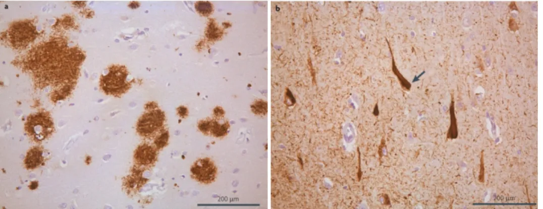
In the description of the first case of AD, some typical features were found, both macroscopic and microscopic features. Macroscopically, in the brains obtained from patients with AD, it is possible to identify a clear cerebral cortical atrophy, specially in the primary motor, sensory and visual areas. Also, a symmetrical dilation of the lateral ventricles (hydrocephalus ex vacuo) is observed, caused by a encephalic volume loss (Perl 2010). Although these changes are associated with AD, they may also be present in the brains of elder persons without AD, the difference being the extent and distribution of the changes, which are increased in AD patients (Terry 1986). Microscopically, AD is characterized by the presence of extracellular aggregated Aβ peptide constituting the senile plaques (SP), and intracellular aggregates of hyperphosphorylated tau protein creating the neurofibrillary tangles (NFTs) (figure 8) (Tiraboschi, Hansen et al. 2004, Kolarova, Garcia-Sierra et al. 2012).
2.2.1.1. Neurofibrillary tangles
When Alois Alzheimer described the first case of AD, he noted the presence of some abnormal fibrils inclusions inside cells, now known to be the neurofibrillary tangles. The NFTs are composed of abnormal fibrils with around 10nm in diameter that occur in pairs and are twisted in a helical fashion with a regular periodicity of 80nm (Perl 2010). These NFTs are mainly constituted by the microtubule-associated protein tau which presents an abnormal hyperphosphorylation (Lee, Balin et al. 1991), but it is possible to find other proteins, such as ubiquitin (Perry, Friedman et al. 1987) and cholinesterases (Mesulam and Asuncion Moran 1987). Relatively to NFTs distribution, it is possible to find them in the layer II neurons of the entorhinal cortex, the CA1 and subicular regions of the hippocampus, the amygdala, and the deeper layers (layers III, V, and superficial VI) of the neocortex (Morrison and Hof 1997). Tau is a cytosolic protein whose physiologic function is the promotion of the assembly of tubulin into microtubules and its stabilization (Weingarten, Lockwood et al. 1975). Moreover, tau is involved in more than 20 clinicopathological entities, besides AD (Williams 2006). The process which leads to hyperphosphorylation of tau is unclear, but studies suggest to be a consequence of an upregulation of tau kinase or a downregulation of tau phosphatase. Furthermore, these abnormal tau phosphorylations modulate tau aggregation by disrupting its binding to microtubules and leading to its aggregation, resulting in the increase of soluble tau. Next, soluble tau dimerize and forms oligomers. In the end, oligomers originate protomers which will form paired helical filaments and NFTs (figure 9) (Martin, Latypova et al. 2011).
Figure 9. Representation of microscopic hallmarks of AD. (a) Senile plaques and (b) neurofibrillary
2.2.1.2. Senile Plaques
Senile plaques, also called neuritic plaques, are extracellular amyloid deposits found in the brain of AD patients, but it is also possible to find in normal aging, being mostly constituted by Aβ (Cras, Kawai et al. 1991). These plaques present a variable morphology and size, and are located specifically in the hippocampus and the cortex (Costa, Ferreira-da-Silva et al. 2008). Also, it is possible to divide amyloid plaques in different subtypes
Figure 10. Representation of NFTs formation
depending on the morphology, including diffuse SP (pre-amyloid), primitive SP (neuritic), classic SP (dense-core) and compact SP (burnt-out) (Armstrong 2009). The last subtype is characterized by an increase of neurite curvature and dytrophic neurites (axons and dendrites), synaptic loss, neuron loss, and recruitment and activation of both astrocytes and microglial cells (Serrano-Pozo, Frosch et al. 2011).
2.3. Biochemistry of Aβ
As referred above, Aβ is the major component of senile plaques in AD. With approximately 4 kDa, Aβ is a small peptide originated by proteolytic processing of APP, a type I transmembrane protein essential for normal brain (Shariati and De Strooper 2013), where the region that corresponds to the Aβ includes the exon 16 and 17 of APP. Aβ was first isolated and sequenced by Glenner and Wong (Glenner and Wong 1984), and is found in the plasma, brain and CSF. Despite being always associated with its negative effect in AD, Aβ has been linked with several physiological functions such as ion channel modulation (Plant, Webster et al. 2006), regulation of cholesterol transport (Yao and Papadopoulos 2002) and more recently suggested as an antimicrobial peptide (Kumar, Choi et al. 2016).
APP is a protein abundantly expressed in the brain but is possible to find it in other tissues. The gene codifying for APP is located in the chromosome 21 in humans, and contains 18 exons (Zheng and Koo 2011). Interestingly, since APP gene duplication alone is capable to cause early-onset AD with cerebral amyloid angiopathy, it explains the increased risk for Down’s syndrome (trisomy 21) patients to develop AD (Thinakaran and Koo 2008). APP processing can occur in two different ways: the non-amyloidogenic pathway and the amyloidogenic pathway, being Aβ produced by the later (figure 10).
2.3.1. APP processing: non-amyloidogenic and amyloidogenic
pathways
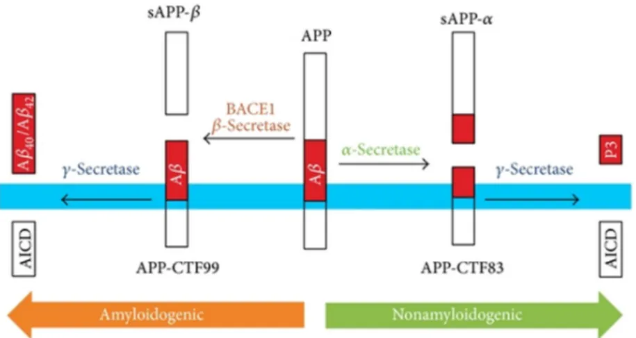
Depending on the proteolysis pathway, APP can originate different peptides which have different functions. In the non-amyloidogenic pathway, two enzymes are responsible for the cleavage of APP. Firstly, α-secretase, an enzyme of the ADAM family (Rossner 2004), cuts the peptide bond between the Lys687-Leu688 of APP770, within the residues 16 and 17 of Aβ sequence, abolishing its production (Esch, Keim et al. 1990). This cleavage originates a large soluble fragment (sAPPα, ~100kDa) and a carboxyl-terminal fragment (CTF83, ~10kDa). The following cleavage is made by γ-secretase which cuts the CTF83, generating the p3 peptide and an APP intracellular domain (AICD, ~6kDA) fragment (Multhaup, Huber et al. 2015). Importantly, all peptides resulting from both cleavages are non-amyloidogenic. Also relevant, γ-secretase is a complex of four proteins, where the PSEN1 and PSEN2 are involved. Mutations in the genes coding these two proteins lead to an increase of Aβ production (enhances Aβ42 production compared to Aβ40), being one of the explications for the importance of these mutations being a cause of early-onset AD.
In a very similar process, in the amyloidogenic pathway, APP is also cleaved by two enzymes. Here, the difference is in the enzyme responsible for the first cut, the β-secretase instead of the α-secretase, generating the soluble ectodomain sAPPβ and the CTF99 fragment after the cleavage (Zhang, Thompson et al. 2011). The CTF99 fragment is then cleaved by
Figure 11. Representation of APP processing. The transmembrane APP can be processed
by two different pathways: in the left it is represented the amyloidogenic pathway and in the right the non-amyloidogenic pathway (Pajak, Kania et al. 2016).
the γ-secretase, releasing the AICD fragment and the Aβ peptide (Multhaup, Huber et al. 2015). This Aβ peptide can have different sizes and, consequently, it has different propensity to oligomerize and form the amyloid plaques (Haass and Selkoe 2007). But, it needs to be clear that APP processing occurs in the normal cellular metabolism through life, being possibly to find Aβ and NFTs in the brain of non-AD elderly people (Terry 1986). But then, how Aβ is related as a cause of AD?
2.3.2. Amyloid cascade hypothesis
The etiology of AD is still unclear, and different theories try to explain this disorder, all based in different molecular mechanisms to back them up, but not absolutely consistents.
Among these theories, the “amyloid cascade hypothesis” is the most known and stronger. It was formalized by Hardy and Higgins (Hardy and Higgins 1992). The beginning of this cascade is when changes in Aβ metabolism occur, such as increase in total Aβ production, increase in the Aβ42 specie compared to Aβ40, since the first is more toxic, or reduction in Aβ degradation/clearance (Selkoe 1991). Then, Aβ assembly occurs (figure 11), starting with its oligomerization and formation of non-fibrillar deposits. These oligomers are
already capable to do severe changes in synaptic function, leading to neuronal dysfunction. In fact, soluble Aβ oligomers represent the most toxic form of this protein (Kumar and Walter 2011). Over time, the non-fibrillar deposits evolve to Aβ fibrils, and several negative events occur: local inflammatory responses (as microgliosis and astrocytosis), synaptic spine loss, neuritic dystrophy, oxidative stress and altered ionic homeostasis. Finally, oligomerization and hyperphosphorylation of tau is observed. All changes together lead to a widespread neuronal dysfunction, cell dead and culminates in dementia with senile plaques and NFTs
(Haass and Selkoe 2007). Nevertheless, this hypothesis is not accepted by everyone, since the accumulation of new studies show data contradicting this theory. For example, it was shown a poor correlation between the degree of deposition and the degree of dementia in AD patients. It was also observed that some mouse models of AD show behavioral deficits prior to amyloid deposition (Pimplikar 2009).
All together, it is clear that the pathogenesis of AD is a complex process and several factors are involved, and it can not be explained by a single theory.
2.3.3. Aβ clearance
It is known that in the brains of AD patients, Aβ is highly present, but how it happens if the majority of patients with sporadic AD do not present an increase of Aβ production? The unbalance between Aβ production and clearance is determinant for Aβ accumulation. Since dysfunction in its clearance is crucial in this process, it is also important to understand the mechanisms behind it (Wang, Zhou et al. 2006).
The clearance of Aβ from the brain can occur by different pathways: receptor-mediated transport across the BBB, enzyme-receptor-mediated degradation and anti-Aβ autoantibodies.
The transport of Aβ across the BBB is possible to happen in both direction: brain-to-blood and brain-to-blood-to-brain. From the brain to the brain-to-blood, it can be removed via interstitial fluid bulk flow into the bloodstream (Crossgrove, Li et al. 2005) or be transported across the BBB via receptor mediated, the principal mechanism of Aβ transport across this barrier. Moreover, removal of Aβ via interstitial fluid occurs very slowly and it is responsible for the clearance of only 10-15% of the total brain Aβ (Wang, Zhou et al. 2006).
During receptor-mediate brain efflux across the BBB, Aβ removal starts with its binding to the Low-density lipoprotein receptor-related protein 1 (LRP1) at the abluminal side of the endothelial membrane, crossing the BBB by endocytosis or transcytosis, and reaching the blood. There, the free Aβ circulating in the blood is sequestered by soluble LRP (sLRP) and guided to its degradation organs, the liver and the kidney (systemic clearance) (Bell and Zlokovic 2009). In addition to LRP1, other transporters are involved in efflux of Aβ, such as LRP2 (also called megalin) and P-gp (Jeynes and Provias 2013).
As said before, Aβ can also enter in the brain from the periphery via receptor-mediated transport. Here, the receptor for advanced glycation end products (RAGE) is the major “helper”, being located on the luminal membrane of the endothelium.
In conclusion, the balance between influx and efflux of Aβ, mediated by LRP1/2 and P-gp or RAGE, is important to maintain a reasonable Aβ level in the brain. Alterations on the levels of these receptors is enough to lead to an impaired clearance of Aβ and consequently, to all the negative effects expected in the amyloid cascade hypothesis.
2.4. BBB dysfunction and AD
In several diseases, such as AD, BBB dysfunction has been described as an important part in both early and late steps of disease progression (Weiss, Miller et al. 2009). In fact, important features of the BBB described here are negatively altered, contributing for the exacerbation of the AD (figure 12).
First, numerous studies demonstrated that Aβ is capable to decrease the levels of tight junction proteins and, consequently, increase the permeability of BBB (Tai, Holloway et al. 2010, Kook, Hong et al. 2012).
Several alterations are also observed in the Aβ influx and efflux, mostly due to alterations in the levels of the transporters. LRP1, the major mediator of Aβ efflux, was
Figure 13. Alterations leading to BBB dysfunction in aging and in AD (Marques, Sousa et al. 2013).
demonstrated to be decreased in AD (Shibata, Yamada et al. 2000). Moreover, it was shown that P-gp, the other protein involved in Aβ efflux is lower in AD (Vogelgesang, Cascorbi et al. 2002, Wijesuriya, Bullock et al. 2010). On the other hand, several studies showed that RAGE is found to be upregulated in AD (Srikanth, Maczurek et al. 2011). All together, these alterations contribute to decreased Aβ efflux and increased influx, and consequently to disease progression.
Other alterations can be found, such as accumulation of extracellular matrix components and stiffening of the vessel wall, increased pinocytotic vesicles and decrease of endothelial mitochondrial density (Marques, Sousa et al. 2013).
2.4.1. Cerebral amyloid angiopathy – Aβ type
Cerebral amyloid angiopathy (CAA) is a disorder caused by the accumulation of amyloid in the walls of the brain’s blood vessels, being classified according to the amyloid protein involved (Yamada 2015). The most common form of CAA is the accumulation of Aβ (figure 13), and it is commonly found in brains of AD patients and also in elder brains without AD (Vinters 1987), however CAA can be an independent pathogenic factor contributing to
dementia. Similar to AD, the prevalence of CAA increases with age, and mutations in the APP gene are associated with this disease. Some studies suggested that Aβ found in vessels is derived from the brain cells, being transported through periarterial interstitial fluid drainage pathways until reach the blood vessels for its clearance (Weller, Massey et al. 1998). Since the mechanism of clearance is compromised, Aβ starts to progressively accumulate.
Figure 14. Representation of brain’s blood vessels in CAA. Deposits of amyloid are observed (a) using histological
Aβ accumulation can also result from an increase of the BM in vessels: the thickness of the BM can lead to a formation of a barrier, preventing Aβ to reach the blood. Thus, Aβ slowly accumulates, creating all the negative effects already described. Moreover, it is known that Aβ has affinity to proteoglycans present in the BM (Snow, Kinsella et al. 1995, Weller, Massey et al. 2000). The presence of these proteoglycans in the BM might sequester Aβ from the interstitial fluid, leading also to accumulation of Aβ (Tian, Shi et al. 2006). Alterations in the BM are present in AD and CAA and, in fact, several studies evaluated the levels of collagen IV in AD brains. In most of the studies performed with mice, it was observed an increase in collagen IV in AD brain vessels compared to control littermates (Bourasset, Ouellet et al. 2009, Mehta, Short et al. 2013). Moreover, similar results were obtained when comparing human brains of AD patients with controls (Kalaria and Pax 1995, Lepelletier, Mann et al. 2017). Interstingly, aging is also associated with increased collagen IV content in human microvessels (Uspenskaia, Liebetrau et al. 2004). This last result is specially important, since CAA can occur in elder brains without AD.
3. Transthyretin
3.1. TTR – Structure and functions
TTR, previously called prealbumin due to the ability to migrate slightly faster than albumin on an electrophoresis of a plasma sample, is a protein mainly synthesized in the liver and in the choroid plexus, being secreted into the bloodstream and the CSF, respectively (Soprano, Herbert et al. 1985). The TTR gene is localized in the chromosome 18 (Wallace, Naylor et al. 1985) and codifies for the TTR-monomer originating a polypeptide with 147 aminoacids. TTR is a homotetrameric protein, having four identical subunits assembled, originating a protein with ~55 kDa (Kanda, Goodman et al. 1974). It is possible to find TTR (or a homologous protein) in many species, including mammals, birds, reptiles and amphibians (Schreiber and Richardson 1997, Power, Elias et al. 2000).
The main known physiological role of TTR is the transport of thyroid hormone thyroxine (T4) (Palha 2002) as well as of retinol (vitamin A) that is bound to retinol binding protein (RBP) (Butler, Chan et al. 2016). TTR in blood transports about 15% of plasma T4